Which of the following is NOT a cause of acute pancreatitis?
Pancreatic pseudocyst most commonly occurs after which condition?
What is the most common genetic mutation found in pancreatic cancer?
What is the commonest cyst to arise in the pancreas after an attack of acute pancreatitis or pancreatic trauma?
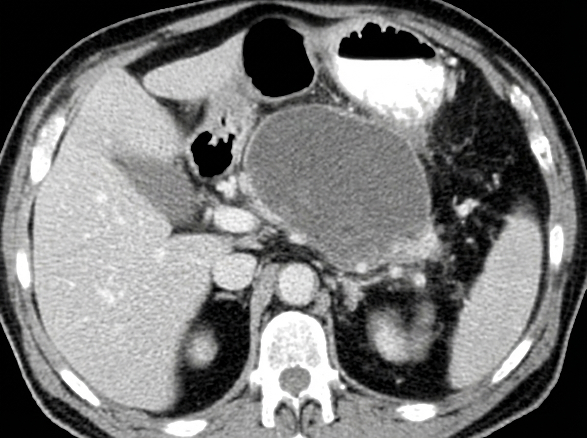
Insulinoma is most commonly located in which part of the pancreas?
True about Insulinoma is:
What is the treatment of choice for carcinoma of the head of the pancreas?
What is the most common cause of chronic pancreatitis?
What is the treatment of choice for annular pancreas?
Which branch of the portal vein is involved in chronic pancreatitis?
Explanation: **Explanation:** The correct answer is **D. Magnetic resonance cholangiopancreatography (MRCP)**. MRCP is a non-invasive diagnostic imaging technique that uses MRI to visualize the biliary and pancreatic ducts. Unlike **ERCP (Endoscopic Retrograde Cholangiopancreatography)**, which involves cannulation of the ampulla and injection of contrast under pressure—a known trigger for post-procedure pancreatitis—MRCP does not involve physical instrumentation of the ducts and therefore **cannot** cause acute pancreatitis. **Analysis of Incorrect Options:** * **A. Hypercalcemia:** High calcium levels lead to the activation of trypsinogen to trypsin within the pancreatic parenchyma and can cause the formation of calcium stones in the ducts, leading to inflammation. * **B. Thrombotic Thrombocytopenic Purpura (TTP):** TTP causes microvascular thrombosis. Ischemia resulting from these microthrombi in the pancreatic vasculature can trigger acute pancreatitis. * **C. Cystic Fibrosis:** This condition leads to thick, inspissated secretions that obstruct the pancreatic ducts, causing premature enzyme activation and chronic/recurrent acute pancreatitis. **Clinical Pearls for NEET-PG:** * **Most Common Cause:** In India and globally, Gallstones (1st) and Alcohol (2nd) are the leading causes. * **Iatrogenic Cause:** ERCP is a high-yield cause of acute pancreatitis (occurring in ~3-5% of cases), whereas MRCP is safe. * **Drug-induced:** Common culprits include Azathioprine, Sulfonamides, Valproate, and Thiazides. * **Scoring:** Remember the **Modified Glasgow** or **Ranson’s Criteria** for predicting severity. * **Investigation of Choice:** **CECT abdomen** (performed after 72 hours) is the gold standard for assessing pancreatic necrosis.
Explanation: **Explanation:** A **pancreatic pseudocyst** is a localized collection of fluid, pancreatic enzymes, and debris, walled off by a rim of fibrous or granulation tissue. Unlike true cysts, it lacks an epithelial lining. **1. Why Pancreatitis is the Correct Answer:** Pancreatitis is the most common cause of pseudocysts, accounting for approximately **70-80% of cases**. It occurs as a complication of both acute and chronic pancreatitis. Following an episode of acute pancreatitis, peripancreatic fluid collections that persist for more than **4 weeks** develop a mature wall of granulation tissue, officially becoming a pseudocyst. In chronic pancreatitis, they often form due to ductal obstruction or leakage. **2. Analysis of Incorrect Options:** * **Trauma (Option A):** While blunt abdominal trauma (e.g., handlebar injuries in children) can cause ductal disruption leading to pseudocysts, it is a much less frequent cause than inflammatory pancreatitis. * **Pancreatic Surgery (Option C):** Post-operative collections are usually classified as pancreatic fistulas or localized abscesses. While a pseudocyst can technically form, it is a rare complication compared to the inflammatory etiology. * **Pancreatic Malignancy (Option D):** Malignancy typically presents with solid masses or cystic neoplasms (like Mucinous Cystadenoma). While a tumor can obstruct a duct and cause secondary pancreatitis, the primary cause remains the inflammatory process. **High-Yield Clinical Pearls for NEET-PG:** * **Location:** Most commonly found in the **lesser sac**. * **Diagnosis:** **CECT** is the gold standard for diagnosis and assessing wall maturity. * **Management:** Most (up to 50%) resolve spontaneously. Intervention (Endoscopic cystogastrostomy or surgical drainage) is indicated only if the cyst is **symptomatic**, infected, or causing complications (e.g., gastric outlet obstruction). * **Rule of 6:** Historically, cysts >6 cm and persisting >6 weeks were considered for surgery, though modern management is now primarily **symptom-driven**.
Explanation: **Explanation:** Pancreatic ductal adenocarcinoma (PDAC) is characterized by a specific sequence of genetic alterations. The **KRAS** gene, a proto-oncogene located on chromosome 12p, is the most frequently mutated gene in pancreatic cancer, occurring in **>90-95%** of cases. It is considered an early event in the neoplastic progression (found even in low-grade PanIN lesions), leading to constitutive activation of signaling pathways that promote cell survival and proliferation. **Analysis of Options:** * **KRAS (Option A):** The correct answer. It is the most common mutation and the earliest genetic hit in the progression from Pancreatic Intraepithelial Neoplasia (PanIN) to invasive cancer. * **p16/CDKN2A (Option B):** This is the most common **tumor suppressor gene** inactivated in pancreatic cancer (found in ~95% of cases), but KRAS remains the most common overall mutation/oncogene. * **TP53 (Option C):** Inactivated in approximately 50-75% of cases. It is typically a late-stage event in the development of pancreatic cancer. * **COX-II (Option D):** While overexpressed in many pancreatic cancers and a target for potential chemoprevention, it is not a primary driver mutation. **High-Yield Clinical Pearls for NEET-PG:** * **SMAD4 (DPC4):** Inactivated in ~55% of cases. Its loss is relatively specific to pancreatic cancer and is associated with poor prognosis and distant metastasis. * **Order of Mutations:** KRAS (Early) → p16 (Intermediate) → TP53/SMAD4 (Late). * **Risk Factors:** Smoking (strongest environmental factor), Chronic pancreatitis, and Diabetes Mellitus. * **Tumor Marker:** CA 19-9 is the most commonly used marker for monitoring response to therapy (not for screening).
Explanation: **Explanation:** **1. Why Pseudocyst is Correct:** A **Pseudocyst** is the most common cystic lesion of the pancreas, accounting for approximately 75–80% of all pancreatic cysts. It typically develops as a complication of **acute pancreatitis** (occurring in about 10% of cases) or **pancreatic trauma**. Pathologically, it is a collection of amylase-rich fluid enclosed by a wall of **granulation or fibrous tissue**. Crucially, it lacks an epithelial lining, which is why it is termed a "pseudo" cyst. It usually takes 4–6 weeks to form after the initial inflammatory insult. **2. Why Other Options are Incorrect:** * **Serous Cyst (Serous Cystadenoma):** These are true primary neoplastic cysts lined by glycogen-rich cuboidal epithelium. They are usually benign ("grandmother tumor") and are not caused by inflammation or trauma. * **Mucinous Cyst (Mucinous Cystadenoma):** These are neoplastic cysts with malignant potential, typically found in the tail of the pancreas in middle-aged women. They are not secondary to pancreatitis. * **Malignant Cyst:** While some cysts (like Mucinous Cystadenocarcinoma) are malignant, they arise from neoplastic transformation rather than as a direct sequela of acute trauma or inflammation. **3. Clinical Pearls for NEET-PG:** * **Timing:** A pseudocyst requires **>4 weeks** to develop. Collections occurring <4 weeks are termed "Acute Fluid Collections." * **Location:** Most common in the **lesser sac**. * **Management:** Most resolve spontaneously. Intervention (Endoscopic or Surgical Cystogastrostomy) is indicated if the cyst is symptomatic, infected, or >6 cm and persisting for >6 weeks. * **Gold Standard Investigation:** Contrast-Enhanced CT (CECT) is the investigation of choice.
Explanation: **Explanation:** The correct answer is **D. Equally distributed**. **Understanding the Concept:** Insulinomas are the most common functional neuroendocrine tumors (NETs) of the pancreas. Unlike many other pancreatic pathologies (such as ductal adenocarcinoma, which predominantly occurs in the head), insulinomas arise from the beta cells of the islets of Langerhans. These islets are scattered throughout the entire parenchyma of the gland. Consequently, insulinomas occur with **equal frequency** in the head, body, and tail of the pancreas. **Why other options are incorrect:** * **A, B, and C:** While specific tumors like Gastrinomas are more common in the "Gastrinoma Triangle" (often involving the head/duodenum), and pancreatic adenocarcinomas favor the head (approx. 60-70%), insulinomas do not show a predilection for any specific anatomical segment. Selecting any single part of the pancreas as the "most common" site is a frequent distractor in exams. **High-Yield Clinical Pearls for NEET-PG:** 1. **The Rule of 90s:** Approximately 90% of insulinomas are solitary, 90% are benign, 90% are <2 cm in size, and 90% are intrapancreatic. 2. **Whipple’s Triad:** Essential for diagnosis—(1) Symptoms of hypoglycemia during fasting, (2) Low blood glucose (<50 mg/dL), and (3) Relief of symptoms after glucose administration. 3. **Localization:** Since they are small and equally distributed, **Endoscopic Ultrasound (EUS)** is the most sensitive preoperative imaging, while **Intraoperative Ultrasound (IOUS)** combined with palpation is the gold standard for localization during surgery. 4. **Association:** While most are sporadic, multiple insulinomas are often associated with **MEN-1 syndrome**.
Explanation: **Explanation:** **Insulinoma** is the most common functional neuroendocrine tumor (NET) of the pancreas, arising from the **beta cells** of the Islets of Langerhans. **Why Option C is correct:** Insulinomas autonomously secrete insulin. Endogenous insulin production involves the cleavage of proinsulin into equimolar amounts of **insulin and C-peptide**. Therefore, in a patient with an insulinoma, both serum insulin and C-peptide levels are characteristically **elevated**. This is a crucial diagnostic marker used to differentiate endogenous hyperinsulinism (Insulinoma) from factitious hypoglycemia (exogenous insulin injection), where C-peptide levels would be suppressed. **Analysis of Incorrect Options:** * **A. Alpha cell tumor:** Insulinomas arise from **Beta cells**. Glucagonomas arise from Alpha cells. * **B. Most of them are malignant:** Approximately **90% of insulinomas are benign**, solitary, and small (<2 cm). Only about 10% are malignant. * **D. Not associated with MEN 1:** Insulinoma is the second most common pancreatic NET associated with **MEN 1 syndrome** (after Gastrinoma). While most insulinomas are sporadic, 5-10% occur in the context of MEN 1. **Clinical Pearls for NEET-PG:** * **Whipple’s Triad:** 1. Symptoms of hypoglycemia during fasting, 2. Low blood glucose (<50 mg/dL), 3. Relief of symptoms after glucose administration. * **Diagnosis:** The gold standard is the **72-hour supervised fast**. * **Localization:** Intraoperative ultrasound (IOUS) combined with surgical palpation is the most sensitive method for localization. * **Treatment:** Surgical enucleation is usually sufficient for benign, solitary tumors. Medical management includes **Diazoxide**, which inhibits insulin release.
Explanation: **Explanation:** The treatment of choice for resectable carcinoma of the head of the pancreas is **Whipple’s Surgery (Pancreaticoduodenectomy)**. This is because the head of the pancreas shares a common blood supply (gastroduodenal and pancreaticoduodenal arteries) with the duodenum and the distal common bile duct, necessitating an en-bloc resection of these structures to achieve oncological clearance (R0 resection). **Why other options are incorrect:** * **Radiotherapy & Chemotherapy (Options A & B):** Pancreatic adenocarcinoma is relatively chemo-resistant and radio-resistant. While used in neoadjuvant (to shrink tumors) or adjuvant (post-surgery) settings, they are not curative primary treatments for resectable disease. * **Total Pancreatectomy (Option D):** Resecting the entire pancreas was once thought to improve outcomes, but it does not increase survival compared to Whipple’s and leads to "brittle diabetes" and severe exocrine insufficiency. It is only reserved for multi-centric tumors or when a safe anastomosis cannot be performed. **High-Yield Clinical Pearls for NEET-PG:** * **Whipple’s Procedure involves removal of:** Head of pancreas, duodenum (up to the first 10cm of jejunum), gallbladder, distal CBD, and antrum of the stomach (in the classic Whipple’s). * **PPPD (Pylorus-Preserving Pancreaticoduodenectomy):** A modification where the stomach and pylorus are preserved to improve nutritional status. * **Most common complication:** Delayed gastric emptying. * **Most dreaded complication:** Pancreatic fistula (leakage from the pancreatico-jejunostomy). * **Tumor Marker:** CA 19-9 is used for monitoring recurrence but not for primary screening.
Explanation: **Explanation:** **1. Why Alcohol is Correct:** Alcohol consumption is the **most common cause of chronic pancreatitis** worldwide (accounting for 60–70% of cases). The underlying pathophysiology involves alcohol-induced secretion of protein-rich pancreatic juice. These proteins precipitate to form **protein plugs** within the small pancreatic ducts, which later calcify (forming stones). This leads to chronic ductal obstruction, progressive inflammation, and irreversible fibrosis of the pancreatic parenchyma, eventually resulting in exocrine and endocrine insufficiency. **2. Why Other Options are Incorrect:** * **Gallstones:** This is the **most common cause of acute pancreatitis**, but it rarely leads to chronic pancreatitis because the obstruction is usually transient. * **Trauma:** While abdominal trauma can cause acute pancreatitis or pancreatic pseudocysts, it is a rare cause of the diffuse, progressive fibrosis seen in chronic pancreatitis. * **ERCP:** Post-ERCP pancreatitis is a well-known iatrogenic cause of **acute pancreatitis**, occurring in about 5–10% of procedures, but it does not lead to chronic disease. **3. Clinical Pearls for NEET-PG:** * **Most common cause in children:** Cystic Fibrosis. * **Tropical Pancreatitis:** A specific form of juvenile chronic calcific pancreatitis seen in parts of India (Kerala), often associated with the *SPINK1* gene mutation. * **Classic Triad:** Pancreatic calcifications (most specific), steatorrhea, and diabetes mellitus. * **Investigation of Choice:** **MRCP** is the best non-invasive test; **CT scan** is excellent for detecting calcifications. * **Surgical Management:** **Frey’s Procedure** (Local resection of the head with lateral pancreaticojejunostomy) is frequently asked as the surgery of choice for head-dominant disease with a dilated duct.
Explanation: **Explanation:** **Annular pancreas** is a congenital anomaly where a ring of pancreatic tissue encircles the second part of the duodenum, leading to extrinsic duodenal obstruction. **1. Why Duodenoduodenostomy is the Correct Choice:** The gold standard treatment for annular pancreas is a **bypass procedure**, specifically **Duodenoduodenostomy** (Kimura’s procedure). This involves creating an anastomosis between the proximal dilated duodenum and the distal collapsed duodenum. * **Crucial Concept:** The pancreatic tissue itself should **never be divided or resected**. Doing so carries a high risk of pancreatic fistula, pancreatitis, and damage to the underlying common bile duct or accessory pancreatic duct. **2. Why Other Options are Incorrect:** * **Gastrojejunostomy (B):** While it bypasses the obstruction, it is associated with long-term complications like stomal ulcers (due to gastric acid) and is generally reserved for cases where duodenoduodenostomy is technically impossible. * **Vagotomy and Gastrojejunostomy (C):** This was historically used in adults to prevent marginal ulcers, but it is unnecessarily invasive and not the primary treatment for the mechanical obstruction of annular pancreas. * **Billroth II (D):** This involves a partial gastrectomy and is an overly aggressive procedure for a benign congenital duodenal obstruction. **Clinical Pearls for NEET-PG:** * **Embryology:** Caused by the failure of the **ventral pancreatic bud** to rotate properly. * **Radiology:** Classic "Double Bubble" sign on X-ray (similar to duodenal atresia). * **Associations:** Frequently associated with **Down Syndrome**, duodenal atresia, and malrotation. * **Presentation:** In neonates, it presents as non-bilious or bilious vomiting; in adults, it may present with peptic ulcers or pancreatitis.
Explanation: **Explanation:** The correct answer is **D. Splenic vein**. **1. Why the Splenic Vein is Correct:** The splenic vein runs along the posterior surface of the body and tail of the pancreas. In chronic pancreatitis, persistent inflammation and peripancreatic fibrosis lead to compression or thrombosis of this vein (Splenic Vein Thrombosis). This results in **"Sinistral" (Left-sided) Portal Hypertension**. The blood is diverted through the short gastric veins into the gastric fundus, leading to isolated **gastric varices** while the liver function and esophageal veins often remain normal. **2. Why the Other Options are Incorrect:** * **A. Superior Mesenteric Vein (SMV):** While the SMV lies posterior to the neck of the pancreas, it is less commonly involved in the inflammatory process of chronic pancreatitis compared to the splenic vein. SMV involvement is more characteristic of pancreatic head malignancy. * **B. Inferior Mesenteric Vein (IMV):** The IMV usually drains into the splenic vein or the junction of the SMV and splenic vein. It is anatomically further from the primary site of pancreatic inflammation. * **C. Left branch of portal vein:** This is an intrahepatic structure or located at the porta hepatis, far from the pancreatic parenchyma affected by chronic pancreatitis. **3. Clinical Pearls for NEET-PG:** * **Classic Triad of Sinistral Hypertension:** Chronic pancreatitis + Splenic vein thrombosis + Gastric varices (with a normal liver). * **Treatment of Choice:** Splenectomy is the definitive treatment for symptomatic gastric varices due to splenic vein thrombosis. * **Puestow Procedure:** Often used in chronic pancreatitis with a dilated main pancreatic duct (>6mm); it involves a lateral pancreaticojejunostomy. * **Chain of Lakes Appearance:** A classic radiological finding in chronic pancreatitis on ERCP/MRCP due to alternating stenosis and dilatation of the pancreatic duct.
Explanation: ### Explanation The management of a **pancreatic abscess** (a circumscribed collection of pus, usually occurring as a late complication of acute necrotizing pancreatitis) requires mandatory evacuation of the infected material. **1. Why External Drainage is Correct:** The gold standard for a pancreatic abscess is **External Drainage**, which can be achieved either surgically (open/laparoscopic) or via **Percutaneous Catheter Drainage (PCD)** under USG or CT guidance. Unlike sterile collections, an abscess contains thick pus and bacteria that must be diverted outside the body to control sepsis and prevent multi-organ failure. **2. Why Other Options are Incorrect:** * **Needle Aspiration (A):** This is primarily a **diagnostic** tool (Fine Needle Aspiration for Gram stain/culture) to differentiate between sterile necrosis and infected collections. It is rarely therapeutic because the needle is too small to drain thick pus effectively, leading to high recurrence rates. * **Gastrocystostomy (B) and Jejunocystostomy (D):** These are forms of **internal drainage**. They are the treatments of choice for **Pseudocysts of the pancreas** (which are non-infected). Performing internal drainage on an abscess is contraindicated as it can lead to worsening sepsis and the formation of internal fistulae. **Clinical Pearls for NEET-PG:** * **Timing:** Pancreatic abscesses typically occur late, usually **>4 weeks** after the onset of pancreatitis. * **Infected Necrosis vs. Abscess:** Infected necrosis (solid debris) requires necrosectomy, whereas an abscess (liquid pus) is managed by drainage. * **Step-up Approach:** Modern management favors the "Step-up approach," starting with minimally invasive PCD (External Drainage) and proceeding to surgery only if the patient fails to improve.
Explanation: **Explanation:** The correct answer is **Hypocalcemia**. In fact, it is **Hypercalcemia** (high serum calcium levels) that is a known cause of acute pancreatitis. Elevated calcium levels lead to the activation of trypsinogen into trypsin within the pancreatic parenchyma, causing autodigestion. Conversely, hypocalcemia is a *consequence* or a prognostic marker of severe acute pancreatitis (as seen in Ranson’s criteria), resulting from the saponification of calcium by fatty acids in areas of fat necrosis. **Analysis of other options:** * **Gallstones (Option B):** The most common cause of acute pancreatitis worldwide (approx. 40%). Obstruction of the ampulla of Vater increases pancreatic ductal pressure, triggering enzyme activation. * **Trauma (Option C):** Blunt abdominal trauma (e.g., handlebar injury) can compress the pancreas against the vertebral column, causing ductal injury and inflammation. * **Mumps (Option A):** A classic viral cause of pancreatitis, especially in children. Other viral causes include Coxsackievirus and Cytomegalovirus. **High-Yield Clinical Pearls for NEET-PG:** * **Mnemonic for Causes:** **I GET SMASHED** (Idiopathic, Gallstones, Ethanol, Trauma, Steroids, Mumps, Autoimmune, Scorpion sting, Hypercalcemia/Hypertriglyceridemia, ERCP, Drugs like Azathioprine/Thiazides). * **Most common cause:** Gallstones (1st), Alcohol (2nd). * **Scorpion sting:** Specifically the *Tityus trinitatis* species causes pancreatitis. * **Hypertriglyceridemia:** Usually occurs when serum levels exceed 1000 mg/dL.
Explanation: ### Explanation The clinical presentation of bizarre behavior, sweating, and tachycardia associated with low blood sugar (40 mg/dL) strongly suggests an **Insulinoma**, the most common functional neuroendocrine tumor of the pancreas. **1. Why Option D is Correct:** The diagnosis of insulinoma is clinically established using **Whipple’s Triad**, which consists of: * Symptoms of hypoglycemia (neuroglycopenic or autonomic). * Low plasma glucose levels (<50 mg/dL) during the episode. * **Relief of symptoms** promptly after the administration of glucose. Option D directly describes this triad, which remains the gold standard for clinical suspicion before biochemical confirmation (via 72-hour fasting test). **2. Why Other Options are Incorrect:** * **Option A:** Insulin antibodies are typically checked to rule out *Factitious Hypoglycemia* (exogenous insulin injection), not to diagnose insulinoma. * **Option B:** Glucagon levels are irrelevant for diagnosing insulinoma; glucagonomas present with necrolytic migratory erythema and diabetes, not hypoglycemia. * **Option C:** While CT scans are used for **localization**, they are not diagnostic of the disease itself. Small insulinomas (<2 cm) are often missed on routine CT; biochemical diagnosis must always precede imaging. **3. NEET-PG High-Yield Pearls:** * **Most common site:** Evenly distributed throughout the head, body, and tail of the pancreas. * **Rule of 10s:** 10% are malignant, 10% are multiple, and 10% are associated with **MEN-1 syndrome**. * **Biochemical Gold Standard:** 72-hour supervised fast showing high insulin (>3 μU/mL), high C-peptide (>0.6 ng/mL), and low plasma glucose. * **Localization:** Endoscopic Ultrasound (EUS) is the most sensitive preoperative modality; Intraoperative Ultrasound (IOUS) is the most sensitive overall.
Explanation: **Explanation:** A pancreatic pseudocyst is a collection of fluid rich in pancreatic enzymes, enclosed by a wall of fibrous or granulation tissue (lacking a true epithelial lining). **Why Option D is the Correct Answer (The False Statement):** Percutaneous aspiration is **not** the treatment of choice because it is associated with a very high recurrence rate (over 70-90%) and a significant risk of introducing infection, converting a sterile pseudocyst into a pancreatic abscess. The treatment of choice for a symptomatic or complicated pseudocyst is **internal drainage** (e.g., cystogastrostomy), provided the cyst wall is "mature" (usually after 6 weeks). **Analysis of Other Options:** * **Option A:** Pseudocysts are frequently associated with persistently **elevated serum amylase** levels. If amylase remains high weeks after an episode of acute pancreatitis, a pseudocyst should be suspected. * **Option B:** The most common clinical presentation is an **epigastric mass** accompanied by pain, early satiety, or nausea due to the cyst's pressure on the stomach. * **Option C:** **Cystogastrostomy** (surgical or endoscopic) is considered an ideal treatment for cysts approximating the posterior wall of the stomach, as it allows internal drainage of the fluid. **NEET-PG High-Yield Pearls:** * **Definition:** A "pseudo" cyst lacks an epithelial lining (unlike true cysts). * **Timing:** Most pseudocysts require **6 weeks** for the wall to mature (become firm enough to hold sutures for internal drainage). * **Indications for Intervention:** Size >6 cm or persistence >6 weeks are traditional markers, but currently, intervention is primarily indicated if the cyst is **symptomatic** or **complicated** (infection, hemorrhage, or rupture). * **Investigation of Choice:** Contrast-Enhanced CT (CECT) scan.
Explanation: **Explanation:** **1. Why Option B is Correct:** Contrast-Enhanced Computed Tomography (CECT) is the **gold standard** for diagnosing acute pancreatitis and its complications (such as necrosis or pseudocysts). While Ultrasound (USG) is often the initial screening tool to look for gallstones, it is frequently limited by overlying bowel gas (ileus), which obscures the pancreas. CECT provides superior anatomical detail and is essential for assessing the severity using the Balthazar score or CT Severity Index (CTSI). **2. Why Other Options are Incorrect:** * **Option A:** Serum amylase is sensitive but **not specific**. It can be elevated in other conditions like cholecystitis, intestinal obstruction, or ectopic pregnancy. Serum lipase is preferred due to its higher sensitivity and longer half-life. * **Option C:** Grey Turner sign (flank ecchymosis) is seen in the **flanks**. Ecchymosis around the **umbilicus** is known as **Cullen’s sign**. Both indicate retroperitoneal hemorrhage and severe necrotizing pancreatitis. * **Option D:** Globally and in India, **Gallstones** are the most common cause of acute pancreatitis, followed by Alcohol. **High-Yield Clinical Pearls for NEET-PG:** * **Best Initial Test:** Serum Lipase (more specific than amylase). * **Most Common Cause:** Gallstones (40%), followed by Alcohol (35%). * **Sentinel Loop Sign:** A localized ileus of the jejunum seen on X-ray, indicating underlying inflammation. * **Timing of CECT:** Ideally performed **72 hours** after symptom onset to accurately assess the extent of pancreatic necrosis. * **Drug-induced causes:** Azathioprine, Thiazides, Valproate, and Estrogen are frequently tested.
Explanation: **Explanation:** Insulinoma is the most common functional neuroendocrine tumor (NET) of the pancreas. The hallmark of this condition is the autonomous secretion of insulin, leading to recurrent episodes of hypoglycemia. **Why Weight Loss is NOT true:** In patients with insulinoma, **weight gain** is a classic clinical feature, not weight loss. This occurs for two reasons: 1. **Anabolic Effect:** Insulin is a potent anabolic hormone that promotes lipogenesis and glucose storage. 2. **Hyperphagia:** Patients frequently overeat or consume high-calorie sugary snacks to prevent or relieve the distressing symptoms of hypoglycemia. **Analysis of Incorrect Options:** * **A. Hypoglycemic attacks:** This is the primary clinical manifestation. It typically presents as **Whipple’s Triad**: (1) Symptoms of hypoglycemia during fasting/exercise, (2) Documented low blood glucose (<50 mg/dL), and (3) Relief of symptoms upon glucose administration. * **C. Usually solitary tumor:** Over 90% of insulinomas occur as a single, isolated lesion. Multiple tumors are rare and usually associated with **MEN-1 syndrome**. * **D. Mostly benign tumor:** Approximately 90% of insulinomas are benign and small (<2 cm). Only about 10% are malignant (indicated by local invasion or metastasis). **NEET-PG High-Yield Pearls:** * **Localization:** Most are distributed equally across the head, body, and tail of the pancreas. * **Diagnosis:** The "Gold Standard" is the **72-hour supervised fast** (showing elevated insulin and C-peptide levels during hypoglycemia). * **Imaging:** Intraoperative ultrasound (IOUS) combined with surgical palpation is the most sensitive method for localization. * **Treatment:** Surgical enucleation is the treatment of choice for benign, solitary tumors. Medical management includes **Diazoxide** (inhibits insulin release).
Explanation: **Explanation:** The most common complication shared by both acute and chronic pancreatitis is a **Pancreatic Pseudocyst**. **Why Pseudocyst is Correct:** A pseudocyst is a collection of pancreatic juice rich in amylase, enclosed by a wall of fibrous or granulation tissue (lacking a true epithelial lining, hence "pseudo"). * In **Acute Pancreatitis**, it typically occurs 4–6 weeks after an episode of acute fluid collection. It affects approximately 10–20% of patients. * In **Chronic Pancreatitis**, it occurs due to ductal obstruction or rupture and is seen in up to 25–40% of cases. Because it is a frequent sequela of both inflammatory processes, it remains the most common overall complication. **Analysis of Incorrect Options:** * **Portal Vein Thrombosis:** While it can occur due to local inflammation (more common in chronic pancreatitis or malignancy), it is significantly less frequent than pseudocyst formation. * **Pancreatic Abscess:** This is a late complication of *acute* necrotizing pancreatitis involving infected fluid collections. It is rare in chronic pancreatitis. * **Pancreatic Necrosis:** This is a feature of severe *acute* pancreatitis. While chronic pancreatitis involves fibrosis and parenchymal loss, "necrosis" as a discrete clinical complication is specific to the acute phase. **High-Yield Clinical Pearls for NEET-PG:** * **Location:** Most common site is the **Lesser Sac**. * **Diagnosis:** Investigation of choice is **CECT**. * **Management:** Most acute pseudocysts (<6cm) resolve spontaneously. Intervention (e.g., Cystogastrostomy) is indicated if the cyst is symptomatic, enlarging, or infected. * **Wait Time:** Traditionally, 6 weeks are allowed for the cyst wall to "mature" before surgical internal drainage.
Explanation: ### Explanation **1. Why Option D is Correct:** **Contrast-Enhanced Computed Tomography (CECT)**, specifically **High-Resolution CT (HRCT)**, is the gold standard investigation for evaluating pancreatic trauma in hemodynamically stable patients. It is highly sensitive for detecting parenchymal lacerations, peripancreatic fluid collections, and associated solid organ injuries. In the context of the AAST (American Association for the Surgery of Trauma) grading, CT helps differentiate between minor contusions and major ductal disruptions. **2. Why the Other Options are Incorrect:** * **Option A:** Most pancreatic injuries are **not iatrogenic**; they are typically caused by external trauma. While iatrogenic injury can occur during splenectomy or gastrectomy, it is less common than traumatic causes. * **Option B:** Globally, **penetrating trauma** (gunshot or stab wounds) is the most common cause of pancreatic injury. Blunt trauma (e.g., "handlebar injury" in children or steering wheel impact) is less frequent but often results in more complex crush injuries against the vertebral column. * **Option C:** Serum or urine amylase levels are **unreliable and non-diagnostic**. Amylase may be normal in up to 40% of patients with significant pancreatic trauma, and elevated levels can occur due to salivary gland injury or simple bowel ischemia. **3. High-Yield Clinical Pearls for NEET-PG:** * **Management Rule:** The integrity of the **Main Pancreatic Duct (MPD)** is the single most important factor determining management. * **MRCP/ERCP:** If CT is equivocal but ductal injury is suspected, MRCP is the non-invasive next step. ERCP remains the gold standard for diagnosing ductal injury and allows for stenting. * **Surgical Landmark:** The **Superior Mesenteric Vein (SMV)** serves as the landmark; injuries to the right of the SMV (Head) are managed with drainage or Whipple’s (if severe), while injuries to the left (Body/Tail) are managed with distal pancreatectomy.
Explanation: **Explanation:** The correct answer is **C. Pancreatic malignancy**. While chronic pancreatitis is a significant risk factor for the eventual development of pancreatic adenocarcinoma, malignancy is considered a **sequela or long-term complication of chronic inflammation**, rather than a direct clinical complication of an episode of pancreatitis itself. **Analysis of Options:** * **Pancreatic Abscess (A):** This is a late complication of acute necrotizing pancreatitis, occurring typically 4+ weeks after the onset. It consists of a circumscribed collection of pus near the pancreas. * **Colonic Necrosis (B):** This is a rare but life-threatening complication. The spread of pancreatic enzymes (proteases and lipases) through the transverse mesocolon can cause fat necrosis, thrombosis of the mesenteric vessels, or direct enzymatic digestion of the colonic wall, leading to necrosis or perforation. * **Pancreatic Fistula (D):** This occurs due to disruption of the pancreatic duct (internal fistula leading to ascites/pleural effusion) or as a result of surgical/percutaneous drainage of a collection (external fistula). **NEET-PG High-Yield Pearls:** * **Most common cause of death in early pancreatitis:** Systemic Inflammatory Response Syndrome (SIRS) and Multi-Organ Dysfunction Syndrome (MODS). * **Most common cause of death in late pancreatitis (after 2 weeks):** Sepsis due to infected pancreatic necrosis. * **Sentinel Loop Sign:** Localized ileus of a jejunal loop seen on X-ray, indicating underlying pancreatitis. * **Cullen’s and Grey Turner’s signs:** Indicate retroperitoneal hemorrhage and are associated with severe necrotizing pancreatitis.
Explanation: **Explanation:** **Why CT scan is the correct answer:** The **Contrast-Enhanced Computed Tomography (CECT)**, specifically using a **Triple-Phase Pancreatic Protocol**, is the gold standard and best initial diagnostic modality for pancreatic adenocarcinoma. It offers high sensitivity (up to 95%) for detecting masses in the head of the pancreas. Its primary clinical value lies in **staging and resectability assessment**; it accurately evaluates vascular invasion (superior mesenteric artery/portal vein involvement), local extension, and distant metastasis (liver/lymph nodes), which determines if the patient is a candidate for a Whipple’s procedure. **Why other options are incorrect:** * **Ultrasound (USG):** Often the first-line screening tool for jaundice, but it is operator-dependent and frequently obscured by overlying bowel gas. It cannot reliably stage the tumor or assess vascular involvement. * **ERCP:** Primarily a therapeutic tool rather than diagnostic. It is used to relieve biliary obstruction (stenting) or obtain brush cytology, but it cannot visualize the tumor's extra-pancreatic extent. * **Angiography:** Historically used to assess vascular anatomy, it has been entirely replaced by non-invasive CT angiography. **High-Yield Clinical Pearls for NEET-PG:** * **Double Duct Sign:** On imaging (CT/MRCP), the simultaneous dilatation of the Common Bile Duct (CBD) and the Pancreatic Duct is highly suggestive of a tumor in the head of the pancreas or periampullary region. * **Tumor Marker:** **CA 19-9** is the most specific marker. It is used for monitoring prognosis and recurrence, not for primary diagnosis. * **Courvoisier’s Law:** In a patient with obstructive jaundice, if the gallbladder is palpable, the obstruction is unlikely to be a stone (suggests malignancy like pancreatic head CA).
Explanation: **Explanation:** The correct answer is **C**. In Pancreatic Carcinoma, the median survival for **locally advanced (Stage III)** disease is typically **8–12 months**, not 3–6 months. The 3–6 month survival window is generally associated with **metastatic (Stage IV)** disease. **Analysis of Options:** * **Option A (P53 Mutation):** This is a true statement. The *TP53* tumor suppressor gene is inactivated in approximately **75%** of pancreatic adenocarcinomas. Other key mutations include *KRAS* (>90%), *CDKN2A/p16* (95%), and *SMAD4/DPC4* (55%). * **Option B (Hereditary Pancreatitis):** This is true. Patients with hereditary pancreatitis (often due to *PRSS1* mutations) have a cumulative risk of pancreatic cancer approaching **40% by age 70**, representing a 50- to 70-fold increase over the general population. * **Option D (5-year survival):** This is true. Despite advances in surgical technique and adjuvant chemotherapy (like FOLFIRINOX), the 5-year survival rate after a successful Whipple’s procedure (pancreaticoduodenectomy) remains modest at **15–25%**, largely due to high rates of recurrence. **High-Yield Clinical Pearls for NEET-PG:** * **Most common site:** Head of the pancreas (70%). * **Courvoisier’s Law:** In a patient with obstructive jaundice, a palpable gallbladder is likely due to malignancy (e.g., pancreatic head CA) rather than gallstones. * **Tumor Marker:** **CA 19-9** is the marker of choice (used for monitoring response, not screening). * **Imaging:** **MDCT (Triple-phase CT)** is the gold standard for diagnosis and staging. * **Genetic Syndrome:** Peutz-Jeghers syndrome carries the highest relative risk (132-fold) for developing pancreatic cancer.
Explanation: **Explanation:** The localization of gastrinomas (Zollinger-Ellison Syndrome) is challenging because they are often small (<2 cm) and frequently located within the "Gastrinoma Triangle." **Why Endoscopic Ultrasound (EUS) is the Correct Answer:** For tumors **less than 5 mm**, EUS is the most sensitive imaging modality (sensitivity >90%). Because the transducer is placed in close proximity to the pancreas and duodenum, it provides high-resolution images that can detect lesions as small as 2–3 mm. It is particularly superior for localizing tumors in the **head of the pancreas**. **Analysis of Incorrect Options:** * **B. Octreotide Scan (Somatostatin Receptor Scintigraphy):** While this is the investigation of choice for **initial global localization** and detecting metastases, its resolution is limited. It often fails to detect lesions smaller than 0.5–1 cm. * **C. CT Scan:** Triple-phase CT is commonly used but has low sensitivity (approx. 30-50%) for small gastrinomas, especially those under 1 cm. * **D. Portal Venous Sampling:** This is an invasive functional study used only when non-invasive imaging fails to localize the tumor. It helps in regionalization rather than precise anatomical localization. **High-Yield Clinical Pearls for NEET-PG:** * **Gastrinoma Triangle (Passaro’s Triangle):** Junction of cystic/common bile duct, junction of 2nd and 3rd parts of the duodenum, and neck/body of the pancreas. 90% of gastrinomas are found here. * **Most common site:** Duodenum (more common than the pancreas). * **Best Initial Test:** Serum Gastrin levels (>1000 pg/mL is diagnostic). * **Screening:** Always screen for **MEN-1 syndrome** in patients with gastrinoma. * **Selective Arterial Secretagogue Injection (SASI) test:** Currently the most sensitive functional test for localization if EUS/SRS are negative.
Explanation: ### Explanation **Correct Answer: C. Ranson Score** The severity of acute pancreatitis is determined by the degree of systemic inflammatory response and organ dysfunction, rather than the magnitude of enzyme elevation. The **Ranson Criteria** is a classic clinical scoring system used to predict the severity and mortality of acute pancreatitis. It assesses 5 parameters at admission and 6 parameters at 48 hours. A score of ≥3 indicates severe pancreatitis, while a score of ≥7 indicates a mortality rate of nearly 50%. **Why other options are incorrect:** * **A. Serial serum amylase levels:** Amylase is a diagnostic marker, not a prognostic one. The absolute level of serum amylase does not correlate with the clinical severity or the extent of pancreatic necrosis. * **B. Stool trypsin levels:** This is primarily used to assess pancreatic exocrine insufficiency (e.g., in chronic pancreatitis or cystic fibrosis), not the acute severity of an inflammatory episode. * **C. Development of ARDS:** While ARDS is a sign of severe systemic involvement (organ failure), it is a *complication* of the disease rather than a comprehensive assessment tool. Scoring systems like Ranson or APACHE II are preferred for overall risk stratification. **High-Yield Clinical Pearls for NEET-PG:** * **Ranson Criteria at Admission (GALAW):** **G**lucose >200, **A**ge >55, **L**DH >350, **A**ST >250, **W**BC >16,000. * **Most sensitive imaging for severity:** Contrast-Enhanced CT (CECT) performed after 72 hours (Balthazar Scoring). * **Most accurate scoring system:** APACHE II (can be used at any time, unlike Ranson). * **Single best laboratory marker for severity:** C-Reactive Protein (CRP) >150 mg/L at 48 hours.
Explanation: The **Duval procedure** is a historical drainage operation for chronic pancreatitis. It involves a **distal pancreatectomy (resection of the tail)** followed by an **end-to-end pancreaticojejunostomy**, where the cut surface of the remaining pancreas is anastomosed to a Roux-en-Y limb of the jejunum (caudal decompression). ### Why Option A is Correct: The procedure was designed based on the "chain of lakes" concept of ductal obstruction. By removing the tail and draining the duct from its distal end, it aimed to relieve pressure. However, it is rarely performed today because it fails to provide adequate decompression if there are proximal strictures in the pancreatic head. ### Why Other Options are Incorrect: * **Option B:** Describes the **Puestow procedure**. Unlike the Duval, the Puestow involves a longitudinal opening of the pancreatic duct but still includes a distal splenopancreatectomy. * **Option C:** Refers to the **Beger procedure**, which is a duodenum-preserving pancreatic head resection (DPPHR) used when the disease is localized to the head. * **Option D:** Refers to the **Frey procedure**, which combines local resection of the pancreatic head with a longitudinal (side-to-side) pancreaticojejunostomy (Partington-Rochelle). ### High-Yield Clinical Pearls for NEET-PG: * **Partington-Rochelle Procedure:** The most commonly performed drainage surgery today; it is a **longitudinal pancreaticojejunostomy** without any resection. * **Indication for Surgery:** Chronic pancreatitis with a dilated duct (>6 mm) and intractable pain. * **Whipple Procedure:** Reserved for chronic pancreatitis when malignancy cannot be ruled out in the pancreatic head. * **Summary of Evolution:** Duval (End-to-end) → Puestow (Lateral + Tail resection) → Partington-Rochelle (Lateral only).
Explanation: **Explanation:** Pancreatic trauma is relatively rare due to the organ's retroperitoneal location, but it carries high morbidity. **Why Option D is Correct:** Serum amylase is a sensitive but non-specific marker in pancreatic trauma. While it may be normal initially (in about 20-40% of cases), serial monitoring shows that **amylase levels eventually become elevated in approximately 90% of patients**. It is important to note that the degree of elevation does not necessarily correlate with the severity of the ductal injury. **Why Other Options are Incorrect:** * **Option A:** Solitary involvement is **rare**. Because of the pancreas's central location, 90% of cases involve associated injuries to adjacent organs like the liver, spleen, duodenum, or major vessels. * **Option B:** While blunt trauma (e.g., "handlebar injury" in children or steering wheel impact) is a classic cause, globally, **penetrating trauma** (gunshot or stab wounds) is the more frequent cause of pancreatic injury in many trauma centers. * **Option C:** Surgery is **not always needed**. Grade I and II injuries (minor contusions or lacerations without ductal involvement) are often managed conservatively. Surgery is reserved for Grade III+ injuries involving ductal disruption. **NEET-PG High-Yield Pearls:** * **Investigation of Choice:** Contrast-Enhanced CT (CECT) is the gold standard for stable patients. * **Management Gold Standard:** The integrity of the **Main Pancreatic Duct** is the most important factor determining management. * **Surgical Approach:** Distal to the SMV (body/tail) with duct injury → Distal Pancreatectomy. Proximal to the SMV (head) with duct injury → Wide drainage or Whipple’s (if severe). * **Common Complication:** Pancreatic fistula is the most common complication following trauma.
Explanation: **Explanation:** A pancreatic pseudocyst is a collection of fluid rich in pancreatic enzymes, enclosed by a wall of fibrous or granulation tissue (lacking an epithelial lining). Treatment is indicated for cysts that are symptomatic, enlarging, or complicated (e.g., infection, hemorrhage, or gastric outlet obstruction). **Why "All of the above" is correct:** The management of a pseudocyst depends on its location, the thickness of its wall (maturation), and its relationship to adjacent organs. * **Cystogastrostomy:** The preferred procedure for cysts firmly adherent to the posterior wall of the stomach. It allows internal drainage into the gastric lumen. * **Cystojejunostomy:** Indicated for large cysts or those located in the head/body of the pancreas that cannot be easily drained into the stomach. A Roux-en-Y loop of the jejunum is used for drainage. * **Excision of the cyst:** While less common due to the risk of injury to surrounding structures, excision (or distal pancreatectomy including the cyst) is a definitive option for cysts located in the **tail of the pancreas**. **Clinical Pearls for NEET-PG:** * **Wait and Watch:** Most pseudocysts (<6 cm) resolve spontaneously. Observation is recommended for at least **6 weeks** to allow the cyst wall to "mature" (thicken) before surgical intervention. * **Gold Standard Investigation:** Contrast-Enhanced CT (CECT) is the investigation of choice. * **Internal Drainage:** This is the mainstay of surgical treatment. Percutaneous external drainage is generally avoided due to the high risk of forming a pancreaticocutaneous fistula. * **EUS-guided drainage:** Currently the preferred minimally invasive approach for internal drainage in many centers.
Explanation: **Explanation:** Pancreatic ductal adenocarcinoma (PDAC) follows a well-defined genetic progression model involving the activation of oncogenes and the inactivation of tumor suppressor genes. **1. Why K-ras is correct:** The **K-ras** oncogene (located on chromosome 12p) is the most frequently mutated gene in pancreatic cancer, present in **>90-95% of cases**. It is considered an "initiating" mutation because it occurs very early in the progression from Pancreatic Intraepithelial Neoplasia (PanIN-1) to invasive carcinoma. K-ras mutations lead to constitutive activation of signaling pathways that promote uncontrolled cell proliferation and survival. **2. Why the other options are incorrect:** * **p16 (CDKN2A):** This is the most common **tumor suppressor gene** inactivated in pancreatic cancer (found in ~95% of cases). However, K-ras remains the most common overall mutation and the hallmark initiating event. * **p53 (TP53):** This tumor suppressor gene is mutated in about 50-75% of cases. It typically occurs later in the progression (PanIN-3 stage) compared to K-ras. * **BRAF:** While common in melanoma and colorectal cancers, BRAF mutations are rare in pancreatic adenocarcinoma, though they may be seen in a small subset of K-ras wild-type tumors. **High-Yield Clinical Pearls for NEET-PG:** * **Sequence of mutations:** K-ras (Early/PanIN-1) → p16 (Intermediate/PanIN-2) → p53 & SMAD4/DPC4 (Late/PanIN-3). * **SMAD4 (DPC4):** Inactivation is relatively specific to pancreatic cancer and is often associated with poor prognosis and distant metastasis. * **Tumor Marker:** CA 19-9 is the most common marker used for monitoring, but it is not used for screening. * **Risk Factors:** Smoking is the most consistent environmental risk factor.
Explanation: **Explanation:** **1. Why Isotonic Crystalloid is Correct:** The cornerstone of early management in acute pancreatitis is **aggressive fluid resuscitation**. Acute pancreatitis leads to significant fluid loss due to "third-spacing" (sequestration of fluid into the retroperitoneum and peritoneal cavity), vomiting, and diaphoresis. **Isotonic crystalloids** (specifically **Ringer’s Lactate** or Normal Saline) are the fluids of choice because they are iso-osmotic to plasma, effectively expanding the intravascular volume without causing major electrolyte shifts. Ringer’s Lactate is often preferred over Normal Saline as it may reduce the risk of hyperchloremic metabolic acidosis and has been shown to decrease systemic inflammation (SIRS). **2. Why Other Options are Incorrect:** * **B. Hypertonic crystalloid:** These fluids (e.g., 3% NaCl) draw water out of the cells into the vascular space. In pancreatitis, they can lead to severe cellular dehydration and hypernatremia, worsening the metabolic profile. * **C. Hypotonic crystalloid:** These fluids (e.g., 0.45% NaCl) shift fluid from the intravascular space into the cells/interstitium. This would worsen the third-spacing and exacerbate hypotension and organ hypoperfusion. * **D. Vasopressin:** This is a vasoconstrictor. Using vasopressors before adequate fluid resuscitation is contraindicated as it further compromises pancreatic microcirculation, potentially turning interstitial pancreatitis into necrotizing pancreatitis. **3. High-Yield Clinical Pearls for NEET-PG:** * **Fluid of Choice:** Ringer’s Lactate (RL) is superior to Normal Saline (NS) in reducing SIRS. * **Rate:** Initial resuscitation is usually 250–500 mL/hr unless cardiovascular comorbidities exist. * **Monitoring:** The goal is to maintain a urine output of **>0.5 mL/kg/hr** and a reduction in Hematocrit/BUN levels. * **Timing:** The "Golden Period" for fluid resuscitation is the first **12–24 hours**; delayed resuscitation increases the risk of pancreatic necrosis.
Explanation: **Explanation:** The management of pancreatic pseudocysts has evolved significantly. The correct answer is **Conservative management** because the majority of asymptomatic pseudocysts, regardless of size, can be managed with observation and serial imaging. **Why Conservative Management is Correct:** Historically, the "6-week, 6-cm rule" dictated mandatory drainage. However, modern evidence shows that many pseudocysts resolve spontaneously or remain stable without causing complications. In the absence of symptoms (such as pain, gastric outlet obstruction, or biliary obstruction) or complications (infection, hemorrhage, or rupture), the risks associated with intervention outweigh the benefits. **Analysis of Incorrect Options:** * **Marsupialization (A):** This is an obsolete surgical technique involving opening the cyst and suturing its edges to the abdominal wall. It is rarely performed today due to high morbidity and the risk of external pancreatic fistulae. * **Drainage (C):** This includes percutaneous or endoscopic drainage. It is indicated only for **symptomatic** or **infected** pseudocysts. Percutaneous drainage is generally avoided for sterile pseudocysts due to the high risk of secondary infection. * **Cystogastrostomy (D):** This is a form of internal drainage (surgical or endoscopic). While it is the gold standard for symptomatic pseudocysts with a mature wall, it is not indicated for asymptomatic cases. **High-Yield Clinical Pearls for NEET-PG:** * **Definition:** A pseudocyst is a collection of pancreatic juice enclosed by a wall of **granulation/fibrous tissue**, lacking an epithelial lining (unlike true cysts). * **Timing:** It typically takes **4–6 weeks** for the wall to mature (become thick enough to hold sutures for internal drainage). * **Indications for Intervention:** Symptoms (pain, early satiety), complications (infection, pseudoaneurysm, rupture), or rapid increase in size. * **Gold Standard Treatment:** Endoscopic Ultrasound (EUS)-guided cystogastrostomy is now preferred over open surgery for symptomatic cases.
Explanation: **Explanation:** The clinical presentation of severe epigastric pain radiating to the back, associated with a history of binge drinking and signs of systemic inflammatory response (tachycardia and hypotension), is a classic hallmark of **Acute Pancreatitis**. **Why Acute Pancreatitis is correct:** Alcohol is the second most common cause of acute pancreatitis (after gallstones). The pain is typically sudden, intense, and radiates to the back due to the retroperitoneal location of the pancreas. The presence of hypotension (90/60 mmHg) and tachycardia (120 bpm) suggests severe pancreatitis with systemic involvement or "third-spacing" of fluids. Contrast-Enhanced CT (CECT) is the gold standard for diagnosing pancreatic necrosis and assessing severity. **Why other options are incorrect:** * **Peptic Ulcer:** While it causes epigastric pain, it rarely causes such profound hemodynamic instability unless perforated (which would show free air under the diaphragm) or bleeding. * **Chronic Calcific Pancreatitis:** This presents with recurrent, chronic pain, steatorrhea, and diabetes. It is a permanent structural change rather than an acute inflammatory episode with systemic shock. * **Acute Cholecystitis:** Pain is typically located in the Right Upper Quadrant (RUQ) and radiates to the right shoulder/scapula (Boas' sign), not the back. **High-Yield Clinical Pearls for NEET-PG:** * **Diagnosis:** Requires 2 of 3: (1) Typical abdominal pain, (2) Serum amylase/lipase >3x normal, (3) Characteristic findings on imaging. * **Lipase vs. Amylase:** Lipase is more specific and remains elevated longer than amylase. * **Prognostic Markers:** Ranson’s Criteria, APACHE II, and elevated **BUN/Hematocrit** (indicators of hemoconcentration) are used to predict severity. * **Imaging Timing:** CECT is most accurate for assessing necrosis 72–96 hours after symptom onset.
Explanation: ### Explanation **Core Concept: Management of Infected Pancreatic Necrosis** The clinical scenario describes a patient with **Infected Pancreatic Necrosis (IPN)**, confirmed by CT-guided Fine Needle Aspiration (FNA) showing *E. coli*. In the context of necrotizing pancreatitis, the development of infection is a critical turning point. While stable patients are increasingly managed with a "step-up" approach, the classic surgical teaching (and the standard for NEET-PG) remains that **infected necrosis is a definitive indication for surgical debridement (Necrosectomy)**, typically via exploratory laparotomy. **Analysis of Options:** * **Exploratory Laparotomy (Correct):** Open necrosectomy is the traditional gold standard for IPN. It allows for the thorough removal of devitalized, infected tissue and the placement of large-bore drains. * **Culture-appropriate antibiotic therapy (Incorrect):** While antibiotics are started immediately, they cannot penetrate the necrotic "sequestrum" effectively. Surgery is required to source-control the infection. * **ERCP with sphincterotomy (Incorrect):** This is indicated for gallstone pancreatitis with biliary obstruction/cholangitis, not for managing parenchymal necrosis. * **CT guided placement of drain (Incorrect):** While part of the "step-up" approach (minimally invasive), percutaneous drainage alone often fails to remove thick, solid necrotic debris. In many exam patterns, if "Laparotomy/Necrosectomy" is an option for confirmed IPN, it is the preferred definitive answer. **NEET-PG High-Yield Pearls:** 1. **Indication for FNA:** Performed usually in the 2nd–3rd week if the patient shows signs of sepsis (fever, leukocytosis) to differentiate sterile from infected necrosis. 2. **Timing of Surgery:** Ideally delayed until **3–4 weeks** after onset. This allows the necrosis to "organize" or "wall off," making surgical planes safer and reducing bleeding. 3. **Step-up Approach (PANTER Trial):** Modern practice starts with percutaneous drainage, followed by Video-Assisted Retroperitoneal Debridement (VARD) if the patient doesn't improve. However, **Open Necrosectomy** remains the definitive surgical answer for infected collections.
Explanation: **Explanation:** **CA 19-9 (Carbohydrate Antigen 19-9)** is the most sensitive and specific tumor marker for **exocrine pancreatic adenocarcinoma**. It is a sialylated Lewis (a) blood group antigen. In clinical practice, it is primarily used for monitoring treatment response and detecting recurrence rather than primary screening, as it can also be elevated in benign conditions like obstructive jaundice and pancreatitis. **Analysis of Options:** * **CA 19-9 (Correct):** It is elevated in approximately 70-80% of patients with pancreatic cancer. A key prerequisite for its elevation is that the patient must be **Lewis antigen positive** (approx. 90-95% of the population); Lewis-negative individuals cannot produce CA 19-9. * **CA 125:** Primarily used as a marker for **Ovarian cancer** (epithelial type). While it can rise in various peritoneal irritations, it is not specific to the pancreas. * **CEA (Carcinoembryonic Antigen):** A non-specific marker most commonly associated with **Colorectal cancer**. While it can be elevated in pancreatic cancer, CA 19-9 has superior sensitivity and specificity. * **CA 15-3:** A classic tumor marker used for monitoring **Breast cancer**. **High-Yield Clinical Pearls for NEET-PG:** * **Cut-off value:** Usually >37 U/mL is considered abnormal. * **Prognostic Value:** Very high pre-operative levels (>1000 U/mL) often correlate with occult metastatic disease (Stage IV). * **The "Jaundice Caveat":** Always interpret CA 19-9 levels cautiously in the presence of biliary obstruction, as cholestasis itself can cause false-positive elevations. * **Best Imaging:** For diagnosis and staging, **Contrast-Enhanced CT (CECT)** with a pancreatic protocol (dual-phase) is the gold standard.
Explanation: The question tests your knowledge of the **Ranson Criteria**, which is a classic scoring system used to predict the severity and prognosis of acute pancreatitis. ### **Explanation of the Correct Answer** **Option D (Prothrombin time > 2 seconds above control)** is the correct answer because it is **not** a component of the Ranson Criteria. While coagulation abnormalities can occur in severe pancreatitis (like DIC), a PT elevation of only 2 seconds is not a validated prognostic indicator in this specific scoring system. ### **Analysis of Incorrect Options (Ranson Criteria Components)** The Ranson Criteria are divided into two timeframes: **At Admission** and **At 48 Hours**. * **Option A (TLC > 16,000 cells/mm³):** This is a valid prognostic sign **at admission**. It indicates a significant systemic inflammatory response. * **Option B (Serum Calcium < 8 mg/dL):** This is a valid prognostic sign **at 48 hours**. Hypocalcemia occurs due to "saponification" (calcium binding to fatty acids in necrotic tissue) and is a hallmark of severe disease. * **Option C (Serum Glucose > 200 mg/dL):** This is a valid prognostic sign **at admission**. Hyperglycemia results from stress-induced catecholamine release and reduced insulin production due to islet cell injury. ### **Clinical Pearls for NEET-PG** To master Ranson Criteria, remember the mnemonic **GAWOT** (at admission) and **C HOBBS** (at 48 hours): 1. **At Admission (GAWOT):** * **G**lucose > 200 mg/dL * **A**ge > 55 years * **W**BC (TLC) > 16,000/mm³ * **O**ST (AST) > 250 U/L * **T**LDH > 350 U/L 2. **At 48 Hours (C HOBBS):** * **C**alcium < 8 mg/dL * **H**ematocrit drop > 10% * **O**xygen (PaO₂) < 60 mmHg * **B**UN increase > 5 mg/dL * **B**ase deficit > 4 mEq/L * **S**equestration (Fluid) > 6 L **Note:** For **Gallstone-induced pancreatitis**, the thresholds differ (e.g., Age > 70, Glucose > 220). If a score is $\geq$ 3, it indicates severe pancreatitis.
Explanation: **Explanation:** The diagnosis of acute pancreatitis is primarily clinical and biochemical, requiring two out of three criteria: characteristic abdominal pain, serum amylase or lipase levels ≥3 times the upper limit of normal, and/or characteristic findings on cross-sectional imaging. **Why Serum Lipase is the Correct Answer:** Serum Lipase is considered the **most sensitive and specific** biochemical marker for acute pancreatitis. Unlike amylase, lipase is produced almost exclusively by the pancreas. It rises within 4–8 hours of onset, peaks at 24 hours, and remains elevated for 7–14 days. Its longer half-life makes it superior for patients who present late. **Analysis of Incorrect Options:** * **Serum Amylase:** While commonly used, it is less specific as it can be elevated in salivary gland disorders, ectopic pregnancy, and perforated ulcers. It also returns to normal quickly (within 3–5 days), leading to potential false negatives in late presentations. * **Hyperglycemia:** This is a common metabolic consequence of pancreatic endocrine dysfunction during inflammation but is non-specific and used more for prognostic scoring (e.g., Ranson’s criteria) rather than diagnosis. * **Hypercalcemia:** This is actually a *cause* of pancreatitis, not a diagnostic test. Conversely, **hypocalcemia** is a poor prognostic sign in acute pancreatitis (due to saponification of fat). **High-Yield Clinical Pearls for NEET-PG:** * **Gold Standard Imaging:** Contrast-Enhanced CT (CECT) is the investigation of choice for assessing severity and complications (best done after 72 hours). * **Most Common Cause:** Gallstones (overall), followed by Alcohol. * **Cullen’s/Grey Turner’s Sign:** Indicators of retroperitoneal hemorrhage in necrotizing pancreatitis. * **P-Amylase:** If lipase is unavailable, the pancreatic isoenzyme of amylase (P-type) is more specific than total amylase.
Explanation: **Explanation:** **Correct Answer: A. Alcohol** Alcohol consumption is the **most common cause of chronic pancreatitis** worldwide, accounting for approximately 70-80% of cases. The underlying pathophysiology involves alcohol-induced oxidative stress and the activation of pancreatic stellate cells, leading to collagen deposition and progressive fibrosis. This results in irreversible structural damage, ductal calcifications, and loss of exocrine and endocrine functions. **Incorrect Options:** * **B. Gallstones:** While gallstones are the **most common cause of acute pancreatitis**, they rarely lead to chronic pancreatitis. Chronic pancreatitis requires long-term, repetitive injury rather than a transient biliary obstruction. * **C. Trauma:** Acute trauma (e.g., handle-bar injury) can cause acute pancreatitis or pancreatic pseudocysts, but it is a rare cause of generalized chronic fibrotic changes. * **D. Smoking:** Smoking is a significant **independent risk factor** that accelerates the progression of chronic pancreatitis and increases the risk of pancreatic cancer, but it is generally considered a synergistic factor rather than the primary etiology. **High-Yield Clinical Pearls for NEET-PG:** * **TIGAR-O Classification:** Used to remember etiologies (Toxic-metabolic, Idiopathic, Genetic, Autoimmune, Recurrent/Severe acute, Obstructive). * **Classic Triad:** Steatorrhea, Diabetes Mellitus, and Pancreatic Calcifications (seen in advanced stages). * **Investigation of Choice:** **MRCP** is the non-invasive gold standard for structural changes; **CT scan** is best for detecting calcifications. * **Most common cause in children:** Cystic Fibrosis. * **Tropical Pancreatitis:** A specific form seen in young adults in developing countries (like India), often associated with the *SPINK1* gene mutation and large ductal calculi.
Explanation: **Explanation:** **Serum amylase** is the most commonly performed screening test for acute pancreatitis because it is widely available, inexpensive, and provides a rapid result. In acute pancreatitis, amylase levels typically rise within 2–12 hours of symptom onset and remain elevated for 3–5 days. A level greater than three times the upper limit of normal is highly suggestive of the diagnosis. **Analysis of Options:** * **Serum Lipase (Option B):** While lipase is **more specific** and stays elevated longer (7–14 days) than amylase, it is not as universally used as the primary "screening" tool in many emergency settings due to cost or availability in smaller centers. However, current guidelines often prefer it over amylase when available. * **Serum Trypsin (Option C):** This is the most sensitive and specific biochemical marker for pancreatitis, but it is not used for screening because it is technically difficult to measure and not routinely available in clinical laboratories. * **NBT-PABA (Option D):** This is a tubeless test used to assess **exocrine pancreatic function** (chronic pancreatitis), not for the diagnosis of acute inflammation. **Clinical Pearls for NEET-PG:** * **Most Specific Enzyme:** Serum Lipase. * **Earliest Marker to Rise:** Urinary Trypsinogen-2. * **Best Prognostic Marker:** C-Reactive Protein (CRP) at 48 hours (levels >150 mg/L indicate necrosis). * **Imaging of Choice:** Contrast-Enhanced CT (CECT) is the gold standard for assessing severity and complications, usually performed 48–72 hours after onset. * **Note:** The degree of amylase elevation does **not** correlate with the severity of the disease.
Explanation: ### Explanation The clinical presentation of **obstructive jaundice** (painless and progressive) combined with significant **weight loss** in an older patient strongly suggests a malignancy. **1. Why Periampullary Carcinoma is correct:** The key radiological finding here is the **"Double Duct Sign"**—the simultaneous dilatation of both the Common Bile Duct (CBD) and the Main Pancreatic Duct (MPD). This occurs when a lesion obstructs the **Ampulla of Vater** or the head of the pancreas, where both ducts converge. Since the CT scan describes the pancreas as appearing "normal," the tumor is likely small and localized to the ampulla or the terminal CBD (periampullary region), rather than a large mass in the pancreatic head. **2. Why the other options are incorrect:** * **Choledocholithiasis:** While it causes CBD dilatation and jaundice, it rarely causes significant weight loss or a persistent double duct sign unless a stone is impacted at the very end of the ampulla. * **Carcinoma of the Gallbladder:** This typically presents with a mass in the gallbladder fossa and local invasion. It does not cause MPD dilatation unless there is secondary nodal compression at the porta hepatis, which would not explain the "lower end" CBD obstruction. * **Hilar Cholangiocarcinoma (Klatskin Tumor):** This occurs at the junction of the right and left hepatic ducts. It causes intrahepatic biliary dilatation, but the **CBD and MPD remain small/normal.** **3. NEET-PG High-Yield Pearls:** * **Double Duct Sign:** Most commonly associated with **Carcinoma Head of Pancreas** and **Periampullary Carcinoma**. * **Courvoisier’s Law:** In a patient with obstructive jaundice, if the gallbladder is palpable, the jaundice is unlikely to be due to stones (suggests malignancy). * **Investigation of Choice:** **CECT** (Triple phase) is the initial investigation; **ERCP/EUS** may be used for biopsy or stenting. * **Treatment:** The definitive surgical procedure for resectable periampullary tumors is **Whipple’s Pancreaticoduodenectomy.**
Explanation: **Explanation:** Hereditary pancreatitis is a rare genetic condition characterized by recurrent episodes of acute pancreatitis starting in childhood, which frequently progresses to chronic pancreatitis. **1. Why Option A is the Correct Answer (The "Except"):** Hereditary pancreatitis follows an **Autosomal Dominant** inheritance pattern, not autosomal recessive. The most common genetic defect involves a mutation in the **PRSS1 gene** (cationic trypsinogen gene) on chromosome 7. This mutation prevents the deactivation of prematurely activated trypsin within the pancreas, leading to autodigestion and chronic inflammation. **2. Analysis of Other Options:** * **Option B (Increased risk of pancreatic cancer):** This is a true statement. Patients with hereditary pancreatitis have a significantly elevated lifetime risk of developing pancreatic adenocarcinoma (estimated at 40–50% by age 70), especially in those who smoke. * **Option C (80% penetrance):** This is true. While the inheritance is autosomal dominant, the gene has an incomplete penetrance of approximately 80%, meaning not everyone with the mutation will manifest clinical symptoms. * **Option D (Increased risk of chronic pancreatitis):** This is true. Recurrent bouts of inflammation lead to permanent structural damage, calcifications, and exocrine/endocrine insufficiency, hallmarks of chronic pancreatitis. **High-Yield Clinical Pearls for NEET-PG:** * **Gene Mutation:** PRSS1 (most common); others include SPINK1 and CFTR. * **Clinical Presentation:** Early onset (usually <20 years) of epigastric pain and recurrent pancreatitis. * **Diagnosis:** Suggested by a family history of pancreatitis in two or more generations. * **Management:** Primarily supportive; screening for pancreatic cancer is recommended starting at age 40.
Explanation: **Explanation:** The primary goal in managing acute pancreatitis is pancreatic rest, fluid resuscitation, and pain control. **Why Anticholinergics are avoided:** Anticholinergics (Option C) were historically used to decrease gastric acid secretion and pancreatic exocrine function. However, they are now strictly avoided because they **exacerbate paralytic ileus**, a common complication of acute pancreatitis. By further decreasing gut motility, they can lead to severe abdominal distension and make it difficult to distinguish between disease progression and drug side effects. **Analysis of other options:** * **Antibiotics (Option A):** While not used routinely for mild interstitial pancreatitis, they are indicated in cases of infected necrosis or suspected sepsis. They are not "best avoided" but rather used selectively. * **Nasogastric suction (Option B):** Though no longer routine for every patient, NG suction is indicated in patients with severe vomiting, gastric distension, or significant ileus to prevent aspiration and provide symptomatic relief. * **Morphine (Option D):** Classically, Morphine was avoided due to the theoretical risk of causing **spasm of the Sphincter of Oddi**. However, modern clinical evidence suggests this effect is clinically insignificant. While Pethidine was traditionally preferred, Morphine is no longer contraindicated and is frequently used for potent analgesia. **NEET-PG High-Yield Pearls:** * **Most common cause:** Gallstones (Global/India), Alcohol (Men). * **Drug of choice for pain:** Traditionally Pethidine (due to less sphincter spasm), but NSAIDs and Morphine are acceptable in modern practice. * **Initial Management:** Aggressive fluid resuscitation (Isotonic crystalloids like Ringer’s Lactate) is the most crucial step. * **Feeding:** Early enteral nutrition (within 24-48h) is preferred over parenteral nutrition to maintain the gut barrier and prevent bacterial translocation.
Explanation: ### Explanation The clinical presentation of profuse watery diarrhea, significant weight loss, and severe hypokalemia (2.8 mEq/L) is classic for **WDHA Syndrome**, also known as **Verner-Morrison Syndrome** or **VIPoma**. **1. Why Option A is Correct:** WDHA syndrome is caused by a pancreatic neuroendocrine tumor (NET) that hypersecretes **Vasoactive Intestinal Peptide (VIP)**. * **W (Watery Diarrhea):** VIP stimulates intestinal secretion of water and electrolytes, leading to "pancreatic cholera" (often >3L/day). * **H (Hypokalemia):** Massive fecal loss of potassium occurs due to the high volume of diarrhea. * **A (Achlorhydria/Hypochlorhydria):** VIP inhibits gastric acid secretion. * **Other features:** Hyperglycemia and hypercalcemia are also frequently associated. **2. Why Other Options are Incorrect:** * **B. Somatostatinoma:** Characterized by the "inhibitory triad": Steatorrhea (not watery diarrhea), Diabetes Mellitus, and Cholelithiasis. * **C. Glucagonoma:** Presents with the "4 Ds": Diabetes, Dermatitis (**Necrolytic Migratory Erythema**), Deep vein thrombosis, and Depression. * **D. Insulinoma:** Presents with **Whipple’s Triad**: Symptoms of hypoglycemia, low blood glucose (<50 mg/dL), and relief of symptoms after glucose administration. **3. NEET-PG High-Yield Pearls:** * **Localization:** Most VIPomas are located in the **tail of the pancreas**. * **Diagnosis:** Elevated fasting serum VIP levels (>200 pg/mL). * **Management:** Initial stabilization requires aggressive fluid and potassium replacement. **Octreotide** (somatostatin analogue) is highly effective in controlling diarrhea by inhibiting VIP release. * **Rule of 10s:** Approximately 10% of VIPomas are associated with **MEN-1 syndrome**.
Explanation: **Explanation:** **1. Why Option C is the correct answer (The "Except" statement):** Historically, gastrinomas were thought to be primarily pancreatic. However, current surgical literature (including Bailey & Love and Sabiston) confirms that the **most common site for gastrinomas is the duodenum (specifically the first part)**, accounting for approximately 60–85% of cases. Pancreatic gastrinomas are less common and are often larger and more aggressive than duodenal ones. **2. Analysis of other options:** * **Option A (Gastrinoma Triangle):** This is **true**. Over 90% of gastrinomas are found within the **Passaro’s Triangle**, defined by the junction of the cystic and common bile ducts (superiorly), the junction of the 2nd and 3rd parts of the duodenum (inferiorly), and the neck/body of the pancreas (medially). * **Option B (Increases acid production):** This is **true**. Gastrinomas secrete excessive gastrin, which stimulates parietal cells to produce massive amounts of hydrochloric acid, leading to **Zollinger-Ellison Syndrome (ZES)** and refractory peptic ulcers. * **Option D (Lymphadenectomy and survival):** This is **true**. While lymph node involvement is common in gastrinoma, studies have shown that routine radical lymphadenectomy does not significantly improve long-term survival compared to simple tumor excision, especially in MEN-1 associated cases. **Clinical Pearls for NEET-PG:** * **MEN-1 Association:** 25% of gastrinomas are associated with Multiple Endocrine Neoplasia Type 1 (3Ps: Pituitary, Parathyroid, Pancreas). * **Diagnosis:** Best initial test is **fasting serum gastrin** (>1000 pg/mL is diagnostic). The gold standard provocative test is the **Secretin Stimulation Test** (gastrin rises >200 pg/mL). * **Localization:** **Somatostatin Receptor Scintigraphy (SRS/Octreoscan)** is the most sensitive imaging modality. * **Malignancy:** Most gastrinomas (up to 60-90%) are considered malignant or have malignant potential.
Explanation: ### Explanation **Correct Answer: D. Acute pancreatitis** **Why it is correct:** The clinical presentation of **epigastric pain radiating to the back**, accompanied by systemic inflammatory signs (fever, tachycardia, and vomiting), is a classic hallmark of **Acute Pancreatitis**. The radiation to the back occurs because the pancreas is a retroperitoneal organ; the pain is often described as "boring" in nature and may be partially relieved by leaning forward (the "Mohammedan prayer position"). The presence of tachycardia (100/min) and fever (39°C) indicates a systemic inflammatory response syndrome (SIRS), which is common in acute pancreatic inflammation. **Why the other options are incorrect:** * **A. Acute appendicitis:** Typically presents with periumbilical pain migrating to the Right Iliac Fossa (RIF). While it causes fever and vomiting, radiation to the back is highly unusual. * **B. Acute cholecystitis:** Pain is localized to the Right Upper Quadrant (RUQ) and typically radiates to the **right scapula** or shoulder (due to phrenic nerve irritation), not the mid-back. Murphy’s sign would likely be positive. * **C. Acute diverticulitis:** Often referred to as "left-sided appendicitis," it presents with pain in the Left Lower Quadrant (LLQ) and changes in bowel habits, rather than epigastric pain radiating to the back. **NEET-PG High-Yield Pearls:** * **Most common causes:** Gallstones (overall) and Alcohol (second most common). * **Diagnosis:** Requires 2 out of 3 criteria: (1) Characteristic abdominal pain, (2) Serum Amylase/Lipase >3x upper limit, (3) Characteristic findings on imaging (CECT is the gold standard but usually done after 72 hours). * **Lipase vs. Amylase:** Lipase is more specific and remains elevated longer than amylase. * **Cullen’s Sign & Grey Turner’s Sign:** Periumbilical and flank ecchymosis, respectively; they indicate hemorrhagic pancreatitis (poor prognosis).
Explanation: ### Explanation The direction of pancreatic ductal leakage is determined by the site of disruption and the path of least resistance for the enzyme-rich fluid. **1. Why Pancreaticopleural Fistula is Correct:** When the pancreatic duct is disrupted **posteriorly**, the fluid leaks into the **retroperitoneum**. From here, the fluid tracks superiorly through the esophageal or aortic hiatus into the mediastinum. If the mediastinal pleura ruptures, a **pancreaticopleural fistula** forms, typically presenting as a large, recurrent, amylase-rich pleural effusion (more common on the left side). **2. Analysis of Incorrect Options:** * **Pancreatic Ascites (Option B):** This occurs due to **anterior** ductal disruption. The fluid leaks directly into the peritoneal cavity. It is characterized by high-protein, high-amylase ascitic fluid. * **Pseudocyst of Pancreas (Option C):** A pseudocyst forms when the leak is walled off by a fibrous capsule (granulation tissue) rather than tracking into a distant cavity. It usually takes 4–6 weeks to develop. * **Walled-off Pancreatic Necrosis (Option D):** This is a late complication of necrotizing pancreatitis (not just a simple ductal leak) where a collection of solid and liquid debris becomes encapsulated. **Clinical Pearls for NEET-PG:** * **Diagnostic Gold Standard:** MRCP is the investigation of choice to visualize the ductal anatomy and site of leak. * **Biochemical Marker:** Pleural fluid amylase levels >1,000 U/L are highly suggestive of a pancreaticopleural fistula. * **Management:** Initial management is conservative (NPO, TPN, Octreotide). If it fails, ERCP with stenting across the disruption is the preferred intervention.
Explanation: **Explanation:** The correct answer is **A (Microcystic adenoma)** because this term refers to **Serous Cystadenoma**, which is a distinct clinical entity from Mucinous Cystadenoma. **1. Why Option A is correct (The False Statement):** Mucinous Cystadenomas are typically **macrocystic** (large cysts, usually < 6 in number, > 2 cm in size). In contrast, **Microcystic adenoma** is a synonym for Serous Cystadenoma, which is characterized by numerous tiny cysts (honeycomb appearance) and is almost always benign. **2. Analysis of Incorrect Options (True Statements):** * **Option B (Lined by columnar epithelium):** Mucinous cystadenomas are pathologically defined by a lining of tall, mucin-producing columnar epithelium. * **Option C (Premalignant):** Unlike serous lesions, mucinous cystadenomas have significant malignant potential and are considered precursors to invasive cystadenocarcinoma. Surgical resection is generally recommended for this reason. * **Option D (Presence of ovarian-like stroma):** This is a **pathognomonic feature** of mucinous cystic neoplasms. They occur almost exclusively in women (95%), and the dense, spindle-cell stroma resembles ovarian tissue. **Clinical Pearls for NEET-PG:** * **Demographics:** Mucinous cystadenomas are most common in the **body and tail** of the pancreas in middle-aged women ("Mother" tumor). * **Imaging:** Look for "peripheral eggshell calcification" on CT, which is highly suggestive of malignancy. * **Cyst Fluid Analysis:** High CEA levels (>192 ng/mL) and high viscosity (mucin) are characteristic, while amylase is typically low (unlike pseudocysts). * **Rule of Thumb:** Serous = Benign/Microcystic; Mucinous = Premalignant/Macrocystic.
Explanation: In pancreatic surgery, determining the resectability of a tumor is critical. The goal of a pancreaticoduodenectomy (Whipple procedure) is an **R0 resection** (microscopically negative margins). ### **Why Ascites is the Correct Answer** While the presence of malignant ascites is a contraindication, **ascites itself is not an absolute contraindication** for resection. Ascites can be caused by non-malignant conditions such as portal hypertension, cirrhosis, or cardiac failure. If the ascites is found to be non-malignant (cytology negative) and the tumor is otherwise localized, resection may still be considered. ### **Analysis of Incorrect Options (Absolute Contraindications)** * **A. Liver Metastasis:** The presence of distant organ metastasis (M1 disease) signifies systemic spread. Resection of the primary tumor does not offer a survival benefit in the presence of hematogenous spread. * **C. Retroperitoneal Seedings:** Peritoneal or retroperitoneal implants indicate Stage IV disease. Any evidence of peritoneal carcinomatosis is an absolute contraindication to surgery. * **D. Involvement of Major Artery:** According to NCCN guidelines, involvement of the **Celiac axis, Superior Mesenteric Artery (SMA), or Common Hepatic Artery** (encasement >180°) classifies the tumor as "Unresectable." While venous involvement (SMV/Portal vein) can often be reconstructed, major arterial involvement usually precludes an R0 resection. ### **High-Yield Clinical Pearls for NEET-PG** * **Resectability Criteria:** Focuses on the absence of distant metastasis and the relationship with major vasculature. * **Borderline Resectable:** Defined by limited contact with the SMA (≤180°) or reconstructible venous involvement. * **Investigation of Choice:** **Triphasic Contrast-Enhanced CT (CECT)** is the gold standard for staging and assessing resectability. * **CA 19-9:** While used for monitoring, very high levels (>1000 U/mL) often correlate with occult metastatic disease even if imaging is negative.
Explanation: **Explanation:** **Whipple’s Triad** is the classic clinical diagnostic hallmark of **Insulinoma**, the most common functioning islet cell tumor of the pancreas. The triad consists of: 1. **Symptoms of hypoglycemia** (e.g., sweating, palpitations, confusion, or seizures) triggered by fasting or exercise. 2. **Low blood glucose levels** (<50 mg/dL) measured during the symptomatic episode. 3. **Relief of symptoms** immediately following the administration of glucose. **Why other options are incorrect:** * **Glucagonoma:** Characterized by the "4Ds": Diabetes mellitus, Dermatitis (Necrolytic Migratory Erythema), Deep vein thrombosis, and Depression. It causes hyperglycemia, not hypoglycemia. * **Gastrinoma (Zollinger-Ellison Syndrome):** Presents with refractory peptic ulcers and secretory diarrhea due to excessive gastric acid secretion. * **Carcinoma of the Pancreas:** Typically presents with painless progressive jaundice (if in the head), weight loss, and Courvoisier’s sign. It does not typically cause hypoglycemia. **High-Yield Clinical Pearls for NEET-PG:** * **Localization:** Most insulinomas are small (<2 cm), solitary, and benign (90%). * **Diagnosis:** The gold standard for diagnosis is the **72-hour supervised fast** (showing elevated insulin and C-peptide levels during hypoglycemia). * **Imaging:** Endoscopic Ultrasound (EUS) is the most sensitive preoperative localization study. * **Treatment:** Surgical enucleation is the treatment of choice for most benign insulinomas. * **Association:** If multiple, consider **MEN-1 syndrome** (Pituitary, Parathyroid, Pancreas).
Explanation: ### **Explanation** The question asks for the statement that is **not true** regarding **Mucinous Cystadenoma (MCA)** of the pancreas. **1. Why "Microcystic adenoma" is the correct answer (False statement):** Microcystic adenoma is a synonym for **Serous Cystadenoma**, which is a completely different entity. Serous cystadenomas are characterized by a "honeycomb" appearance (multiple small cysts <2cm), contain clear serous fluid, and are almost always **benign**. In contrast, Mucinous Cystadenomas are **macrocystic** (fewer, larger cysts >2cm). **2. Analysis of other options (True statements):** * **Lined by columnar epithelium:** MCAs are histologically characterized by a lining of tall, mucin-producing columnar epithelium. * **Premalignant:** Unlike serous adenomas, MCAs have significant malignant potential and are considered precursors to invasive mucinous cystadenocarcinoma. Therefore, surgical resection (e.g., Distal Pancreatectomy) is usually recommended. * **Focus of ovarian stroma:** This is a **pathognomonic** feature of MCA. The presence of dense, spindle-cell "ovarian-like" stroma is required for the diagnosis and explains why this tumor occurs almost exclusively in **females** (95% of cases). --- ### **High-Yield Clinical Pearls for NEET-PG** * **Demographics:** Classically seen in the "Mother" (middle-aged women, 40–50s), whereas Serous Cystadenoma is seen in the "Grandmother" (elderly). * **Location:** 80% of MCAs occur in the **body and tail** of the pancreas. * **Imaging:** Look for a "thick-walled unilocular or multilocular cyst" with possible **peripheral (eggshell) calcification** (highly suggestive of malignancy). * **Cyst Fluid Analysis:** * **MCA:** High CEA (>192 ng/mL), High Viscosity, Low Amylase. * **Serous:** Low CEA, Low Viscosity. * **IPMN:** High CEA, High Amylase.
Explanation: **Explanation:** In the context of acute pancreatitis, complications are categorized into systemic and local. **Hypoxemia** is the most common systemic complication, occurring in approximately 30-50% of patients, often within the first 24-48 hours. **Why Hypoxemia is the Correct Answer:** The underlying mechanism involves the systemic release of pancreatic enzymes (like phospholipase A2) and inflammatory cytokines. These mediators damage the alveolar-capillary membrane and degrade pulmonary surfactant, leading to ventilation-perfusion mismatch, atelectasis, and pleural effusion. This spectrum of lung injury ranges from mild hypoxemia to full-blown **Acute Respiratory Distress Syndrome (ARDS)**, which is also the leading cause of death in the early phase of the disease. **Analysis of Incorrect Options:** * **A. Chronic pancreatitis:** This is a separate disease process characterized by permanent structural damage. While recurrent acute attacks can lead to chronic pancreatitis (the "necrosis-fibrosis" sequence), it is not a common immediate complication of a single acute episode. * **B. Abscess:** Pancreatic abscess (infected walled-off necrosis) is a late local complication, usually occurring 4-6 weeks after the onset. It is less frequent than systemic respiratory issues. * **C. Pseudocyst of pancreas:** This is the **most common local complication** of acute pancreatitis. However, since the question asks for the most common complication overall, systemic hypoxemia takes precedence due to its higher incidence in the early phase. **NEET-PG High-Yield Pearls:** * **Most common systemic complication:** Hypoxemia/Pulmonary complications. * **Most common local complication:** Pseudocyst (requires 4 weeks to form). * **Most common cause of early death (<1 week):** Multi-organ failure (primarily Respiratory/ARDS). * **Most common cause of late death (>1 week):** Pancreatic infection/Sepsis.
Explanation: **Explanation:** The distinction between **Cushing’s Syndrome** and **Cushing’s Disease** is a frequent high-yield topic in NEET-PG. Cushing’s Syndrome is the broad clinical state resulting from chronic exposure to excessive glucocorticoids. **Cushing’s Disease** specifically refers to the subset of cases (approximately 70% of endogenous causes) where the hypercortisolism is caused by an **ACTH-secreting pituitary adenoma** (Option A). **Analysis of Options:** * **Option A (Correct):** Cushing’s Disease is defined by a pituitary microadenoma (usually <10mm) that secretes excess ACTH, leading to bilateral adrenal hyperplasia and elevated cortisol. * **Option B (Incorrect):** Adrenal tumors (adenomas or carcinomas) cause ACTH-independent Cushing’s syndrome. In these cases, ACTH levels are suppressed due to negative feedback. * **Option C (Incorrect):** Ectopic ACTH production (often from Small Cell Lung Cancer or Bronchial Carcinoid) causes Cushing’s syndrome, not "Disease." These patients often present with rapid onset, severe hypokalemia, and hyperpigmentation. * **Option D (Incorrect):** Adrenal hyperplasia is a *consequence* of excess ACTH (either pituitary or ectopic), not the primary cause of Cushing’s Disease itself. **High-Yield Clinical Pearls for NEET-PG:** 1. **Screening Test:** 24-hour urinary free cortisol or Low-Dose Dexamethasone Suppression Test (LDDST). 2. **Confirmatory/Localization:** High-Dose Dexamethasone Suppression Test (HDDST) will suppress cortisol in Cushing’s **Disease** but *not* in ectopic ACTH or adrenal tumors. 3. **Gold Standard for Localization:** Inferior Petrosal Sinus Sampling (IPSS) is used if MRI is inconclusive to differentiate pituitary vs. ectopic sources. 4. **Treatment of Choice:** Transsphenoidal Surgery (TSS).
Explanation: **Explanation:** A pancreatic pseudocyst is a localized collection of fluid, rich in amylase and other pancreatic enzymes, surrounded by a wall of fibrous or granulation tissue. Crucially, it lacks a true epithelial lining, which is why it is termed a "pseudo" cyst. **Why Carcinomatous Change is the Correct Answer:** Since a pseudocyst is composed of non-epithelialized fibrous tissue (inflammatory in origin), it has **no malignant potential**. It cannot undergo carcinomatous change because there are no epithelial cells to undergo neoplastic transformation. In contrast, true cystic neoplasms of the pancreas (like Mucinous Cystadenoma) do have an epithelial lining and carry a risk of malignancy. **Analysis of Incorrect Options:** * **Hemorrhage:** A common and life-threatening complication. It occurs when pancreatic enzymes erode into adjacent major vessels (most commonly the **splenic artery**), leading to a pseudoaneurysm or bleeding into the cyst. * **Infection:** A pseudocyst can become secondary infected, transforming into a pancreatic abscess, which requires drainage and antibiotics. * **Torsion:** While rare, torsion can occur if the pseudocyst is pedunculated or lacks firm adhesions to surrounding structures (though it is still more likely than malignant change). **NEET-PG High-Yield Pearls:** * **Most common site:** Lesser sac (behind the stomach). * **Most common vessel involved in hemorrhage:** Splenic artery. * **Wait-and-Watch Rule:** Most pseudocysts resolve spontaneously. Intervention is generally considered if the cyst is symptomatic, enlarging, or >6 cm after 6 weeks of observation (allowing the wall to "mature"). * **Gold Standard Treatment:** Internal drainage (e.g., Cystogastrostomy or Roux-en-Y Cystojejunostomy).
Explanation: **Explanation:** The key to solving this clinical scenario lies in analyzing the **bilirubin profile**. The patient has a total bilirubin of 6 mg/dL, of which 5.6 mg/dL is **indirect (unconjugated) bilirubin**. 1. **Why Alcoholic Hepatitis is correct:** In a patient with a history of heavy alcohol consumption ("large amounts of rum"), jaundice characterized by a predominant rise in indirect bilirubin suggests hepatocellular dysfunction or hemolysis rather than biliary obstruction. Alcoholic hepatitis causes hepatic parenchymal inflammation, impairing the liver's ability to conjugate bilirubin, leading to unconjugated hyperbilirubinemia. 2. **Why other options are incorrect:** * **Pancreatic Pseudocyst & Carcinoma of the Pancreas:** Both these conditions cause jaundice via **extrahepatic biliary obstruction** (compression of the common bile duct). Obstructive jaundice characteristically presents with a rise in **direct (conjugated) bilirubin** and alkaline phosphatase. * **Intrahepatic Cyst:** While these can cause localized issues, they rarely cause significant jaundice unless they are massive or multiple (e.g., Polycystic Liver Disease), and even then, the pattern would likely be obstructive. **Clinical Pearls for NEET-PG:** * **Bilirubin Rule:** Direct hyperbilirubinemia = Post-hepatic/Obstructive cause; Indirect hyperbilirubinemia = Pre-hepatic (Hemolysis) or Hepatic (Conjugation failure). * **Pseudocyst Management:** Most pseudocysts in chronic pancreatitis are managed conservatively unless symptomatic or complicated (infection, hemorrhage, or obstruction). * **Alcoholic Hepatitis Ratio:** Look for an **AST:ALT ratio > 2:1** in labs, which is highly suggestive of alcoholic liver disease.
Explanation: ### Explanation **Correct Answer: A. Pancreatic Pseudocyst** The clinical presentation is classic for a **pancreatic pseudocyst** complicating chronic pancreatitis. The patient has a long-standing history of alcohol abuse (the most common cause of chronic pancreatitis) and presents with the triad of **abdominal pain, weight loss, and a palpable epigastric mass** (epigastric fullness). A pseudocyst is a collection of pancreatic juice encapsulated by a wall of granulation tissue (lacking an epithelial lining, hence "pseudo") that typically forms 4–6 weeks after an episode of acute pancreatitis or in the setting of chronic pancreatitis. The absence of high fever and a normal WBC count (10,000/mm³) makes an infectious process like an abscess less likely. **Why the other options are incorrect:** * **B. Subhepatic abscess:** This would typically present with high-grade fever, significant leukocytosis, and localized right upper quadrant pain, often following biliary surgery or perforated cholecystitis. * **C. Biliary pancreatitis:** While it causes epigastric pain, it is an acute presentation. It would not explain a palpable epigastric mass or significant weight loss over three weeks without acute inflammatory markers. * **D. Cirrhosis:** While alcohol is a risk factor, cirrhosis presents with stigmata of portal hypertension (ascites, splenomegaly, caput medusae) rather than a localized epigastric mass and acute-on-chronic pain. **High-Yield Clinical Pearls for NEET-PG:** * **Definition:** A pseudocyst requires **>4 weeks** to develop a mature wall. * **Location:** Most commonly found in the **lesser sac**, posterior to the stomach. * **Diagnosis:** **CECT** is the gold standard investigation. * **Management:** Asymptomatic cysts are managed conservatively. Symptomatic or complicated cysts (>6cm or persisting >6 weeks) require drainage. **Cystogastrostomy** is the preferred surgical treatment. * **Complication:** The most dreaded complication is hemorrhage into the cyst due to erosion of the **splenic artery** (pseudoaneurysm).
Explanation: ### Explanation **Diagnosis:** The patient presents with **Whipple’s Triad** (symptoms of hypoglycemia, low blood glucose <50 mg/dL, and relief of symptoms upon glucose administration), suggesting an **Insulinoma**. The CT findings of a small (8 mm), solitary, hyper-enhancing lesion in the pancreatic head are classic for this neuroendocrine tumor. **Why Enucleation is Correct:** Insulinomas are typically small (<2 cm), solitary, and benign (90%). Because they are usually encapsulated and do not involve the main pancreatic duct, **enucleation** (simple excision of the tumor) is the treatment of choice. This approach preserves pancreatic parenchyma and function, avoiding the morbidity of major resections. **Why Other Options are Incorrect:** * **Whipple’s Operation (Pancreaticoduodenectomy):** This is a radical resection reserved for malignant lesions or large tumors in the head of the pancreas that involve the pancreatic duct. It is unnecessarily morbid for a small, benign 8 mm insulinoma. * **Enucleation with Radiotherapy:** Insulinomas are not radiosensitive. Surgery is curative for benign lesions; radiotherapy has no role here. * **Streptozotocin:** This is a chemotherapeutic agent used for metastatic or unresectable malignant pancreatic neuroendocrine tumors. It is not indicated for a localized, resectable insulinoma. **Clinical Pearls for NEET-PG:** * **Most common** islet cell tumor: Insulinoma. * **Rule of 10s:** 10% are malignant, 10% are multiple, 10% are associated with MEN-1 syndrome. * **Localization:** If CT/MRI is negative, **Endoscopic Ultrasound (EUS)** is the most sensitive preoperative investigation. Intraoperative USG is the gold standard for localization during surgery. * **Medical Management:** Diazoxide can be used preoperatively to inhibit insulin release and control hypoglycemia.
Explanation: **Explanation:** Ranson’s Criteria is a classic clinical prediction rule used to predict the severity and mortality of acute pancreatitis. The correct answer is **S. amylase > 350 I.U.** because serum amylase levels, while essential for diagnosis, have **no prognostic value** and do not correlate with the severity of the disease. **Analysis of Options:** Ranson’s criteria are divided into two timeframes: at admission and during the first 48 hours. * **At Admission (Mnemonic: GAWOT):** 1. **G**lucose > 200 mg/dL 2. **A**ge > 55 years (Option B is a criterion) 3. **W**BC count > 16,000 cells/mm³ (Option A is a criterion) 4. **A**ST > 250 U/L (Option C is a criterion) 5. **L**DH > 350 IU/L * **During Initial 48 Hours:** Includes Hct drop > 10%, BUN rise > 5 mg/dL, Calcium < 8 mg/dL, PaO₂ < 60 mmHg, Base deficit > 4 mEq/L, and Fluid sequestration > 6L. **Clinical Pearls for NEET-PG:** * **Amylase vs. Lipase:** Lipase is more specific and stays elevated longer, but neither indicates prognosis. * **Modified Glasgow Score (PANCREAS):** Similar to Ranson’s but can be calculated for both biliary and alcoholic pancreatitis and uses a 48-hour window. * **APACHE II:** The most accurate scoring system that can be used at any time (on admission and daily), though it is more complex. * **BISAP Score:** A simplified bedside tool (BUN, Impaired mental status, SIRS, Age > 60, Pleural effusion). * **CRP:** A level > 150 mg/L at 48 hours is a reliable indicator of pancreatic necrosis.
Explanation: **Explanation:** **Cullen’s sign** is defined as superficial edema and bruising (ecchymosis) in the subcutaneous fatty tissue around the **umbilicus**. 1. **Why Hemorrhagic Pancreatitis is correct:** In severe acute pancreatitis, pancreatic enzymes cause retroperitoneal hemorrhage. This blood tracks forward from the retroperitoneum along the **falciform ligament** to the umbilicus, manifesting as periumbilical discoloration. It typically takes 24–72 hours to appear and indicates severe necrotizing disease with a high mortality rate. 2. **Analysis of Incorrect Options:** * **Liver trauma:** While it causes intraperitoneal bleeding (hemoperitoneum), it does not typically track specifically to the periumbilical subcutaneous tissue unless associated with specific ligamentous tracking, making it a less characteristic finding than in pancreatitis. * **Ectopic pregnancy:** A ruptured ectopic pregnancy can cause a positive Cullen’s sign due to hemoperitoneum; however, in the context of standard surgical teaching and NEET-PG patterns, it is most classically associated with **Hemorrhagic Pancreatitis**. * **Cirrhosis:** This leads to *Caput Medusae* (dilated veins around the umbilicus due to portal hypertension), not ecchymosis. **High-Yield Clinical Pearls for NEET-PG:** * **Grey Turner’s Sign:** Ecchymosis of the **flanks**; caused by blood tracking from the posterior pararenal space to the lateral abdominal wall. * **Fox’s Sign:** Ecchymosis involving the **inguinal ligament** (seen in hemorrhagic pancreatitis). * **Bryant’s Sign:** Scrotal ecchymosis. * **Significance:** These signs are not pathognomonic (can occur in any massive retroperitoneal bleed) but are highly specific for predicting **severity** in pancreatitis.
Explanation: **Explanation:** A pancreatic pseudocyst is a localized collection of fluid with high enzyme concentrations, enclosed by a wall of fibrous or granulation tissue. Unlike true cysts, it lacks an epithelial lining. **1. Why Option A is correct:** According to the **Revised Atlanta Classification**, a pseudocyst typically takes **4 to 6 weeks** to develop. This time is required for the inflammatory fluid collection (Acute Peripancreatic Fluid Collection) to mature and form a well-defined granulation tissue wall. Identifying this timeline is crucial because a mature wall is necessary to hold sutures if surgical drainage is required. **2. Why other options are incorrect:** * **Option B:** While Cystogastrostomy is a common procedure, it is not the universal "surgery of choice." The choice depends on the location of the cyst. If the cyst is in the tail, **Cystojejunostomy** (Roux-en-Y) is often preferred. Furthermore, many pseudocysts (up to 50%) resolve spontaneously and are managed conservatively unless they are symptomatic or complicated. * **Option C:** Pseudocysts can occur after **both acute and chronic pancreatitis**, as well as following pancreatic trauma. In chronic pancreatitis, they often result from ductal obstruction. * **Option D:** Since B and C are incorrect, "All of the above" is invalid. **Clinical Pearls for NEET-PG:** * **Most common site:** Lesser sac (behind the stomach). * **Most common symptom:** Epigastric pain; most common sign: Palpable tender mass. * **Investigation of choice:** Contrast-Enhanced CT (CECT). * **Indication for intervention:** Symptoms (pain, gastric outlet obstruction), complications (infection, hemorrhage), or rapid increase in size. * **Internal drainage:** Should only be performed after the wall has matured (usually >6 weeks).
Explanation: **Explanation:** The prognosis of periampullary and hepatobiliary tumors is generally poor; however, when comparing these specific malignancies, **Hepatoma (specifically Fibrolamellar Hepatocellular Carcinoma)** or localized hepatocellular carcinomas often carry a better 5-year survival rate compared to the aggressive nature of pancreatic and biliary ductal cancers. **Why Hepatoma is the correct answer:** While "Hepatoma" is a broad term, in the context of competitive exams comparing these four options, it is recognized that localized hepatocellular carcinomas (especially the fibrolamellar variant) have a significantly better prognosis following surgical resection or transplant than pancreatic or biliary tract cancers. Pancreatic and bile duct cancers often present late and have extremely high recurrence rates even after "curative" surgery. **Analysis of Incorrect Options:** * **Carcinoma of the Pancreas:** This has the worst prognosis among the options. The 5-year survival rate is typically less than 5-10% because it is biologically aggressive and usually metastatic at the time of diagnosis. * **Cholangiocarcinoma of the CBD:** While slightly better than pancreatic head cancer, it still carries a poor prognosis due to early local invasion and technical difficulties in achieving clear surgical margins (R0 resection). * **Periampullary Tumors (General):** If the question compared different periampullary tumors, **Ampullary carcinoma** would have the best prognosis (approx. 40-60% 5-year survival) because it presents early with jaundice. However, among the specific choices provided, Hepatoma is statistically superior. **NEET-PG High-Yield Pearls:** 1. **Best prognosis among periampullary tumors:** Ampullary Carcinoma (due to early symptoms). 2. **Worst prognosis among periampullary tumors:** Pancreatic Head Carcinoma. 3. **Courvoisier’s Law:** In a patient with obstructive jaundice, if the gallbladder is palpable, the cause is unlikely to be gallstones (points towards malignancy like pancreatic head CA). 4. **Tumor Marker:** CA 19-9 is the most common marker for pancreatic and cholangiocarcinoma, while AFP is for Hepatoma.
Explanation: **Explanation:** The core of this question lies in distinguishing between the **etiology** (cause) and the **complications** (consequences) of acute pancreatitis. **Why Gallbladder Stone is the correct answer:** Gallbladder stones are a primary **cause** (etiology) of acute pancreatitis, not a complication of it. In biliary pancreatitis, a gallstone migrates from the gallbladder into the common bile duct, obstructing the Ampulla of Vater and triggering pancreatic inflammation. While pancreatitis is a result of the stone, the stone is not caused by the pancreatitis. **Analysis of Incorrect Options (Complications):** * **Pleural Effusion:** A common systemic complication. Inflammatory exudate and pancreatic enzymes can track into the thorax via lymphatics or trans-diaphragmatic pathways. It is typically left-sided and exudative (high amylase). * **Pseudocyst:** A local complication occurring ≥4 weeks after the onset. It is a collection of fluid rich in pancreatic enzymes walled off by granulation tissue (lacking an epithelial lining). * **Pancreatic Necrosis:** A severe local complication involving non-viable pancreatic parenchyma or peripancreatic fat, often identified by a lack of enhancement on Contrast-Enhanced CT (CECT). **NEET-PG High-Yield Pearls:** * **Most common cause of Acute Pancreatitis:** Gallstones (followed by Alcohol). * **Most common site of Pseudocyst:** Lesser sac. * **Atlanta Classification:** Used to classify severity (Mild, Moderate, Severe) based on organ failure and local/systemic complications. * **Imaging Gold Standard:** CECT is the investigation of choice for diagnosing necrosis and local complications (ideally performed after 72–96 hours).
Explanation: ### Explanation **Pancreatic fistula** is a common complication following pancreatic surgery or as a result of chronic pancreatitis. Understanding its management is crucial for NEET-PG. **Why Option B is the Correct Answer (The "Except" statement):** While **Somatostatin** and its analogues (Octreotide) are widely used to decrease the volume of pancreatic secretions, multiple randomized controlled trials and meta-analyses have shown that they **do not significantly increase the rate of fistula closure** or reduce the time to closure. They are primarily effective in reducing the output volume, making management easier, but they are not a definitive "cure" for closure. **Analysis of Other Options:** * **Option A:** Most pancreatic fistulae (especially low-output ones) are managed conservatively with NPO (nothing by mouth), TPN (total parenteral nutrition), and skin care. Approximately **70-80% resolve spontaneously** within 4–6 weeks. * **Option C:** Surgical literature emphasizes that while soft pancreas texture is a risk factor, **meticulous surgical technique** (e.g., tension-free anastomosis, good blood supply) is the most critical modifiable factor in preventing leaks, outweighing the specific type of anastomosis (Invagination vs. Duct-to-mucosa). * **Option D:** Conservative management is only for stable patients. If a fistula is complicated by **hemorrhage** (often due to pseudoaneurysm erosion) or **uncontrolled sepsis**, immediate radiological or surgical intervention is mandatory. --- ### High-Yield Clinical Pearls for NEET-PG: * **Definition:** A Postoperative Pancreatic Fistula (POPF) is defined by the ISGPS as a drain output of any measurable volume of fluid on or after postoperative day 3, with an **amylase level >3 times the upper limit of normal serum amylase**. * **Classification:** * **Grade A (Biochemical Leak):** No clinical impact. * **Grade B:** Requires change in management (e.g., keeping drains longer, antibiotics). * **Grade C:** Requires major intervention (re-operation, organ failure). * **Most common cause of death** after Whipple’s procedure is complications arising from a pancreatic fistula (hemorrhage/sepsis).
Explanation: **Explanation:** The prognosis of acute pancreatitis is largely determined by the etiology and the ability to remove the inciting factor. **1. Why Gallstone Pancreatitis is the correct answer:** Gallstone pancreatitis generally has a **better prognosis** because the underlying cause is mechanical. Once the offending gallstone passes into the duodenum or is removed (via ERCP), the inflammatory process typically resolves rapidly. Furthermore, definitive treatment (cholecystectomy) prevents recurrence, leading to lower long-term morbidity compared to other types. **2. Why the other options are incorrect:** * **Postoperative Pancreatitis:** This carries a **poor prognosis** and high mortality rate. It often occurs after major abdominal or cardiac surgeries, where factors like ischemia, hypotension, and multi-organ dysfunction complicate the clinical picture. * **Alcoholic Pancreatitis:** While the initial acute episode may be manageable, it has a high rate of recurrence and a significant tendency to progress to **chronic pancreatitis**, leading to permanent endocrine and exocrine insufficiency. * **Chronic Pancreatitis:** This is a progressive, irreversible inflammatory condition. It is not "acute" with a good prognosis; rather, it leads to chronic pain, malabsorption, diabetes, and an increased risk of pancreatic adenocarcinoma. **Clinical Pearls for NEET-PG:** * **Most common cause of Acute Pancreatitis:** Gallstones (followed by Alcohol). * **Ranson’s Criteria:** Used to predict severity; a high score at 48 hours indicates a poor prognosis. * **Sentinel Loop & Colon Cut-off Sign:** Classic X-ray findings in acute pancreatitis. * **Best Initial Test:** Serum Lipase (more specific than Amylase). * **Gold Standard Imaging:** Contrast-Enhanced CT (CECT) scan (best done after 72 hours to assess necrosis).
Explanation: **Explanation:** The patient presents with a **pancreatic abscess** (or infected walled-off necrosis), a complication occurring typically 2–4 weeks after an episode of acute pancreatitis. The clinical triad of fever, high leukocytosis (23,000/mm³), and a rim-enhancing fluid collection on CT scan strongly suggests an infected collection rather than a simple pseudocyst. **Why Option A is Correct:** The standard management for infected pancreatic collections has shifted from early open surgery to a **"step-up approach."** The first definitive step in an unstable or symptomatic patient with an infected collection is **Percutaneous Catheter Drainage (PCD)** combined with broad-spectrum **antibiotics**. This stabilizes the patient, drains the pus, and in many cases, obviates the need for formal surgical necrosectomy. **Why Other Options are Incorrect:** * **Options B & C:** Simple needle aspiration (FNA) is primarily a diagnostic tool to confirm infection via Gram stain/culture. It is not a therapeutic "definitive management" because the thick, infected fluid will re-accumulate rapidly without continuous drainage. * **Option D:** Antibiotics alone are insufficient for a localized collection of pus (abscess). Source control via drainage is mandatory to resolve the infection and prevent sepsis. **High-Yield Clinical Pearls for NEET-PG:** * **Timing:** Pancreatic abscesses usually occur >4 weeks after onset; however, infected necrosis can appear earlier (2 weeks). * **Step-up Approach:** Start with PCD/Endoscopic drainage $\rightarrow$ if no improvement in 72 hours $\rightarrow$ Minimally invasive necrosectomy (VARD) $\rightarrow$ Open necrosectomy (last resort). * **Imaging:** "Gas bubbles" within a collection on CT is a pathognomonic sign of infection. * **Pseudocyst vs. Abscess:** Pseudocysts are usually non-infected and present with pressure symptoms (early satiety) but *without* high fever or extreme leukocytosis.
Explanation: **Explanation:** The cornerstone of initial management in acute pancreatitis is **aggressive fluid resuscitation**. The ideal fluid of choice is an **isotonic crystalloid** (Option A). **Why Isotonic Crystalloids?** Acute pancreatitis leads to significant fluid loss due to "third-spacing" (inflammation-induced capillary leak), vomiting, and diaphoresis. This causes intravascular volume depletion and hemoconcentration, which impairs pancreatic microcirculation and can lead to pancreatic necrosis. Isotonic crystalloids (specifically **Ringer’s Lactate** or Normal Saline) effectively restore intravascular volume, maintain organ perfusion, and prevent systemic complications like Acute Kidney Injury (AKI). Ringer’s Lactate is often preferred over Normal Saline as it may reduce systemic inflammation and the risk of hyperchloremic metabolic acidosis. **Analysis of Incorrect Options:** * **Hypertonic crystalloids (B):** These cause rapid fluid shifts from the intracellular to the extracellular space, potentially worsening cellular dehydration and electrolyte imbalances. * **Hypotonic crystalloids (C):** These (e.g., 0.45% Saline) exit the intravascular space quickly, leading to cellular edema and failing to provide adequate volume expansion for the circulatory system. * **Vasopressin (D):** This is a vasoconstrictor. In pancreatitis, the goal is to improve microcirculation; vasopressors can actually worsen pancreatic ischemia and are only used in refractory septic shock, not as initial fluid therapy. **High-Yield Clinical Pearls for NEET-PG:** * **Fluid of Choice:** Ringer’s Lactate (RL) is superior to Normal Saline (NS) in reducing SIRS. * **Rate:** Typically 250–500 mL/hr initially, unless contraindicated by cardiac or renal issues. * **Monitoring:** The most reliable indicator of successful resuscitation is a **reduction in Hematocrit** and a steady **Urine Output** (>0.5 mL/kg/hr). * **Goal:** Resuscitation is most effective when started within the first 12–24 hours of onset.
Explanation: **Explanation:** **Multiple Endocrine Neoplasia type 1 (MEN-1)**, also known as Wermer’s syndrome, is characterized by the triad of the "3 Ps": **P**ituitary adenoma, **P**arathyroid hyperplasia (the most common initial manifestation), and **P**ancreatic neuroendocrine tumors (pNETs). **Why Gastrinoma is the correct answer:** Among the functional pancreatic neuroendocrine tumors associated with MEN-1, **Gastrinoma** is the most common. It typically presents as multiple small tumors located within the "Gastrinoma Triangle" (Passaro’s triangle). These patients often present with Zollinger-Ellison Syndrome (ZES), characterized by refractory peptic ulcers and diarrhea. Notably, while Gastrinoma is the most common *functional* tumor in MEN-1, non-functioning pNETs are actually the most frequent overall. **Why other options are incorrect:** * **B. Insulinoma:** This is the second most common functional pNET in MEN-1. In the general population, insulinoma is the most common pNET overall, but in the specific context of MEN-1, it ranks behind gastrinomas. * **C. Glucagonoma:** These are rare in MEN-1. They typically present with the classic triad of necrolytic migratory erythema, diabetes mellitus, and weight loss. * **D. Somatostatinoma:** These are very rare and often associated with neurofibromatosis type 1 (NF1) rather than MEN-1. They present with the inhibitory syndrome (diabetes, cholelithiasis, and steatorrhea). **High-Yield Clinical Pearls for NEET-PG:** * **Most common cause of death in MEN-1:** Pancreatic neuroendocrine tumors (specifically malignant progression). * **Most common component of MEN-1:** Hyperparathyroidism (seen in >90% of patients). * **MEN-1 Gene:** Located on **Chromosome 11q13**, encoding the protein Menin. * **ZES in MEN-1:** Unlike sporadic gastrinomas, those in MEN-1 are usually multiple and have a higher risk of malignancy.
Explanation: **Explanation:** Acute pancreatitis follows a **bimodal distribution** of mortality. Understanding the timeline is crucial for NEET-PG: 1. **Early Phase (First 1–2 weeks):** The primary cause of death is **Multiorgan Dysfunction Syndrome (MODS)**. This is driven by a massive systemic inflammatory response syndrome (SIRS) triggered by the release of pancreatic enzymes and inflammatory cytokines into the systemic circulation. This leads to early organ failure, most commonly involving the lungs (ARDS), kidneys (AKI), and cardiovascular system. 2. **Late Phase (>2 weeks):** Mortality in this phase is typically due to **sepsis** and infectious complications, such as infected pancreatic necrosis or peripancreatic abscesses. **Analysis of Options:** * **A. Sepsis:** This is the leading cause of death *after* the first two weeks (late phase). * **B. Shock:** While shock is a component of MODS, it is the collective failure of multiple systems (MODS) that is the definitive cause of early mortality. * **D. Massive Bleeding:** This is a rare, late complication usually resulting from a ruptured pseudoaneurysm (most commonly the splenic artery) or erosion into major vessels. **NEET-PG High-Yield Pearls:** * **Most common cause of death overall:** Sepsis (due to infected necrosis). * **Most common cause of death in the first week:** MODS/SIRS. * **Single most important prognostic factor:** The presence and persistence of organ failure. * **Atlanta Classification:** Defines "Severe Acute Pancreatitis" by the presence of persistent organ failure (>48 hours).
Explanation: ### **Explanation** **Correct Answer: A. Isotonic crystalloid** In acute pancreatitis, the primary goal of initial management is aggressive fluid resuscitation. The disease causes significant **third-space fluid loss** due to systemic inflammation, increased capillary permeability, and vomiting. **Isotonic crystalloids** (specifically **Ringer’s Lactate** or Normal Saline) are the fluids of choice because they are most effective at restoring intravascular volume and improving pancreatic microcirculation, which helps prevent pancreatic necrosis. *Note:* Recent guidelines (e.g., IAP/APA) often favor **Ringer’s Lactate** over Normal Saline as it may reduce systemic inflammation and the risk of hyperchloremic metabolic acidosis. **Why other options are incorrect:** * **B. Hypertonic crystalloids:** These can cause rapid fluid shifts from the intracellular to the extracellular space, potentially leading to cellular dehydration and electrolyte imbalances (e.g., hypernatremia). * **C. Hypotonic crystalloids:** These fluids (e.g., 0.45% NS) leave the intravascular space quickly and enter the cells, making them ineffective for volume expansion and increasing the risk of cerebral edema. * **D. Vasopressin:** This is a vasoconstrictor. In pancreatitis, the goal is to improve perfusion to the pancreas; vasopressors can actually worsen pancreatic ischemia and are only used as a last resort in refractory septic shock. --- ### **High-Yield Clinical Pearls for NEET-PG** * **Rate of Resuscitation:** The most critical period for fluid resuscitation is the first **12–24 hours**. * **Monitoring:** The best non-invasive indicator of adequate resuscitation is **Urine Output** (target >0.5 ml/kg/hr). * **Hematocrit:** A rising hematocrit despite fluid therapy suggests inadequate resuscitation and is a predictor of pancreatic necrosis. * **Goal-Directed Therapy:** Over-hydration should be avoided as it can lead to abdominal compartment syndrome and pulmonary edema.
Explanation: **Explanation:** A pancreatic pseudocyst is a localized collection of fluid rich in pancreatic enzymes, walled off by granulation tissue (not epithelium). While many pseudocysts resolve spontaneously, complications can occur if they persist. **1. Why Infection is the Correct Answer:** **Infection** is the most frequent complication of a pancreatic pseudocyst. It occurs when the stagnant fluid within the cyst becomes seeded with bacteria (commonly *E. coli*, *Klebsiella*, or *Pseudomonas*), transforming the pseudocyst into a **pancreatic abscess**. Clinically, this manifests as high-grade fever, leukocytosis, and worsening abdominal pain. **2. Analysis of Incorrect Options:** * **B. Rupture:** This is a serious but less common complication. It can be sudden (causing generalized peritonitis) or slow (leading to pancreatic ascites). * **C. Hemorrhage:** This occurs due to the erosion of the cyst into adjacent major vessels (most commonly the **splenic artery**), leading to a pseudoaneurysm. While life-threatening, it is statistically less common than infection. * **D. Compression:** Large cysts can compress the stomach (early satiety), duodenum (obstruction), or bile duct (jaundice). While frequent in large cysts, it is often considered a clinical presentation rather than a "complication" in the same category as infection. **Clinical Pearls for NEET-PG:** * **Definition:** A pseudocyst lacks an epithelial lining (unlike true cysts). * **Timing:** It typically takes **4–6 weeks** to develop after an episode of acute pancreatitis. * **Management Rule:** Small, asymptomatic cysts are observed. Intervention (Endoscopic or Surgical internal drainage) is indicated for cysts that are symptomatic, enlarging, or >6 cm persisting for >6 weeks. * **Most common site:** Lesser sac. * **Most common vessel involved in hemorrhage:** Splenic artery.
Explanation: **Explanation:** **Contrast-Enhanced Computed Tomography (CECT)**, specifically using a dedicated **multidetector-row CT (MDCT) pancreatic protocol**, is the gold standard for assessing the resectability of pancreatic adenocarcinoma. **Why CECT is the best investigation:** The resectability of a pancreatic tumor is primarily determined by its relationship with major peripancreatic vessels (Celiac axis, Superior Mesenteric Artery, and Portal vein/SMV). CECT provides high-resolution, thin-slice images during the late arterial (pancreatic) and venous phases. This allows for precise evaluation of **vascular encasement, abutment, or occlusion**, as well as the detection of distant metastases (liver, peritoneum), which are the key criteria for categorizing tumors as Resectable, Borderline Resectable, or Unresectable. **Why other options are incorrect:** * **MRI Abdomen:** While excellent for characterizing small liver metastases or "isointense" tumors, it is generally reserved for patients with iodine allergies or as a problem-solving tool. It is not the primary initial modality for resectability. * **Ultrasound (USG):** Often the first-line screening tool for jaundice, but it lacks the resolution to assess vascular involvement and is limited by bowel gas. * **ERCP:** This is a therapeutic tool used for biliary drainage (stenting) or obtaining tissue. It cannot assess the extraluminal extent of the tumor or vascular involvement. **High-Yield Clinical Pearls for NEET-PG:** * **Double Duct Sign:** Seen on imaging when there is simultaneous dilatation of the Common Bile Duct and Pancreatic Duct (highly suggestive of head of pancreas or periampullary CA). * **Resectability Criterion:** A tumor is generally considered **unresectable** if there is >180° involvement (encasement) of the SMA or Celiac axis. * **CA 19-9:** The most specific tumor marker for monitoring recurrence, but not used for primary diagnosis or resectability assessment.
Explanation: **Explanation:** Zollinger-Ellison Syndrome (ZES) is caused by a gastrin-secreting neuroendocrine tumor (gastrinoma), leading to gastric acid hypersecretion and refractory peptic ulcers. **1. Why Duodenum is Correct:** Historically, the pancreas was thought to be the primary site. However, modern studies and surgical series have established that the **duodenum** is the most common site of origin for gastrinomas (approx. 50–70%), particularly in cases of sporadic ZES and MEN-1 associated ZES. Most of these tumors are located within the **"Gastrinoma Triangle"** (Passaro’s Triangle). **2. Analysis of Incorrect Options:** * **Pancreas (D):** While the pancreas is the second most common site, it is no longer considered the most frequent primary location. Pancreatic gastrinomas are often larger and have a higher malignant potential compared to duodenal ones. * **Lymph Nodes (B):** Gastrinomas frequently metastasize to peripancreatic lymph nodes. While "primary" nodal gastrinomas exist, they are rare and usually represent metastatic spread from an occult duodenal primary. * **Spleen (C):** The spleen is not a site of origin for gastrinomas. It may only be involved secondary to local invasion or distal pancreatectomy during surgery. **3. High-Yield Clinical Pearls for NEET-PG:** * **Gastrinoma Triangle (Passaro’s Triangle):** Boundaries are (1) Junction of cystic and common bile duct, (2) Junction of 2nd and 3rd parts of the duodenum, and (3) Neck of the pancreas. * **MEN-1 Association:** About 25% of ZES cases occur as part of Multiple Endocrine Neoplasia Type 1 (3Ps: Parathyroid, Pancreas/Duodenum, Pituitary). * **Diagnosis:** Best initial test is **Fasting Serum Gastrin** (>1000 pg/mL is diagnostic). The gold standard provocative test is the **Secretin Stimulation Test** (gastrin rises >200 pg/mL). * **Localization:** **Somatostatin Receptor Scintigraphy (SRS)** or Gallium-68 DOTATATE PET/CT are the imaging modalities of choice.
Explanation: **Explanation:** In chronic pancreatitis (CP), the fundamental pathophysiology is the progressive, irreversible destruction of pancreatic parenchyma and its replacement by fibrous tissue. **Why Option D is the Correct Answer (The Exception):** Serum amylase and lipase levels are typically **normal or even low** in chronic pancreatitis. This is because the extensive fibrosis and atrophy of the acinar cells lead to a "burnt-out" pancreas with significantly diminished enzyme production capacity. While amylase may rise during an acute exacerbation of CP, it is never "always raised." **Analysis of Other Options:** * **Option A:** This describes the classic **"Chain of Lakes"** appearance. Chronic inflammation leads to alternating segments of ductal strictures and ectasia (dilation), which is a hallmark morphological feature seen on MRCP or ERCP. * **Option B:** Most patients (60-80%) have a clinical history of recurrent attacks of acute pancreatitis (often alcohol-induced) before the disease progresses to the chronic, permanent stage. * **Option C:** Pancreatic calcification is considered **pathognomonic** for chronic pancreatitis. While CT is highly sensitive, a plain X-ray showing calcifications in the distribution of the pancreas is also a high-yield diagnostic sign. **Clinical Pearls for NEET-PG:** * **Triad of CP:** Steatorrhea, Diabetes Mellitus, and Pancreatic Calcification (seen in advanced stages). * **Most Common Cause:** Alcohol (Worldwide/India); Malnutrition (Tropical Pancreatitis). * **Investigation of Choice:** **MRCP** is the non-invasive gold standard for ductal anatomy; **CT scan** is best for detecting calcifications. * **Surgery:** Indicated primarily for intractable pain. **Puestow Procedure** (Lateral Pancreaticojejunostomy) is done if the main pancreatic duct is dilated (>6mm).
Explanation: **Explanation:** **1. Why Jaundice is Correct:** Carcinoma of the head of the pancreas accounts for approximately 75% of all pancreatic cancers. Due to its anatomical location, a tumor in the head frequently compresses the **intrapancreatic portion of the Common Bile Duct (CBD)**. This leads to obstructive jaundice, which is the most common presenting symptom (seen in 80-90% of patients). Classically, this jaundice is described as **painless and progressive**, often associated with a palpable gallbladder (Courvoisier’s Law). **2. Analysis of Incorrect Options:** * **Pain (Option B):** While pain (epigastric radiating to the back) is a very common symptom, it is more characteristic of tumors in the **body and tail** of the pancreas. In the head of the pancreas, jaundice usually precedes or overshadows significant pain. * **Vomiting (Option A):** This occurs late in the disease course if the tumor grows large enough to cause gastric outlet obstruction by compressing the duodenum. * **Malabsorption (Option D):** Steatorrhea and malabsorption occur due to exocrine insufficiency (blockage of the main pancreatic duct), but these are typically secondary features rather than the primary presenting complaint. **3. Clinical Pearls for NEET-PG:** * **Courvoisier’s Law:** In a patient with obstructive jaundice, if the gallbladder is palpable, the obstruction is unlikely to be due to a stone (as stones cause a fibrotic, non-distensible gallbladder). It is likely due to malignancy (e.g., Pancreatic head CA). * **Double Duct Sign:** On ERCP/MRCP, simultaneous dilatation of both the CBD and the Pancreatic duct is highly suggestive of a pancreatic head mass. * **Tumor Marker:** **CA 19-9** is the most specific marker for monitoring and prognosis. * **Surgery of Choice:** Whipple’s Procedure (Pancreaticoduodenectomy).
Explanation: **Explanation:** **Zollinger-Ellison Syndrome (ZES)** is characterized by the triad of non-beta islet cell tumors (gastrinomas), gastric acid hypersecretion, and severe peptic ulceration. 1. **Why Option A is Correct:** Traditionally, the pancreas was thought to be the primary site. However, modern studies and surgical series have confirmed that the **duodenum (specifically the duodenal loop/wall)** is the most common site for gastrinomas (approx. 50–70%). These tumors are usually found within the **"Gastrinoma Triangle"** (Passaro’s Triangle), bounded by the junction of the cystic and common bile ducts, the junction of the second and third portions of the duodenum, and the neck of the pancreas. 2. **Why Other Options are Incorrect:** * **Option B & C:** ZES is an **endocrine** disease, not exocrine. It arises from neuroendocrine cells (G-cells) that secrete the hormone gastrin. While Option C is technically a true statement, Option A is the more specific "best answer" in the context of surgical anatomy questions for NEET-PG. * **Option D:** Approximately **60–90%** of gastrinomas are **malignant**, often metastasizing to regional lymph nodes and the liver. The 50% figure is an underestimate. **High-Yield Clinical Pearls for NEET-PG:** * **MEN-1 Association:** About 25% of ZES cases occur as part of Multiple Endocrine Neoplasia Type 1 (3Ps: Parathyroid, Pancreas, Pituitary). * **Diagnosis:** Best initial test is **fasting serum gastrin** (>1000 pg/mL is diagnostic). The gold standard provocative test is the **Secretin Stimulation Test** (gastrin rises >200 pg/mL). * **Localization:** **Somatostatin Receptor Scintigraphy (SRS)** or Octreoscan is the most sensitive imaging modality for locating the primary tumor and metastases.
Explanation: ### Explanation **Correct Option: D. ERCP with sphincterotomy and stone removal.** The patient presents with the classic **Charcot’s Triad** (RUQ pain, jaundice, and fever/rigors), which is diagnostic of **Acute Cholangitis**. This condition is a surgical emergency caused by biliary stasis and infection, in this case, due to choledocholithiasis (distal CBD stones). The primary goal of management is **biliary decompression**. While antibiotics are the initial step to control sepsis, definitive treatment requires the mechanical removal of the obstruction. **ERCP (Endoscopic Retrograde Cholangiopancreatography)** with sphincterotomy is the gold standard because it is minimally invasive, allows for immediate drainage of infected bile, and enables stone extraction. **Why other options are incorrect:** * **Options A & B:** Acute cholangitis is life-threatening. Observation (with or without antibiotics) is insufficient as the underlying obstruction remains, leading to a high risk of septic shock and multi-organ failure (Reynolds' Pentad). * **Option C:** Surgical CBD exploration is generally reserved for cases where ERCP fails or is unavailable. It carries higher morbidity and mortality compared to endoscopic intervention in an acutely ill patient. ### Clinical Pearls for NEET-PG * **Charcot’s Triad:** Fever, Jaundice, RUQ pain (seen in 50-70% of cases). * **Reynolds’ Pentad:** Charcot’s Triad + Hypotension + Altered Mental Status (indicates obstructive suppurative cholangitis). * **Management Algorithm:** 1st: Fluid resuscitation + IV Antibiotics; 2nd: Urgent biliary decompression via ERCP (within 24-48 hours). * **Post-Cholecystectomy Stones:** Stones appearing >2 years after surgery are usually **primary CBD stones** (typically brown pigment stones) formed due to stasis or infection within the duct.
Explanation: ### Explanation **Grey Turner sign** is the correct answer. It refers to ecchymosis (bruising) or bluish discoloration of the **flanks**. In the context of acute pancreatitis, this occurs when pancreatic enzymes cause retroperitoneal hemorrhage. The blood tracks from the retroperitoneum, through the posterior pararenal space, to the lateral abdominal wall. It is a sign of severe, necrotizing pancreatitis and carries a poor prognosis. **Analysis of Incorrect Options:** * **Cullen sign:** This is superficial bruising/edema around the **umbilicus**. Like Grey Turner sign, it indicates retroperitoneal hemorrhage (blood tracking along the falciform ligament), but the location is different. * **Kehr sign:** This refers to **referred pain to the left shoulder** caused by diaphragmatic irritation, typically due to a ruptured spleen or intra-abdominal blood. * **Klein sign:** This is a pediatric surgical sign where a shift in the point of maximum tenderness occurs when the patient moves from a supine to a lateral position, suggesting **mesenteric lymphadenitis** rather than appendicitis. **High-Yield Clinical Pearls for NEET-PG:** * **Fox’s Sign:** Ecchymosis over the inguinal ligament (seen in hemorrhagic pancreatitis). * **Timing:** These signs typically take **48–72 hours** to appear after the onset of acute pancreatitis. * **Prognostic Value:** While these signs are highly specific for retroperitoneal hemorrhage, they are not sensitive (seen in <3% of cases). Their presence is associated with increased mortality. * **Other Causes:** Besides pancreatitis, these signs can be seen in ruptured ectopic pregnancy, ruptured AAA, or blunt abdominal trauma.
Explanation: **Explanation:** The diagnosis of pancreatic carcinoma relies on understanding the difference between "sensitivity" (ability to detect the presence of disease) and "investigation of choice" (the best initial or staging tool). **1. Why ERCP is the correct answer:** Endoscopic Retrograde Cholangiopancreatography (ERCP) is considered the **most sensitive** investigation (sensitivity >90-95%) for detecting pancreatic carcinoma, particularly for lesions in the head of the pancreas. It allows for the direct visualization of the ductal system. The classic finding is the **"Double Duct Sign"** (simultaneous dilatation of the common bile duct and the pancreatic duct). Furthermore, ERCP provides the opportunity for brush cytology or biopsy, which increases its diagnostic yield. **2. Why other options are incorrect:** * **CT Scan (Contrast-Enhanced):** While CECT is the **investigation of choice** for staging and assessing resectability (evaluating vascular invasion), it may miss very small tumors (<2 cm) that ERCP can detect via ductal changes. * **Ultrasound:** This is usually the **first-line/initial investigation** for a patient presenting with jaundice. However, its sensitivity is low due to interference from overlying bowel gas and its inability to detect small lesions. * **Angiography:** Historically used to assess vascular encasement, it has been entirely replaced by CT angiography and is neither sensitive nor specific for primary diagnosis. **Clinical Pearls for NEET-PG:** * **Investigation of choice for staging:** CECT (Pancreatic Protocol). * **Most sensitive imaging for small tumors (<2cm):** Endoscopic Ultrasound (EUS). Note: If EUS is not an option, ERCP is traditionally cited as the most sensitive for ductal changes. * **Tumor Marker:** CA 19-9 (primarily used for monitoring prognosis and recurrence, not for screening). * **Courvoisier’s Law:** In a patient with obstructive jaundice, if the gallbladder is palpable, the obstruction is unlikely to be a gallstone (points towards periampullary or pancreatic head carcinoma).
Explanation: **Explanation:** The prognosis of acute pancreatitis is largely determined by the etiology and the ability to eliminate the triggering factor. **Why Gallstone Pancreatitis is the correct answer:** Gallstone pancreatitis generally has the **best prognosis** because the underlying cause is mechanical and definitive. Once the offending gallstone passes into the duodenum or is removed (via ERCP), and the gallbladder is subsequently removed (cholecystectomy), the inflammatory stimulus is eliminated. This prevents recurrent attacks and allows for rapid recovery, provided there is no extensive necrosis. **Analysis of Incorrect Options:** * **Alcoholic Pancreatitis:** This carries a poorer prognosis compared to gallstone etiology because it is often associated with chronic, repetitive injury to the acinar cells. Patients frequently have underlying chronic changes, and recidivism (continued alcohol use) leads to high recurrence rates and progression to chronic pancreatitis. * **Postoperative Pancreatitis:** This is associated with a **very high mortality rate** (often cited as the worst prognosis). It usually occurs after major abdominal or cardiac surgeries, and the diagnosis is often delayed. The patient is already in a catabolic, stressed state, making the systemic inflammatory response more lethal. * **Idiopathic Pancreatitis:** Since the trigger is unknown, it cannot be specifically treated or prevented. This leads to a higher risk of recurrence and potential progression to complications compared to a treatable cause like gallstones. **High-Yield Clinical Pearls for NEET-PG:** * **Most common cause of Acute Pancreatitis:** Gallstones (Global/India), followed by Alcohol. * **Worst Prognosis:** Postoperative pancreatitis. * **Ranson’s Criteria:** Used for prognosis; a score of <3 indicates a low mortality rate (<1%), while >6 indicates high mortality (>50%). * **Sentinel Loop Sign:** A localized ileus of the jejunum seen on X-ray, suggestive of acute pancreatitis.
Explanation: This question tests your knowledge of the **Ranson Criteria**, a classic scoring system used to predict the severity and prognosis of acute pancreatitis. ### **Explanation of the Correct Option** **D. Base deficit < 4 mEq/L** is the correct answer because it does not meet the threshold for a poor prognosis. According to Ranson’s criteria (measured at 48 hours), a **base deficit > 4 mEq/L** (meaning a more negative value, indicating metabolic acidosis) is a sign of severe pancreatitis. A deficit of less than 4 is considered a relatively stable finding. ### **Analysis of Incorrect Options** The other options are established indicators of poor prognosis as per Ranson’s Criteria: * **A. TLC > 16,000/mm³:** This is one of the five criteria assessed **at admission** (GAWOT). High leukocytosis indicates a significant systemic inflammatory response. * **B. Calcium < 8 mg/dL:** Measured **at 48 hours**, hypocalcemia occurs due to saponification of fat in the retroperitoneum and is a marker of severe disease. (Note: The option says mmol/dL, but in clinical exams, this is a common typo for mg/dL). * **C. BUN elevation > 5 mg/dL:** An increase in BUN **at 48 hours** despite fluid resuscitation suggests worsening renal perfusion or ongoing third-spacing, signaling a poor prognosis. ### **High-Yield Clinical Pearls for NEET-PG** To master Ranson’s Criteria, remember the mnemonics: 1. **At Admission (GAWOT):** **G**lucose (>200), **A**ge (>55), **W**BC (>16k), **A**ST (>250), **L**DH (>350). 2. **At 48 Hours (C HOBBS):** **C**alcium (<8), **H**ematocrit drop (>10%), **O**xygen/PaO2 (<60), **B**UN increase (>5), **B**ase deficit (>4), **S**equestration of fluid (>6L). * **Score Interpretation:** 0-2 (1% mortality), 3-4 (15%), 5-6 (40%), >6 (100%). * **Modern Trend:** While Ranson is high-yield for exams, **APACHE II** is the most accurate (but complex) score, and **BISAP** is the preferred bedside tool in modern practice.
Explanation: **Explanation:** **ERCP (Endoscopic Retrograde Cholangiopancreatography)** is an invasive procedure that involves cannulating the Ampulla of Vater and injecting contrast into the pancreatic and biliary ducts. **Post-ERCP Pancreatitis (PEP)** is the most common complication of this procedure, occurring in approximately 3–10% of cases. The underlying mechanism involves mechanical trauma to the papilla, hydrostatic pressure from contrast injection, or chemical/allergic reactions to the dye, all of which can trigger premature activation of pancreatic enzymes. **Analysis of Incorrect Options:** * **MRCP (Magnetic Resonance Cholangiopancreatography):** This is a non-invasive MRI technique that uses the body’s natural fluids (bile and pancreatic juice) as contrast. Since no instrumentation or exogenous dye is injected into the ducts, it does not cause pancreatitis. * **CECT Abdomen:** While intravenous contrast can rarely cause nephrotoxicity or allergic reactions, it does not involve the pancreatic ductal system and is not a cause of acute pancreatitis. In fact, CECT is the gold standard for diagnosing complications of pancreatitis. * **PTC (Percutaneous Transhepatic Cholangiography):** This involves entering the biliary tree through the liver. While it carries risks of bleeding, bile leak, or sepsis, it bypasses the pancreatic duct and is not typically associated with pancreatitis. **High-Yield Clinical Pearls for NEET-PG:** * **Risk Factors for PEP:** Female gender, young age, Sphincter of Oddi dysfunction, and difficult cannulation. * **Prevention:** Prophylactic **Rectal Indomethacin** or Diclofenac (NSAIDs) and temporary pancreatic duct stenting are proven to reduce the incidence of PEP. * **Diagnosis:** PEP is defined as new-onset upper abdominal pain with amylase levels >3 times the upper limit of normal, 24 hours after the procedure.
Explanation: **Explanation:** **Pseudocyst of the pancreas** is the most common complication shared by both acute and chronic pancreatitis. It is a collection of pancreatic juice enriched with enzymes, enclosed by a wall of fibrous or granulation tissue (lacking a true epithelial lining, hence "pseudo"). * In **Acute Pancreatitis**, it typically develops 4 weeks after the initial insult due to ductal disruption or peripancreatic fluid collection. * In **Chronic Pancreatitis**, it occurs due to ductal hypertension or progressive ductal damage. **Analysis of Incorrect Options:** * **A. Portal vein thrombosis:** While it can occur (more commonly as splenic vein thrombosis leading to left-sided portal hypertension), it is less frequent than pseudocyst formation. * **B. Pancreatic abscess:** This is a late complication of *infected* necrosis in acute pancreatitis but is not a standard feature of chronic pancreatitis. * **C. Pancreatic head mass:** While chronic pancreatitis can present with an inflammatory head mass (mimicking malignancy), it is a specific morphological variant rather than a universal complication of both types. **High-Yield Clinical Pearls for NEET-PG:** * **Definition:** A pseudocyst requires **>4 weeks** to form (maturation of the wall). * **Location:** Most commonly found in the **lesser sac**. * **Management:** Most acute pseudocysts resolve spontaneously. Intervention (Endoscopic Cystogastrostomy) is indicated only if the cyst is symptomatic, infected, or >6 cm and persisting for >6 weeks. * **Gold Standard Investigation:** Contrast-Enhanced CT (CECT) scan.
Explanation: ### Explanation **Correct Answer: B. A cyst that develops after inflammation.** **Why it is correct:** A pancreatic pseudocyst is a localized collection of fluid, pancreatic enzymes, and necrotic debris. The defining feature is that its wall is composed of **granulation tissue and fibrous tissue**, rather than an epithelial lining (hence the term "pseudo"). It most commonly occurs as a delayed complication (usually >4 weeks) of **acute pancreatitis** or in the setting of **chronic pancreatitis**. The inflammatory process leads to ductal disruption or leakage of pancreatic juices, which the body wall off with inflammatory tissue. **Why the other options are incorrect:** * **Option A:** While trauma can lead to a pseudocyst, it is not the primary definition. Most pseudocysts are sequelae of inflammatory processes (pancreatitis). * **Option B vs. A:** In the context of NEET-PG, "inflammation" is the most comprehensive answer as it encompasses both gallstone-induced and alcohol-induced pancreatitis, which are the leading causes globally. * **Option C:** Cysts present from birth are **congenital cysts** (e.g., associated with polycystic kidney disease or von Hippel-Lindau syndrome) and possess a true epithelial lining. * **Option D:** Cysts arising from tumors are **cystic neoplasms** (e.g., Serous Cystadenoma, Mucinous Cystic Neoplasm, or IPMN). These are "true" cysts with epithelial linings and malignant potential. **High-Yield Clinical Pearls for NEET-PG:** * **Timeline:** Requires at least **4–6 weeks** to form a mature wall (important for surgical planning). * **Location:** Most commonly found in the **lesser sac**. * **Diagnosis:** **Contrast-Enhanced CT (CECT)** is the gold standard. * **Management:** Most resolve spontaneously. Intervention (Endoscopic cystogastrostomy or surgical drainage) is indicated only if the cyst is symptomatic, infected, or causing gastric outlet obstruction. * **Biochemical Marker:** High amylase levels in the cyst fluid.
Explanation: **Explanation:** The **Pylorus-Preserving Pancreatoduodenectomy (PPPD)**, also known as the **Traverso-Longmire procedure**, is a modification of the standard Whipple’s procedure. The primary goal of this modification is to preserve the entire stomach, the pylorus, and approximately 2–3 cm of the first part of the duodenum to maintain normal gastric emptying physiology and reduce post-operative complications like "dumping syndrome." 1. **Why Option A is correct:** In a PPPD, the stomach is preserved in its entirety. The resection line occurs distal to the pylorus (at the proximal duodenum). Therefore, the **pyloric antrum** remains intact. 2. **Why Options B and C are incorrect:** Even in the pylorus-preserving variant, the "head" of the pancreas and its surrounding structures must be removed to ensure oncological clearance. This necessitates the resection of the **common bile duct** (to be anastomosed via hepaticojejunostomy) and the **distal duodenum** (along with the jejunum to be reconstructed). 3. **Why Option D is incorrect:** Since the pyloric antrum is explicitly preserved, "None of the above" is invalid. **High-Yield Clinical Pearls for NEET-PG:** * **Standard Whipple vs. PPPD:** The standard Whipple involves a **distal gastrectomy** (removing the antrum), whereas PPPD preserves it. * **Indications:** PPPD is preferred for benign or low-grade malignant tumors of the pancreatic head/periampullary region where there is no evidence of tumor infiltration into the duodenum or stomach. * **Complication:** The most common short-term complication specific to PPPD compared to standard Whipple is **Delayed Gastric Emptying (DGE)**, though long-term nutritional status is often better. * **Blood Supply:** In PPPD, the preservation of the **right gastric** and **right gastroepiploic arteries** is crucial for the viability of the pylorus.
Explanation: **Explanation:** Pancreatic ascites occurs due to a persistent leak from the pancreatic ductal system, usually following chronic pancreatitis or an episode of acute pancreatitis. The underlying mechanism is the formation of an internal pancreatic fistula that drains into the peritoneal cavity. **1. Why "Low protein content" is the correct answer:** Unlike cirrhotic ascites (which is a transudate with low protein), pancreatic ascites is an **exudative** fluid. It is characterized by **high protein content (>2.5 g/dL)** because the fluid consists of pancreatic secretions and inflammatory exudate. Therefore, "low protein content" is a false statement. **2. Analysis of incorrect options:** * **Raised amylase levels:** This is the hallmark of pancreatic ascites. The amylase levels in the ascitic fluid are significantly higher than the serum levels (often >1000 IU/L). * **Communication with the pancreatic duct:** In approximately 80% of cases, the leak originates from a disruption in the main pancreatic duct or a leaking pseudocyst. This can often be visualized via ERCP or MRCP. * **Somatostatin/Octreotide:** These are the medical drugs of choice. They work by decreasing pancreatic exocrine secretions, allowing the ductal disruption to heal spontaneously in about 50-60% of patients. **Clinical Pearls for NEET-PG:** * **SAAG (Serum-Ascites Albumin Gradient):** In pancreatic ascites, the SAAG is typically **low (<1.1 g/dL)**, confirming its non-portal hypertensive (exudative) nature. * **Management:** Initial management is conservative (NPO, TPN, and Octreotide). If medical management fails after 3–4 weeks, surgical intervention or endoscopic stenting is required. * **Differential:** Always differentiate from "Chylous ascites" (milky appearance, high triglycerides). Pancreatic ascites is usually clear or straw-colored.
Explanation: **Explanation:** **ERCP (Endoscopic Retrograde Cholangiopancreatography)** is considered the gold standard for detecting early structural changes in the pancreatic ducts in chronic pancreatitis. While non-invasive imaging like MRCP has largely replaced it for purely diagnostic purposes, ERCP remains the definitive choice when **simultaneous therapeutic intervention** is required. It allows for the visualization of subtle ductal irregularities (the "chain of lakes" appearance) and provides the unique ability to perform interventions such as ductal dilation, stone extraction, or stent insertion to relieve obstruction. **Why other options are incorrect:** * **MRI/MRCP:** While excellent for visualizing ductal anatomy non-invasively, it is purely diagnostic and does not allow for simultaneous therapeutic procedures like stenting. * **Pancreatic Function Tests (e.g., Secretin-CCK test):** These are sensitive for detecting early functional impairment (exocrine insufficiency) but do not provide the anatomical detail or therapeutic capability required by the question. * **Fecal Fat Estimation:** This is a marker of malabsorption (steatorrhea) and only becomes positive when >90% of pancreatic function is lost. It is a late-stage finding and lacks anatomical specificity. **Clinical Pearls for NEET-PG:** * **Cambridge Classification:** Used to grade the severity of chronic pancreatitis based on ERCP findings. * **Investigation of Choice (IOC):** For chronic pancreatitis diagnosis, **CECT** is usually the initial investigation, while **MRCP** is the preferred non-invasive test for ductal anatomy. * **Most Sensitive Test:** The **Secretin Stimulation Test** is the most sensitive for early functional changes, but it is cumbersome and rarely performed clinically. * **Classic Triad:** Pancreatic calcification, steatorrhea, and diabetes mellitus (seen in advanced cases).
Explanation: **Explanation:** The diagnosis of **Acute Respiratory Distress Syndrome (ARDS)** in the context of severe acute pancreatitis is based on the **Berlin Definition** (which updated the older AECC criteria). ARDS is a form of non-cardiogenic pulmonary edema caused by increased alveolar-capillary permeability. **1. Why Option A is Correct:** The hallmark of ARDS is refractory hypoxemia. The severity is categorized by the **PaO2/FiO2 (P/F) ratio** (the ratio of arterial oxygen partial pressure to fractional inspired oxygen). According to the criteria: * **Mild ARDS:** P/F ratio 200–300 mmHg. * **Moderate ARDS:** P/F ratio 100–200 mmHg. * **Severe ARDS:** P/F ratio < 100 mmHg. A P/F ratio of **< 200** (Moderate to Severe) is a classic diagnostic threshold used in exams to define significant ARDS. **2. Why Other Options are Incorrect:** * **Option B:** While a PaO2 < 60 mmHg indicates hypoxemia, it does not account for the amount of supplemental oxygen (FiO2) being delivered. ARDS diagnosis specifically requires the ratio. * **Option C:** ARDS is **non-cardiogenic**. A Wedge Pressure > 18 mmHg suggests left atrial hypertension (heart failure). To diagnose ARDS, the wedge pressure must be **≤ 18 mmHg** (or there must be no clinical evidence of left heart failure). * **Option D:** Response to diuretics is not a formal diagnostic criterion, although ARDS typically does not resolve with diuretics alone as the fluid is inflammatory exudate, not simple hydrostatic transudate. **Clinical Pearls for NEET-PG:** * **Ranson’s Criteria:** A score of ≥ 3 indicates severe pancreatitis; this patient’s score of 6 predicts a mortality rate of ~50%. * **Pancreatitis & ARDS:** Phospholipase A2 release during pancreatitis damages alveolar surfactants, leading to ARDS. * **Radiology:** ARDS requires **bilateral opacities** on CXR/CT not fully explained by effusions or collapse. * **Management:** The mainstay is **Low Tidal Volume Ventilation** (6 mL/kg) to prevent volutrauma.
Explanation: ### Explanation The pancreas is a complex organ containing endocrine cells (Islets of Langerhans) that can give rise to various **Pancreatic Neuroendocrine Tumors (PanNETs)**. These tumors are classified based on the specific hormone they hypersecrete. **Why ACTH is the Correct Answer:** While the pancreas can produce a wide variety of hormones, **ACTH (Adrenocorticotropic Hormone)** is not a physiological product of pancreatic islet cells. ACTH is primarily secreted by the anterior pituitary gland. Although "Ectopic ACTH Syndrome" can occur in certain neuroendocrine tumors (like Small Cell Lung Cancer or Bronchial Carcinoids), it is not a standard or characteristic secretion of primary pancreatic tumors in the context of this classification. **Analysis of Incorrect Options:** * **A. VIP (Vasoactive Intestinal Peptide):** Secreted by **VIPomas**. This leads to **WDHA Syndrome** (Watery Diarrhea, Hypokalemia, and Achlorhydria), also known as Verner-Morrison syndrome. * **B. Insulin:** Secreted by **Insulinomas**, the most common functional PanNET. They present with Whipple’s Triad (symptoms of hypoglycemia, low blood glucose, and relief upon glucose administration). * **C. Somatostatin:** Secreted by **Somatostatinomas**. These rare tumors present with a "inhibitory syndrome" characterized by diabetes mellitus, cholelithiasis, and steatorrhea due to the universal inhibitory nature of somatostatin. **NEET-PG High-Yield Pearls:** * **Most common PanNET:** Insulinoma (usually benign). * **Most common malignant PanNET:** Gastrinoma (Zollinger-Ellison Syndrome). * **Glucagonoma:** Presents with **Necrolytic Migratory Erythema (NME)** and diabetes. * **MEN-1 Syndrome (Wermer’s):** Remember the 3 Ps—**P**ituitary, **P**arathyroid, and **P**ancreas (most commonly Gastrinomas).
Explanation: ### Explanation The diagnosis of acute pancreatitis is primarily clinical, requiring two out of three criteria: characteristic abdominal pain, biochemical evidence (enzymes >3x upper limit), and/or imaging findings. **Why Serum Lipase is the Correct Answer:** Serum lipase is considered the most diagnostic biochemical marker because of its superior **sensitivity and specificity** compared to amylase. 1. **Duration:** Lipase remains elevated for a longer period (7–14 days) than amylase (3–5 days), making it more useful in patients who present late. 2. **Specificity:** Lipase is produced almost exclusively by the pancreas, whereas amylase is also produced by salivary glands, the fallopian tubes, and the lungs. **Analysis of Incorrect Options:** * **A. Serum Amylase:** While commonly used, it has a shorter half-life and lower specificity. Levels can be normal in cases of hypertriglyceridemia-induced pancreatitis or chronic-on-acute episodes. * **C. Serum P-isoamylase:** This is the pancreatic fraction of amylase. While more specific than total amylase, it is not as widely available or as clinically superior as serum lipase for initial diagnosis. * **D. Serum LDH:** LDH is a marker of systemic inflammation and tissue necrosis. In pancreatitis, it is used as a **prognostic marker** (e.g., in Ranson’s Criteria) rather than a diagnostic one. **High-Yield Clinical Pearls for NEET-PG:** * **Gold Standard Imaging:** Contrast-Enhanced CT (CECT) is the best imaging modality, but it is ideally performed after 72 hours to accurately assess necrosis. * **Early Marker:** Urinary Trypsinogen-2 is a highly sensitive early screening marker. * **Prognosis:** The most common cause of death in the first week is **SIRS/Multiorgan failure**; after two weeks, it is **infected pancreatic necrosis**. * **Lipase/Amylase Ratio:** A ratio >2–5 is highly suggestive of **Alcoholic Pancreatitis**.
Explanation: **Explanation:** Pancreatic cholera, also known as **WDHA Syndrome** (Watery Diarrhea, Hypokalemia, Achlorhydria) or **Verner-Morrison Syndrome**, is caused by a **VIPoma**—a non-beta cell islet tumor of the pancreas that hypersecretes Vasoactive Intestinal Peptide (VIP). **Why Hypocalcemia is the Correct Answer:** Hypocalcemia is **not** a feature of pancreatic cholera. In fact, **Hypercalcemia** is frequently observed in these patients. This occurs for two reasons: VIP itself has a direct bone-resorbing effect, and VIPomas are often associated with **MEN-1 syndrome**, where co-existing hyperparathyroidism leads to elevated calcium levels. **Analysis of Incorrect Options:** * **Hypochlorhydria (A):** VIP inhibits gastric acid secretion by the parietal cells. This leads to achlorhydria or hypochlorhydria, distinguishing it from Zollinger-Ellison Syndrome (which has high acid). * **Hypokalemia (B):** Massive secretory diarrhea (often >3L/day) results in significant enteric loss of potassium, leading to profound muscle weakness and cardiac arrhythmias. * **Glucose Intolerance (C):** VIP promotes hepatic glycogenolysis, which can lead to hyperglycemia and impaired glucose tolerance in about 20-50% of patients. **NEET-PG High-Yield Pearls:** * **Classic Presentation:** "Tea-colored" watery diarrhea that persists even during fasting. * **Diagnosis:** Elevated fasting plasma VIP levels (>200 pg/mL). * **Localization:** Most VIPomas are located in the **tail of the pancreas**. * **Management:** Initial stabilization requires aggressive fluid/electrolyte replacement and **Octreotide** (somatostatin analogue) to inhibit VIP release. Surgical resection is the definitive treatment.
Explanation: **Explanation:** **Gastrinomas** (Zollinger-Ellison Syndrome) are neuroendocrine tumors (NETs) that primarily secrete gastrin. However, these tumors are often plurihormonal, meaning they can synthesize and secrete multiple peptide hormones simultaneously. **Why ACTH is the correct answer:** Among the non-gastrin hormones produced by gastrin-secreting islet cell tumors, **ACTH (Adrenocorticotropic Hormone)** is the most common. When a gastrinoma secretes ACTH, it can lead to ectopic Cushing’s syndrome. This is particularly significant in malignant gastrinomas or those associated with **MEN-1 syndrome**. The presence of ACTH secretion is a poor prognostic indicator as it often signifies a more aggressive tumor biology. **Analysis of Incorrect Options:** * **B. Glucagon:** While glucagon is secreted by alpha-cell tumors (Glucagonomas), it is not the most common co-secreted hormone in a primary gastrinoma. * **C. Melanocyte Stimulating Hormone (MSH):** MSH can be elevated in states of high ACTH (due to shared precursor POMC), but it is not the primary hormone of interest in plurihormonal gastrinomas. * **D. Growth Hormone Releasing Factor (GHRF):** GHRF secretion is more typically associated with pancreatic NETs causing acromegaly (Somatotropinomas), but it is less frequent than ACTH in gastrinomas. **High-Yield Clinical Pearls for NEET-PG:** * **Passaro’s Triangle (Gastrinoma Triangle):** Boundaries are the junction of the cystic and common bile duct, the junction of the 2nd and 3rd parts of the duodenum, and the neck of the pancreas. 80-90% of gastrinomas are found here. * **MEN-1 Association:** Approximately 25% of gastrinomas occur as part of MEN-1 (3 Ps: Parathyroid, Pancreas, Pituitary). * **Most common site:** The **duodenum** is now recognized as the most common site for gastrinomas, especially in MEN-1 cases. * **Diagnosis:** Best initial test is fasting serum gastrin (>1000 pg/mL is diagnostic); the most sensitive provocative test is the **Secretin Stimulation Test**.
Explanation: **Explanation:** **Why Pseudocyst is the correct answer:** A pancreatic pseudocyst is a localized collection of fluid, rich in amylase and other pancreatic enzymes, surrounded by a wall of fibrous or granulation tissue (lacking an epithelial lining). It is the **most common complication** of both acute and chronic pancreatitis. * In **acute pancreatitis**, it typically occurs 4–6 weeks after the onset of an attack (acute peripancreatic fluid collection that fails to resolve). * In **chronic pancreatitis**, it occurs due to ductal obstruction or rupture. Approximately 10–25% of patients with chronic pancreatitis develop pseudocysts. **Why the other options are incorrect:** * **A. Portal vein thrombosis:** While it can occur due to local inflammation (especially in the splenic vein, leading to sinistral hypertension), it is much less frequent than pseudocyst formation. * **B. Pancreatic abscess:** Now clinically referred to as "infected walled-off necrosis" (WON), this is a serious but less common complication compared to the high incidence of sterile pseudocysts. * **D. Pancreatic head mass:** While chronic pancreatitis can present as an inflammatory mass in the head (mimicking malignancy), it is a specific morphological variant rather than the most common overall complication. **High-Yield Clinical Pearls for NEET-PG:** * **Definition:** Requires **>4 weeks** to form a mature wall. * **Location:** Most commonly found in the **lesser sac**. * **Management:** Most (up to 50%) resolve spontaneously. Intervention (Endoscopic cystogastrostomy or surgical drainage) is indicated only if the cyst is **symptomatic** (pain, gastric outlet obstruction) or **complicated** (infection, hemorrhage). * **Rule of 6s (Traditional):** Intervention was historically considered if the cyst was >6 cm or persisted for >6 weeks, though modern practice focuses more on symptoms.
Explanation: **Explanation:** **1. Why Diabetes Mellitus is Correct:** The pancreas is a dual-function organ with both exocrine and endocrine roles. The endocrine function is localized in the **Islets of Langerhans**, which contain **Beta cells** (secreting insulin) and **Alpha cells** (secreting glucagon). A total pancreatectomy results in the complete removal of these cells, leading to an absolute deficiency of insulin. This results in **"Pancreatogenic Diabetes" (Type 3c DM)**. Unlike Type 1 DM, these patients also lack glucagon, making them highly sensitive to insulin and prone to severe, "brittle" hypoglycemia. **2. Why Incorrect Options are Wrong:** * **Hypercalcemia:** Pancreatic surgery is more likely to cause *hypocalcemia* (especially in acute pancreatitis due to saponification). There is no physiological mechanism linking total pancreatectomy to elevated calcium levels. * **Hyperphosphatemia:** This is typically associated with renal failure or hypoparathyroidism, not pancreatic resection. * **Constipation:** Total pancreatectomy removes the source of digestive enzymes (lipase, amylase, proteases). This leads to **Exocrine Pancreatic Insufficiency (EPI)**, resulting in malabsorption and **steatorrhea** (foul-smelling, oily stools/diarrhea), rather than constipation. **3. Clinical Pearls for NEET-PG:** * **Post-Pancreatectomy Triad:** Steatorrhea (Exocrine loss), Diabetes (Endocrine loss), and Weight loss. * **Whipple’s Procedure (PD):** Removes the head of the pancreas; total pancreatectomy is reserved for diffuse tumors or IPMN. * **Management:** Patients require lifelong **Insulin** therapy and **PERT** (Pancreatic Enzyme Replacement Therapy). * **High-Yield Fact:** Post-total pancreatectomy diabetes is uniquely dangerous because the loss of Alpha cells eliminates the "counter-regulatory" response to hypoglycemia.
Explanation: ### Explanation **Correct Option: A. Penicillamine as soon as the diagnosis is established** The clinical vignette describes a young patient with liver cirrhosis due to an abnormality in copper metabolism, which is the hallmark of **Wilson’s Disease** (Hepatolenticular degeneration). Wilson’s disease is an autosomal recessive disorder caused by a mutation in the *ATP7B* gene, leading to impaired biliary copper excretion and systemic copper accumulation. The primary goal of management is to prevent irreversible organ damage (cirrhosis, neurological deterioration, and Kayser-Fleischer rings). **Chelation therapy with D-Penicillamine** is the first-line treatment and must be initiated **immediately upon diagnosis**, even in asymptomatic patients. Early treatment can halt disease progression and, in some cases, reverse hepatic and neurological symptoms. **Why other options are incorrect:** * **Option B:** Waiting for variceal bleeding is dangerous. Treatment aims to prevent the complications of portal hypertension, not wait for them to occur. * **Option C:** A portocaval shunt is a surgical intervention for refractory portal hypertension or variceal bleeding. It does not treat the underlying copper overload and is not a primary treatment for Wilson’s disease. * **Option D:** Prophylactic sclerosis (or banding) is generally reserved for high-risk varices in established cirrhosis but does not address the metabolic cause of the disease. ### NEET-PG High-Yield Pearls: * **Diagnosis:** Low serum ceruloplasmin (<20 mg/dL), increased 24-hour urinary copper excretion (>100 μg), and the presence of KF rings on slit-lamp exam. * **Gold Standard Diagnosis:** Liver biopsy showing increased copper content (>250 μg/g dry weight). * **Treatment:** D-Penicillamine (requires Vitamin B6 supplementation). Alternatives include Trientine (better tolerated) and Zinc (blocks intestinal copper absorption). * **Liver Transplant:** Indicated for fulminant hepatic failure or decompensated cirrhosis unresponsive to chelation.
Explanation: A **Pseudopancreatic cyst** is a collection of pancreatic fluid surrounded by a wall of fibrous or granulation tissue (lacking an epithelial lining), typically occurring 4–6 weeks after an episode of acute pancreatitis. ### **Explanation of the Correct Answer** The management of a pseudocyst is highly individualized based on the cyst's size, location, duration, and the presence of complications. While many resolve spontaneously, symptomatic or enlarging cysts require intervention: 1. **Internal Drainage (Surgical/Endoscopic):** This is the **gold standard** for mature cysts (wall >6 weeks old). Options include Cystogastrostomy, Cystoduodenostomy, or Roux-en-Y Cystojejunostomy. 2. **Percutaneous Drainage:** Indicated for **infected pseudocysts** or in patients who are too hemodynamically unstable for surgery. However, it carries a risk of pancreaticocutaneous fistula. 3. **Pancreatectomy:** Reserved for cysts located in the **tail of the pancreas** or those associated with chronic pancreatitis where the pancreatic parenchyma is extensively diseased. Since all three modalities are recognized treatment options depending on the clinical scenario, **Option D (All of the above)** is correct. ### **Clinical Pearls for NEET-PG** * **Wait and Watch:** Most pseudocysts <6 cm and <6 weeks old resolve spontaneously. * **The "6-week rule":** Traditionally, surgeons wait 6 weeks for the cyst wall to "mature" (become firm enough to hold sutures) before attempting internal drainage. * **Most common site:** The **lesser sac** (behind the stomach). * **Most common complication:** Infection; however, **hemorrhage** (due to erosion into the splenic artery causing a pseudoaneurysm) is the most lethal. * **Investigation of choice:** Contrast-Enhanced CT (CECT) scan.
Explanation: **Explanation:** The correct answer is **Retinopathy (Purtscher’s Retinopathy)**. While acute pancreatitis presents with a wide array of systemic complications, retinopathy is considered an extremely rare manifestation. **1. Why Retinopathy is the correct answer:** Purtscher’s Retinopathy in acute pancreatitis is caused by the activation of the complement cascade and the formation of microemboli (leukocyte aggregates, fibrin, or fat) that occlude the retinal arterioles. Clinically, it presents as sudden visual loss with characteristic "cotton wool spots" and hemorrhages on fundoscopy. Its incidence is estimated at **<1%**, making it significantly rarer than the other listed complications. **2. Why other options are incorrect:** * **Hypotension:** Very common. It occurs due to "third-spacing" of fluids, systemic inflammatory response syndrome (SIRS), and kinin release leading to vasodilation. * **Jaundice:** Common (approx. 20-30%). It usually occurs due to compression of the intrapancreatic portion of the common bile duct by an edematous pancreatic head or due to an underlying gallstone (biliary pancreatitis). * **Hypertriglyceridemia:** Both a cause and a common metabolic consequence of acute pancreatitis. It is frequently seen in alcohol-induced cases or patients with underlying Type I, IV, or V hyperlipoproteinemia. **High-Yield Clinical Pearls for NEET-PG:** * **Purtscher’s Retinopathy** is also associated with severe head trauma and chest compression. * **Most common cause of death** in early pancreatitis (<1 week) is **SIRS/Multi-organ failure**; in late stages (>2 weeks), it is **Sepsis/Infected necrosis**. * **Grey Turner’s sign** (flank ecchymosis) and **Cullen’s sign** (periumbilical ecchymosis) indicate retroperitoneal hemorrhage and signify severe necrotizing pancreatitis.
Explanation: **Explanation:** Acute pancreatitis is an inflammatory condition of the pancreas characterized by the premature activation of digestive enzymes (trypsinogen to trypsin) within the acinar cells, leading to autodigestion. **Why Starvation is the Correct Answer:** Starvation is **not** a cause of acute pancreatitis. In fact, "bowel rest" (NPO - *Nil Per Os*) is a cornerstone of management for acute pancreatitis to decrease pancreatic stimulation. Interestingly, it is **Hypertriglyceridemia** (specifically levels >1000 mg/dL) and **Re-feeding syndrome** (rapid reintroduction of food after starvation) that are associated with pancreatic injury, but starvation itself is protective against enzyme secretion. **Analysis of Incorrect Options:** * **Gallstones (Option A):** The most common cause worldwide (approx. 40%). Obstruction of the ampulla of Vater leads to increased pancreatic ductal pressure and bile reflux, triggering enzyme activation. * **Alcohol (Option B):** The second most common cause. Alcohol exerts a direct toxic effect on acinar cells and increases the protein content of pancreatic secretions, leading to "protein plugs" that obstruct small ducts. * **Hypercalcemia (Option C):** High calcium levels (e.g., due to hyperparathyroidism) act as a cofactor for the activation of trypsinogen to trypsin and can cause the deposition of calcium in the pancreatic duct. **High-Yield Clinical Pearls for NEET-PG:** * **Mnemonic for Causes:** **I GET SMASHED** (Idiopathic, Gallstones, Ethanol, Tumors, Scorpion sting, Microbiological (Mumps), Autoimmune, Surgery/ERCP, Hyperlipidemia/Hypercalcemia, Emboli/Ischemia, Drugs). * **Drug-induced:** Azathioprine, Sulfonamides, Valproate, and Thiazides are common culprits. * **Post-ERCP Pancreatitis:** The most common complication of ERCP (approx. 5%). * **Diagnosis:** Requires 2 out of 3: (1) Acute epigastric pain radiating to the back, (2) Serum Amylase/Lipase >3x normal, (3) Characteristic findings on CECT.
Explanation: ### Explanation The risk of pancreatic adenocarcinoma is significantly elevated in several hereditary syndromes. To answer this question correctly, one must compare the **Relative Risk (RR)** or **Cumulative Lifetime Risk** associated with each condition. **1. Why Peutz-Jeghers Syndrome (PJS) is Correct:** PJS, caused by a germline mutation in the **STK11 (LKB1)** gene, carries the highest cumulative lifetime risk for pancreatic cancer among the options provided. Patients with PJS have a **132-fold increased relative risk**, with a cumulative lifetime risk of developing pancreatic cancer estimated at **36% to 52%**. This makes it the strongest syndromic risk factor listed. **2. Analysis of Incorrect Options:** * **Hereditary Pancreatitis (HP):** Caused primarily by **PRSS1** mutations. While it carries a massive lifetime risk (approx. 40%), the relative risk (approx. 50-80 fold) is statistically lower than that of PJS in most surgical literature. * **Hereditary Atypical Multiple Mole Melanoma (FAMMM):** Caused by **CDKN2A** mutations. The lifetime risk of pancreatic cancer is roughly **10-17%**, which is significant but lower than PJS. * **Familial Adenomatous Polyposis (FAP):** Caused by **APC** mutations. While FAP increases the risk of periampullary and duodenal cancers, the specific risk for pancreatic adenocarcinoma is relatively low (less than 5%). **Clinical Pearls for NEET-PG:** * **Highest Relative Risk:** Peutz-Jeghers Syndrome (>100x). * **Most Common Genetic Mutation in Sporadic Pancreatic Cancer:** **K-ras** (90% of cases), followed by CDKN2A, TP53, and SMAD4. * **Screening:** High-risk individuals (like those with PJS) should undergo screening starting at age 30-35 with Endoscopic Ultrasound (EUS) or MRCP. * **Trousseau Sign:** Migratory thrombophlebitis associated with pancreatic visceral malignancy.
Explanation: **Explanation:** **Pancreaticoduodenectomy (Whipple’s Procedure)** is the standard surgical treatment for resectable tumors located in the **head of the pancreas** and the **periampullary region**. 1. **Why Pancreatic Carcinoma is Correct:** The head of the pancreas shares a common blood supply (the pancreaticoduodenal arcade) with the duodenum and the distal common bile duct. Due to this intimate anatomical and vascular relationship, a localized malignancy in the pancreatic head requires the en-bloc resection of the pancreatic head, duodenum, gallbladder, distal CBD, and sometimes the gastric antrum to ensure oncological clearance. 2. **Why Other Options are Incorrect:** * **Duodenal Carcinoma:** While Whipple’s can be used for duodenal tumors (especially those near the ampulla), it is most classically associated with pancreatic head cancer in the context of "treatment of choice" for major pancreatic surgery. * **Gallbladder Carcinoma:** The standard treatment is **Radical Cholecystectomy** (cholecystectomy + wedge resection of the liver bed + lymphadenectomy). Whipple’s is only indicated if there is direct invasion into the duodenum or pancreas. * **Gastric Carcinoma:** The treatment of choice is **Subtotal or Total Gastrectomy** with D2 lymph node dissection. **High-Yield Clinical Pearls for NEET-PG:** * **Periampullary Tumors:** These include tumors of the pancreatic head, ampulla of Vater, distal CBD, and the second part of the duodenum. All are treated with Whipple’s. * **Artery of Concern:** During surgery, the **replaced right hepatic artery** (arising from the SMA) must be identified to avoid accidental ligation. * **Most Common Complication:** **Delayed gastric emptying** is the most frequent complication, while **Pancreatic Fistula** is the most dreaded/serious complication. * **Reconstruction Sequence:** The standard order of anastomosis is Pancreaticojejunostomy (most proximal), followed by Hepaticojejunostomy, and finally Gastrojejunostomy.
Explanation: **Explanation:** The most common cause of acute pancreatitis worldwide, and specifically in India, is **Biliary tract diseases (Gallstones)**, accounting for approximately 40–50% of cases. The underlying mechanism involves the passage of a gallstone into the common bile duct, which leads to temporary or permanent obstruction of the Ampulla of Vater. This causes reflux of bile into the pancreatic duct or increased intraductal pressure, triggering the premature activation of pancreatic enzymes (trypsinogen to trypsin) within the acinar cells, leading to autodigestion of the gland. **Analysis of Options:** * **B. Alcoholism:** This is the second most common cause (approx. 30%). It is more frequently associated with *chronic* pancreatitis. In some Western demographics, it may rival gallstones, but globally, biliary causes remain more prevalent. * **C. Post-traumatic:** This is a less common cause (approx. 1–5%). It typically results from blunt abdominal trauma (e.g., handlebar injury) where the pancreas is crushed against the vertebral column. * **D. Idiopathic:** About 10–20% of cases have no identifiable cause after initial workup, though many are later attributed to biliary sludge or microlithiasis. **High-Yield Clinical Pearls for NEET-PG:** * **Most common cause in children:** Blunt abdominal trauma. * **Most common metabolic cause:** Hypertriglyceridemia (typically when levels exceed 1000 mg/dL). * **Iatrogenic cause:** Post-ERCP pancreatitis (occurs in ~3–5% of procedures). * **Drug-induced:** Azathioprine, Sulfonamides, Valproate, and Thiazides are classic culprits. * **Scoring Systems:** Ranson’s Criteria and APACHE II are frequently tested for predicting severity.
Explanation: **Explanation:** **Mechanism of Nausea and Vomiting:** In patients with carcinoma of the head of the pancreas, the most common cause of nausea and vomiting is **mechanical gastric outlet obstruction (GOO)**. Due to the anatomical proximity, the enlarging tumor mass in the pancreatic head exerts **external compression** on the second part of the duodenum. This extrinsic pressure narrows the lumen, leading to proximal stasis and vomiting. **Analysis of Options:** * **Option A (Correct):** External compression is the primary mechanism. The pancreatic head is nestled in the C-loop of the duodenum; thus, mass effect occurs early in the disease progression. * **Option B:** Portal vein infiltration typically leads to portal hypertension, esophageal varices, or ascites, rather than direct mechanical obstruction of the GI tract. * **Option C:** While tumor infiltration (direct invasion) can occur, it is less common as the initial cause of obstruction compared to simple external compression by the bulk of the tumor. * **Option D:** While chemotherapy causes nausea, the question asks for the mechanism inherent to the disease pathology itself. **Clinical Pearls for NEET-PG:** * **Courvoisier’s Law:** In a patient with obstructive jaundice, if the gallbladder is palpable, the cause is unlikely to be gallstones (usually signifies periampullary or pancreatic head malignancy). * **Double Duct Sign:** On MRCP/ERCP, simultaneous dilatation of the common bile duct and the pancreatic duct is highly suggestive of pancreatic head carcinoma. * **Palliative Management:** For patients with duodenal obstruction, a **gastrojejunostomy** or duodenal stenting is indicated. If biliary obstruction is also present, a "double bypass" (choledochojejunostomy + gastrojejunostomy) is performed.
Explanation: **Explanation:** A **Pseudopancreatic cyst** is a localized collection of fluid, rich in amylase and other pancreatic enzymes, typically occurring as a complication of acute or chronic pancreatitis or pancreatic trauma. **Why Option D is the Correct Answer (The "Except"):** Percutaneous aspiration is **not** the treatment of choice. It is associated with a high failure rate and a high risk of recurrence and secondary infection. The definitive management depends on the symptoms and maturity of the cyst wall. For symptomatic or enlarging pseudocysts, the gold standard is **internal drainage** (e.g., Cystogastrostomy, Cystoduodenostomy, or Roux-en-Y Cystojejunostomy), usually performed after 6 weeks to allow the cyst wall to mature. **Analysis of Other Options:** * **Option A:** True. Pseudocysts most commonly follow an episode of acute pancreatitis (occurring in about 10% of cases) or blunt abdominal trauma. * **Option B:** True. Unlike true cysts, pseudocysts lack an epithelial lining. They are lined by **granulation tissue and fibrosis**, which is why they are termed "pseudo." * **Option C:** True. Approximately 50% of pseudocysts regress spontaneously, especially those <6 cm in size and those that have been present for less than 6 weeks. **Clinical Pearls for NEET-PG:** * **Timing:** Wait **6 weeks** for the wall to mature before surgical intervention. * **Location:** Most common site is the **lesser sac**. * **Investigation of Choice:** **CECT** is the gold standard for diagnosis and monitoring. * **Most common complication:** Infection (forming a pancreatic abscess). * **Most serious complication:** Hemorrhage due to erosion into a vessel (commonly the **splenic artery**), leading to a hemosuccus pancreaticus.
Explanation: **Explanation:** **Hereditary Spherocytosis (HS)** is the correct answer because it is the most common indication for elective splenectomy in children and young adults. In HS, a defect in red blood cell (RBC) membrane proteins (like spectrin or ankyrin) leads to the formation of spherical, rigid cells. These spherocytes are trapped and destroyed by the splenic macrophages in the cords of Billroth. While splenectomy does not cure the underlying genetic defect, it significantly increases RBC lifespan, resolves hemolytic anemia, and reduces the risk of pigment gallstones. **Analysis of Incorrect Options:** * **Hereditary Neurofibromatosis:** This is a neurocutaneous syndrome (NF1/NF2) characterized by tumors of the nerve sheath (neurofibromas), café-au-lait spots, and Lisch nodules. It has no primary hematologic component requiring splenectomy. * **Aplastic Anemia:** This is a bone marrow failure syndrome. Treatment focuses on bone marrow transplantation or immunosuppressive therapy (ATG/Cyclosporine). Splenectomy is not indicated as the pathology is central (production failure), not peripheral destruction in the spleen. * **Pheochromocytoma:** This is a catecholamine-secreting tumor of the adrenal medulla. The management is medical stabilization (alpha/beta-blockade) followed by adrenalectomy, not splenectomy. **Clinical Pearls for NEET-PG:** * **Timing:** In HS, splenectomy is ideally delayed until after age 5–6 to reduce the risk of **OPSI (Overwhelming Post-Splenectomy Infection)**. * **Concomitant Procedure:** Always screen for gallstones via ultrasound before surgery; if present, a cholecystectomy is performed simultaneously. * **Prophylaxis:** Patients must receive vaccinations against *S. pneumoniae*, *H. influenzae*, and *N. meningitidis* at least 2 weeks prior to elective surgery. * **Post-Op Finding:** Look for **Howell-Jolly bodies** on the peripheral smear post-splenectomy.
Explanation: **Explanation:** The prognosis of acute pancreatitis is largely determined by the ability to identify and remove the underlying trigger. **Why Gallstone Pancreatitis is the Correct Answer:** Gallstone pancreatitis (Option A) generally carries the best prognosis because it is caused by a mechanical obstruction (a stone passing through or lodging in the Ampulla of Vater). Once the stone passes into the duodenum—which happens spontaneously in the majority of cases—or is removed via ERCP, the inflammatory stimulus is eliminated. Because the cause is "correctable," these patients have a lower risk of recurrent attacks and chronic progression compared to other etiologies, provided a cholecystectomy is performed during the same admission. **Analysis of Incorrect Options:** * **Alcoholic Pancreatitis (Option B):** This often involves chronic, repetitive insults to the pancreatic parenchyma. It carries a higher risk of progressing to chronic pancreatitis, permanent endocrine/exocrine insufficiency, and recurrent acute episodes due to lifestyle factors. * **Idiopathic Pancreatitis (Option C):** By definition, the cause is unknown. This makes targeted prevention impossible, leading to a higher rate of recurrence and potential progression to severe disease. * **Traumatic Pancreatitis (Option D):** This is often associated with ductal disruption, pseudocyst formation, and concomitant injury to other intra-abdominal organs, leading to higher surgical morbidity and complications. **NEET-PG High-Yield Pearls:** * **Most common cause of Acute Pancreatitis:** Gallstones (Worldwide and in India). * **Most common cause of Chronic Pancreatitis:** Alcohol. * **Sentinel Loop Sign:** A localized ileus of the jejunum seen on X-ray in acute pancreatitis. * **Ranson’s Criteria:** Used to predict severity; "Glucose >200 mg/dL" and "Age >55" are key admission parameters for non-gallstone cases.
Explanation: A **Pseudocyst of the Pancreas** is a localized collection of fluid rich in amylase and other pancreatic enzymes, surrounded by a wall of fibrous or granulation tissue. ### **Explanation of Options** * **Option D (Correct Answer):** Percutaneous aspiration is **not** the treatment of choice. It is associated with a high failure rate and a high risk of recurrence and secondary infection. The gold standard for symptomatic or complicated pseudocysts is **Internal Drainage** (e.g., Cystogastrostomy, Cystoduodenostomy, or Roux-en-Y Cystojejunostomy). Endoscopic drainage is currently the preferred first-line approach in many centers. * **Option A:** Pseudocysts most commonly occur as a complication of **acute pancreatitis** (usually appearing 4–6 weeks after the onset) or chronic pancreatitis. * **Option B:** Unlike true cysts, pseudocysts lack an epithelial lining. They are lined by **granulation tissue and fibrosis**, which is why they are termed "pseudo" (false). * **Option C:** Many pseudocysts, especially those <6 cm in size and present for <6 weeks, **regress spontaneously** with conservative management (bowel rest and nutritional support). ### **Clinical Pearls for NEET-PG** * **Time Factor:** A pseudocyst requires at least **6 weeks** to develop a mature wall (fibrous capsule) thick enough to hold sutures for internal drainage. * **Most Common Site:** The **lesser sac** (behind the stomach). * **Indications for Surgery:** Persistent symptoms (pain, early satiety), increase in size, or complications like infection, hemorrhage (due to pseudoaneurysm), or gastric outlet obstruction. * **Investigation of Choice:** **CECT** is the gold standard for diagnosis and monitoring.
Explanation: **Explanation:** The management of acute pancreatitis (AP) focuses on aggressive fluid resuscitation, analgesia, and nutritional support. **Gabexate mesilate** is a synthetic low-molecular-weight protease inhibitor designed to inhibit trypsin and other inflammatory mediators. While theoretically sound, multiple clinical trials and meta-analyses have shown that it does not significantly improve clinical outcomes, reduce mortality, or prevent complications in routine cases of AP. Therefore, it is not recommended for routine medical treatment. **Analysis of Options:** * **Ringer’s Lactate (RL):** This is the **fluid of choice** for initial resuscitation. RL has been shown to reduce systemic inflammation (measured by CRP levels) and the incidence of Systemic Inflammatory Response Syndrome (SIRS) compared to Normal Saline. * **Normal Saline (NS):** While RL is preferred, NS is a standard isotonic crystalloid used for volume expansion in AP, especially if RL is unavailable or contraindicated (e.g., hypercalcemia). * **Intravenous Opiates:** Pain management is a cornerstone of therapy. IV opioids (e.g., Fentanyl, Hydromorphone) are the standard of care. Contrary to older teaching, Morphine is now considered safe as there is no clinical evidence that it causes significant Sphincter of Oddi dysfunction in AP. **High-Yield Clinical Pearls for NEET-PG:** * **Most common cause of AP:** Gallstones (Global/India), followed by Alcohol. * **Fluid Resuscitation:** The most critical step in the first 12–24 hours. * **Antibiotics:** Routine prophylactic antibiotics are **not** recommended; they are only indicated for infected pancreatic necrosis. * **Feeding:** Early enteral nutrition (within 24 hours) is preferred over parenteral nutrition to maintain the gut barrier and prevent bacterial translocation.
Explanation: ### Explanation **1. Why Option C is the Correct (False) Statement:** The median survival for **locally advanced (Stage III)** pancreatic cancer is typically **8–12 months**, not 3–6 months. A survival of 3–6 months is more characteristic of **metastatic (Stage IV)** disease. While the prognosis for pancreatic adenocarcinoma remains poor, advancements in multi-agent chemotherapy (like FOLFIRINOX or Gemcitabine/Nab-paclitaxel) have slightly extended survival benchmarks for non-metastatic unresectable cases. **2. Analysis of Other Options:** * **Option A (P53 Mutation):** This is a true statement. The **TP53** tumor suppressor gene is mutated in approximately **75%** of pancreatic cancers. Other common mutations include **KRAS** (>90%—the most common), **p16/CDKN2A** (95%), and **SMAD4/DPC4** (55%). * **Option B (Hereditary Pancreatitis):** This is true. Patients with hereditary pancreatitis (often due to **PRSS1** mutations) have a cumulative lifetime risk of pancreatic cancer approaching **40%**, representing a 50- to 70-fold increase over the general population. * **Option D (5-Year Survival):** This is true. Following a successful R0 resection (Whipple’s procedure) and adjuvant chemotherapy, the 5-year survival rate is approximately **15–25%**. Without surgery, the 5-year survival is less than 5%. **High-Yield Clinical Pearls for NEET-PG:** * **Most common site:** Head of the pancreas (70%). * **Courvoisier’s Law:** In a patient with painless jaundice and a palpable gallbladder, the cause is unlikely to be gallstones (more likely a periampullary malignancy). * **Tumor Marker:** **CA 19-9** (used for monitoring recurrence, not for screening). * **Investigation of choice:** Triple-phase Contrast-Enhanced CT (CECT)—often called a "Pancreatic Protocol CT." * **Double Duct Sign:** Dilatation of both the common bile duct and the pancreatic duct on imaging.
Explanation: ### Explanation The medical management of acute pancreatitis primarily focuses on aggressive fluid resuscitation, pain control, and addressing metabolic derangements. **Why Calcium is the Correct Answer:** Hypocalcemia is a common and serious metabolic complication of acute pancreatitis, occurring due to **saponification** (the deposition of calcium soaps in areas of fat necrosis). Low serum calcium levels are a poor prognostic sign and are included in the **Ranson Criteria** (a fall in serum calcium <8 mg/dL within 48 hours). Therefore, intravenous **Calcium gluconate** is administered as part of the medical treatment to correct symptomatic hypocalcemia and prevent tetany or cardiac arrhythmias. **Analysis of Incorrect Options:** * **B. Glucagon:** While glucagon can inhibit pancreatic exocrine secretion, clinical trials have shown it has no significant benefit in improving the outcome or mortality of acute pancreatitis. * **C. Aprotinin:** This is an antifibrinolytic and enzyme inhibitor. It was historically studied to inhibit trypsin and other proteases, but it failed to show clinical efficacy in treating pancreatitis. * **D. Cholestyramine:** This is a bile acid sequestrant used for hyperlipidemia or pruritus in obstructive jaundice; it has no role in the acute management of pancreatitis. **High-Yield Clinical Pearls for NEET-PG:** * **Most common cause:** Gallstones (overall), Alcohol (second most common). * **Ranson Criteria (at 48 hours):** Remember the mnemonic **C H O B A S** (Calcium <8, Hematocrit drop >10%, Oxygen/PaO2 <60, BUN rise >5, Base deficit >4, Sequestration of fluids >6L). * **Drug of Choice for Pain:** **Fentanyl** or Buprenorphine. (Morphine is traditionally avoided due to theoretical Spasm of the Sphincter of Oddi, though this is debated). * **Sentinel Loop:** A localized ileus of the jejunum seen on X-ray, indicating underlying pancreatitis.
Explanation: ### Explanation **1. Why Medical Management is Correct:** The patient presents with Chronic Pancreatitis (CP). The management of CP follows a **step-ladder approach**. According to the ESGE and AGA guidelines, the first line of treatment for pain in CP is always **medical management**. This includes lifestyle modifications (absolute cessation of alcohol and smoking), pancreatic enzyme replacement therapy (PERT), and a step-wise analgesic protocol (NSAIDs, then weak opioids). Since the patient's pain currently **responds to analgesics**, there is no immediate indication for surgical or endoscopic intervention. Surgery is reserved for "refractory pain" that fails medical therapy or for specific complications (e.g., pseudocyst, biliary obstruction). **2. Why Other Options are Incorrect:** * **Pancreatic Tail Resection (Distal Pancreatectomy):** This is an invasive surgical procedure. While stones are in the tail, surgery is only considered if medical and endoscopic treatments fail. Furthermore, for a dilated duct, a drainage procedure (like Frey’s) is usually preferred over resection to preserve endocrine/exocrine function. * **Pancreaticogastrostomy:** This is typically a reconstructive step after a Whipple’s procedure (Pancreaticoduodenectomy), not a primary treatment for ductal stones in the tail. * **Percutaneous removal of stones:** This is not a standard or feasible treatment for pancreatic ductal stones. The standard interventional approach for stone removal is **ERCP with ESWL** (Extracorporeal Shock Wave Lithotripsy). **3. Clinical Pearls for NEET-PG:** * **Gold Standard for Pain in CP:** Medical management (First-line). * **Surgical Procedure of Choice (Dilated Duct >6mm):** Modified Puestow Procedure (Lateral Pancreaticojejunostomy). * **Surgical Procedure of Choice (Head Mass + Dilated Duct):** Frey’s Procedure. * **Most common cause of CP:** Alcohol (Adults), Cystic Fibrosis (Children). * **Classic Triad:** Steatorrhea, Diabetes Mellitus, and Pancreatic Calcifications (seen in only ~30% of cases).
Explanation: **Explanation:** **1. Why the Head of the Pancreas is Correct:** The head of the pancreas is the most common site for primary pancreatic adenocarcinoma, accounting for approximately **60–75% of all cases**. The underlying medical reason for this distribution is not fully understood, but it is clinically significant because tumors in the head often present earlier than those in the body or tail. This is due to their proximity to the common bile duct; even small tumors can cause compression, leading to the classic presentation of **painless, progressive obstructive jaundice**. **2. Why the Other Options are Incorrect:** * **Body (Option D) and Tail (Option C):** These sites account for about 15–20% and 5–10% of cases, respectively. Tumors here are often "clinically silent" for longer periods because they do not obstruct the biliary tree early on. Consequently, they often present at a more advanced stage with systemic symptoms like weight loss or back pain. * **Neck (Option B):** The neck is a small transitional zone between the head and body. While tumors can occur here, it is statistically much less frequent than the head. **3. Clinical Pearls for NEET-PG:** * **Courvoisier’s Law:** In a patient with painless obstructive jaundice, if the gallbladder is palpable, the cause is unlikely to be gallstones (it is usually a periampullary or pancreatic head tumor). * **Double Duct Sign:** On imaging (ERCP/MRCP), the simultaneous dilatation of the common bile duct and the pancreatic duct is highly suggestive of a tumor in the pancreatic head. * **Tumor Marker:** **CA 19-9** is the most specific marker used for monitoring response to treatment and recurrence, though it is not used for primary screening. * **Surgery of Choice:** For tumors of the head, a **Whipple’s procedure** (Pancreaticoduodenectomy) is performed; for body/tail tumors, a distal pancreatectomy with splenectomy is standard.
Explanation: **Explanation:** Drug-induced pancreatitis is a rare but important clinical entity, accounting for approximately 0.1–2% of acute pancreatitis cases. The question asks to identify which drug is **not** a known cause; however, there appears to be a discrepancy in the provided key. In clinical practice and standard surgical textbooks (like Bailey & Love), **all four drugs listed are established causes of acute pancreatitis.** **Why L-asparaginase is often the "intended" answer in specific MCQ contexts:** While L-asparaginase is a notorious cause of pancreatitis (occurring in up to 10% of patients treated for ALL), some examiners classify it separately because its mechanism involves profound protein synthesis inhibition rather than hypersensitivity or toxic metabolite accumulation. However, strictly speaking, it **is** a cause. If this question appeared in NEET-PG with the provided key, it suggests a focus on "Class I" vs. "Class II" drug associations, though this is controversial. **Analysis of Options:** * **A. Azathioprine:** A well-documented **Class Ia** drug (strongest evidence). It is one of the most common causes of drug-induced pancreatitis, often occurring within the first month of treatment for IBD or post-transplant. * **B. Pentamidine:** Frequently used for *Pneumocystis jirovecii*, it is directly toxic to pancreatic islet cells and can cause both pancreatitis and hypoglycemia/diabetes. * **C. Metronidazole:** A recognized cause of acute pancreatitis, likely through the formation of redox-active metabolites that induce oxidative stress in the pancreas. * **D. L-asparaginase:** Highly associated with acute pancreatitis. It causes a decrease in insulin and plasma proteins, leading to necrotizing pancreatitis in severe cases. **NEET-PG High-Yield Pearls:** * **Most common drugs (Class Ia):** Azathioprine, 6-Mercaptopurine, Valproic acid, Estrogens, and Thiazides. * **Steroids:** Their role is controversial; while often listed, recent evidence suggests the underlying disease being treated is more likely the cause. * **Clinical Tip:** Drug-induced pancreatitis is usually mild and self-limiting upon withdrawal of the offending agent. Re-challenge is generally contraindicated.
Explanation: **Explanation:** The management of chronic pancreatitis is dictated by the **anatomical distribution** of the disease and the **diameter of the Main Pancreatic Duct (MPD)**. **Why Distal Pancreatectomy is correct:** In this clinical scenario, the disease is **localized** (limited to the body and tail). When chronic pancreatitis is focal and involves the distal segment, resection of the diseased portion is the definitive treatment. Furthermore, the MPD diameter is **4mm**. For a drainage procedure (like Puestow) to be successful and remain patent, the duct generally needs to be dilated to **≥6mm**. Since the duct is "narrow" (relative to surgical drainage standards) and the disease is localized distally, a **Distal Pancreatectomy** is the ideal choice. **Analysis of Incorrect Options:** * **Stenting (Endotherapy):** This is typically a temporizing measure for dominant strictures in the head or body but does not provide a definitive cure for localized parenchymal disease. * **Puestow’s Operation (Lateral Pancreaticojejunostomy):** This is a drainage procedure indicated for **diffuse** disease with a **dilated MPD (≥6mm)**. It is inappropriate here due to the localized nature and the small duct diameter (4mm). * **Frey’s Operation:** This involves local resection of the pancreatic head combined with longitudinal drainage. It is the procedure of choice for **head-dominant** disease with a dilated duct, which is not the case here. **High-Yield Clinical Pearls for NEET-PG:** * **Chain of Lakes Appearance:** Classic radiological finding in chronic pancreatitis on ERCP/MRCP. * **Surgery for Pain:** The most common indication for surgery in chronic pancreatitis is intractable pain. * **Duct Diameter Rule:** MPD <6mm = Resection (e.g., Distal pancreatectomy or Whipple); MPD ≥6mm = Drainage (e.g., Puestow). * **Beger’s Procedure:** Duodenum-preserving pancreatic head resection (DPPHR).
Explanation: **Explanation:** The core principle in modern surgical nutrition is **"If the gut works, use it."** Enteral nutrition (EN) is generally preferred over parenteral nutrition because it maintains the gut mucosal barrier, prevents bacterial translocation, and reduces the risk of sepsis. **Why "Severe Pancreatitis" is the correct answer:** Historically, patients with severe acute pancreatitis were kept "NPO" (nothing by mouth) to "rest the pancreas." However, current guidelines (IAP/APA) state that **severe pancreatitis is an indication for, rather than a contraindication to, enteral nutrition.** Starting EN (especially via nasojejunal or even nasogastric routes) within 48–72 hours reduces infectious complications, multi-organ failure, and mortality compared to parenteral nutrition. It does not significantly stimulate pancreatic secretions if delivered distally. **Why the other options are contraindications:** * **Severe Diarrhea:** This indicates significant malabsorption or intestinal transit issues where the gut cannot effectively process nutrients, making EN ineffective or harmful. * **Inflammatory Bowel Disease (IBD):** While EN is used in IBD, **severe/fulminant** presentations (like toxic megacolon or complete bowel obstruction) are contraindications. In the context of this question, it represents a state where the bowel may require complete rest. * **Intestinal Fistula:** High-output fistulae (>500ml/day) are relative or absolute contraindications for distal EN because feeding can increase output and prevent the fistula from closing. **NEET-PG High-Yield Pearls:** * **Preferred Route in Pancreatitis:** Nasojejunal (NJ) feeding was traditionally preferred to bypass the cephalic/gastric phases of secretion, but recent studies show **Nasogastric (NG)** feeding is equally safe and effective. * **Total Parenteral Nutrition (TPN)** is reserved only for patients who cannot tolerate EN after 5–7 days. * **Absolute Contraindications to EN:** Mechanical bowel obstruction, severe shock (mesenteric ischemia risk), and intestinal perforation.
Explanation: In the preoperative evaluation of primary hyperparathyroidism, the primary goal is to localize the hyperfunctioning parathyroid gland to facilitate minimally invasive parathyroidectomy. **Why MRI is the Correct Answer:** MRI is considered the **least sensitive** (sensitivity ~40–80%) among the listed modalities for initial localization. While it provides excellent anatomical detail and is useful for detecting ectopic glands (e.g., mediastinal), it is expensive, time-consuming, and prone to motion artifacts. It is typically reserved as a second-line or "rescue" imaging modality when first-line studies are negative or in cases of recurrent/persistent hyperparathyroidism. **Analysis of Incorrect Options:** * **Ultrasound (Option D):** Usually the **first-line** investigation. It is highly sensitive (70–90%) for glands in the typical perithyroidal location and has the advantage of being non-invasive and radiation-free. * **Sestamibi Scintigraphy (Option A):** Uses Technetium-99m (99mTc) methoxyisobutylisonitrile, which is taken up by mitochondria-rich oxyphil cells in adenomas. It has a high sensitivity (70–90%) and is excellent for detecting ectopic glands. * **Sestamibi SPECT (Option B):** Single-photon emission computed tomography (SPECT) provides 3D localization and is **more sensitive** than planar scintigraphy, especially for small adenomas or those deep in the neck/mediastinum. **NEET-PG High-Yield Pearls:** * **Best Initial Investigation:** Ultrasound + Sestamibi scan (Combined sensitivity >95%). * **Most Sensitive Imaging:** **4D-CT** is currently regarded as the most sensitive modality for localizing parathyroid adenomas, especially in re-operative cases. * **Definitive Diagnosis:** Primary hyperparathyroidism is diagnosed **biochemically** (elevated Calcium and PTH), not by imaging. Imaging is only for surgical planning. * **Intraoperative Confirmation:** **Miami Criteria** (a >50% drop in intraoperative PTH levels 10 minutes after excision) confirms successful removal.
Explanation: ### Explanation **1. Why the Correct Answer is Right:** The **Common Bile Duct (CBD)** is the structure most likely obstructed in this scenario. Anatomically, the distal portion of the CBD (intrapancreatic part) passes through or behind the **head of the pancreas** before joining the main pancreatic duct to enter the duodenum at the Ampulla of Vater. A tumor in the pancreatic head causes extrinsic compression or direct invasion of this segment, leading to **obstructive jaundice** (characterized by elevated conjugated bilirubin, pale stools, and dark urine). **2. Why the Incorrect Options are Wrong:** * **Common Hepatic Duct (B):** This duct is formed by the union of the right and left hepatic ducts and is located superior to the pancreas, near the porta hepatis. It would only be involved in very high biliary obstructions (e.g., Klatskin tumors). * **Cystic Duct (C):** This duct drains the gallbladder into the CBD. Obstruction here causes cholecystitis or biliary colic but does **not** cause jaundice, as bile can still flow from the liver through the CBD into the duodenum. * **Accessory Pancreatic Duct (D):** Also known as the Duct of Santorini, it drains the upper part of the pancreatic head. While it may be compressed, its obstruction does not cause jaundice. **3. Clinical Pearls for NEET-PG:** * **Courvoisier’s Law:** In a patient with obstructive jaundice, if the gallbladder is palpable, the cause is unlikely to be gallstones (as the gallbladder is usually fibrotic) and more likely to be a **malignancy of the pancreatic head** or periampullary region. * **Painless Jaundice:** Classically associated with pancreatic head carcinoma (though the question mentions pain, which can occur in later stages). * **Double Duct Sign:** On ERCP/MRCP, simultaneous dilatation of both the CBD and the Main Pancreatic Duct is highly suggestive of a pancreatic head mass.
Explanation: ### Explanation **Why Option B is the Correct Answer (The False Statement):** Serum amylase is a sensitive but transient marker in acute pancreatitis. It typically rises within 2–12 hours of onset and peaks at 24 hours. However, due to its short half-life and rapid renal clearance, **serum amylase levels usually return to normal within 3 to 5 days (48–72 hours).** Therefore, the statement that it *remains* elevated for more than 72 hours is generally incorrect. In contrast, **Serum Lipase** remains elevated for a longer period (7–14 days) and is more specific for pancreatic injury. **Analysis of Other Options:** * **Option A:** Gallstones (most common globally) and alcohol (second most common) account for nearly 80% of all cases of acute pancreatitis. * **Option C:** Acute pancreatitis can cause **exudative pleural effusion** (typically left-sided) due to diaphragmatic inflammation or the formation of a pancreaticopleural fistula. The fluid usually has high amylase content. * **Option D:** Severe acute pancreatitis can lead to **hypovolemic shock** (due to "third-spacing" of fluids) or **septic shock** (due to infected necrosis), often accompanied by Systemic Inflammatory Response Syndrome (SIRS). **High-Yield Clinical Pearls for NEET-PG:** * **Lipase vs. Amylase:** Lipase is the preferred biochemical test due to higher sensitivity and a longer diagnostic window. * **Severity Scoring:** Ranson’s Criteria, APACHE II, and the **BISAP score** are frequently tested for predicting prognosis. * **Imaging:** Contrast-Enhanced CT (CECT) is the gold standard for diagnosing necrosis but is ideally performed **72–96 hours** after symptom onset for maximum accuracy. * **Cullen’s Sign:** Periumbilical ecchymosis indicating retroperitoneal hemorrhage (associated with necrotizing pancreatitis).
Explanation: ### Explanation **1. Why "Pain is early in the course of the disease" is the Correct Answer:** Pancreatic cancer, particularly in the head of the pancreas, is notorious for being a **"silent killer."** The classic clinical presentation is **painless, progressive obstructive jaundice.** Pain typically develops only in the advanced stages when the tumor invades the celiac plexus or causes significant ductal obstruction. Therefore, early-stage disease is usually asymptomatic or presents only with jaundice and weight loss. **2. Analysis of Incorrect Options:** * **A. A palpable gallbladder:** This refers to **Courvoisier’s Law**, which states that in a patient with obstructive jaundice, if the gallbladder is palpable, the obstruction is unlikely to be due to a stone (as stones cause a fibrotic, non-distensible gallbladder). Instead, it suggests a malignant obstruction (e.g., pancreatic head cancer). * **C. Pulmonary metastasis:** Pancreatic cancer is highly aggressive. While it primarily spreads to the liver and peritoneum, the lungs are the most common site of extra-abdominal visceral metastasis. * **D. Thrombocytopenia:** While not a primary symptom, it can occur in advanced pancreatic cancer due to **disseminated intravascular coagulation (DIC)**, hypersplenism from portal/splenic vein thrombosis, or bone marrow infiltration. **3. High-Yield Clinical Pearls for NEET-PG:** * **Courvoisier’s Sign:** Palpable, non-tender gallbladder + Jaundice = Malignancy (Pancreatic head cancer/Periampullary CA). * **Trousseau’s Sign of Malignancy:** Migratory thrombophlebitis (recurrent blood clots in superficial veins) is a classic paraneoplastic syndrome associated with pancreatic cancer. * **Tumor Marker:** **CA 19-9** is the most specific marker for monitoring response to treatment (not for screening). * **Investigation of Choice:** **CECT (Triple-phase CT)** is the gold standard for diagnosis and staging.
Explanation: The Whipple procedure (Pancreaticoduodenectomy) is a complex surgery involving multiple anastomoses. Understanding its complications is high-yield for NEET-PG. **Correct Answer: A. Delayed Gastric Emptying (DGE)** DGE is the **most common** complication following a Whipple procedure, occurring in approximately **15–40%** of patients. It is defined by the inability to tolerate oral intake by the end of the first postoperative week or the need for prolonged nasogastric tube (NGT) decompression. The underlying pathophysiology is multifactorial, involving the loss of neural pathways (vagotomy effect), reduction in motilin levels (due to duodenal resection), and local inflammation/edema at the gastrojejunostomy. **Explanation of Incorrect Options:** * **B. Bleeding:** While life-threatening (especially if secondary to a sentinel bleed from a pseudoaneurysm), it occurs in less than 10% of cases. * **C. Exocrine Insufficiency:** This is a common **long-term/late** sequela rather than an immediate postoperative complication. * **D. Anastomotic Leak:** Specifically, a **Postoperative Pancreatic Fistula (POPF)** is the most feared and "signature" complication of Whipple’s, but its incidence (approx. 10–15%) is lower than that of DGE. **High-Yield Clinical Pearls for NEET-PG:** * **Most common complication:** Delayed Gastric Emptying. * **Most common cause of mortality:** Postoperative Pancreatic Fistula (POPF) leading to sepsis or hemorrhage. * **ISGPS Criteria:** Complications like DGE and POPF are graded (A, B, C) based on clinical impact. * **Management of DGE:** Primarily conservative (NGT suction, prokinetics like Erythromycin, and nutritional support).
Explanation: **Explanation:** Acute pancreatitis is an inflammatory condition of the pancreas characterized by premature activation of digestive enzymes, leading to autodigestion of the gland. The correct answer is **"All of the above"** because it encompasses both the primary etiologies and the hallmark biochemical marker of the disease. * **Gallbladder stones (Option C):** Globally and in India, gallstones are the **most common cause** of acute pancreatitis. A stone obstructing the Ampulla of Vater causes reflux of bile into the pancreatic duct or increases ductal pressure, triggering enzyme activation. * **Alcohol (Option B):** This is the **second most common cause**. Alcohol exerts a direct toxic effect on acinar cells and increases the protein content of pancreatic secretions, leading to the formation of protein plugs that obstruct small ducts. * **Elevated serum amylase (Option A):** This is the classic biochemical marker used for diagnosis. Serum amylase levels typically rise within 2–12 hours of onset. A level **>3 times the upper limit of normal** is highly suggestive of acute pancreatitis. **High-Yield Clinical Pearls for NEET-PG:** * **Diagnosis:** Requires 2 out of 3 criteria: (1) Characteristic abdominal pain, (2) Serum amylase or lipase >3x normal, (3) Characteristic findings on imaging (CECT). * **Lipase vs. Amylase:** Serum lipase is **more specific** and remains elevated longer than amylase, making it the preferred biochemical test. * **Scoring Systems:** Modified Glasgow, Ranson’s, and APACHE II are used to predict severity. **BISAP** is a quick bedside tool. * **Imaging:** CECT is the gold standard for assessing necrosis but is ideally performed **72 hours after** symptom onset to accurately demarcate non-viable tissue.
Explanation: **Explanation:** Zollinger-Ellison Syndrome (ZES) is caused by a gastrin-secreting tumor (gastrinoma), typically located within the **Gastrinoma Triangle** (Passaro’s Triangle). Localization is a two-step process: biochemical diagnosis followed by anatomical localization. **1. Why Endoscopic Ultrasound (EUS) is correct:** EUS is considered the most sensitive imaging modality for localizing small gastrinomas, especially those located in the **pancreas or the duodenal wall**. It can detect lesions as small as 2–5 mm, which are often missed by conventional CT or MRI. While Somatostatin Receptor Scintigraphy (SRS/Octreoscan) or Ga-68 DOTATATE PET-CT are excellent for whole-body screening, EUS remains superior for precise regional localization and guided biopsy. **2. Why other options are incorrect:** * **Secretin Injection Test:** This is a **biochemical provocative test** used to *diagnose* ZES (a rise in serum gastrin >200 pg/mL is diagnostic). It confirms the presence of the disease but does not localize the tumor. * **Basal Gastric Acid Output (BAO):** This is a diagnostic screening tool. A BAO >15 mEq/hr is suggestive of ZES but provides no information regarding the anatomical site of the tumor. * **MRI:** While useful for detecting liver metastases, MRI has lower sensitivity than EUS or SRS for identifying small primary gastrinomas in the duodenum. **Clinical Pearls for NEET-PG:** * **Gastrinoma Triangle Boundaries:** Junction of cystic/common bile duct, junction of 2nd and 3rd parts of the duodenum, and the neck/body of the pancreas. * **Most common site:** Historically the pancreas, but more recent studies show the **duodenum** is the most common site for primary gastrinomas. * **MEN-1 Association:** 25% of ZES cases occur as part of Multiple Endocrine Neoplasia Type 1. * **Best Initial Imaging:** Ga-68 DOTATATE PET-CT (now preferred over Octreoscan). * **Most Sensitive for Pancreatic Lesions:** EUS.
Explanation: ### Explanation The correct diagnosis is **Intraductal Papillary Mucinous Neoplasm (IPMN)**. **1. Why IPMN is correct:** The key diagnostic features in this clinical vignette are the **"bunch of grapes"** appearance and the **grossly dilated pancreatic duct**. IPMNs are mucin-producing epithelial neoplasms that arise within the main pancreatic duct or its side branches. * **Main-duct IPMN:** Characterized by segmental or diffuse dilation of the main pancreatic duct (>5 mm). * **Side-branch IPMN:** Presents as a multicystic lesion (the "bunch of grapes") communicating with the duct. The presence of jaundice suggests involvement of the pancreatic head, potentially causing biliary obstruction. **2. Why other options are incorrect:** * **Serous Cystadenoma:** Typically presents with a "honeycomb" appearance and a **central stellate scar** with calcification. It does not communicate with or cause gross dilation of the pancreatic duct. * **Mucinous Cystadenoma:** Usually occurs in the **body or tail** of the pancreas in middle-aged women. It consists of large, unilocular or macrocystic lesions (usually <6 cysts) and lacks ductal communication. * **Solid Pseudopapillary Epithelial Neoplasm (SPEN):** Typically seen in **young females** ("Frantz tumor"). It appears as a large, well-circumscribed mass with both solid and cystic (hemorrhagic/necrotic) components, rather than a cluster of small cysts. **3. High-Yield Clinical Pearls for NEET-PG:** * **Pathognomonic Sign:** Visualization of mucin extruding from a "fish-mouth" ampulla during ERCP is diagnostic for IPMN. * **Malignant Potential:** Main-duct IPMNs have a much higher risk of malignancy (up to 60-70%) compared to side-branch IPMNs. * **Rule of Gender:** Remember "Mother" (Mucinous - middle-aged female), "Grandmother" (Serous - elderly female), and "Daughter" (SPEN - young female). IPMN affects males and females more equally.
Explanation: **Explanation:** The correct answer is **D. Equally distributed**. **Understanding the Concept:** Insulinoma is the most common functional neuroendocrine tumor (NET) of the pancreas. Unlike many other pancreatic pathologies (such as ductal adenocarcinoma, which predominantly occurs in the head), insulinomas arise from the islet cells (Beta cells). Since the Islets of Langerhans are distributed throughout the pancreatic parenchyma, insulinomas occur with **equal frequency** in the head, body, and tail of the pancreas. **Analysis of Options:** * **A, B, and C (Head, Body, Tail):** While these are common sites for various pancreatic tumors, choosing any single one is incorrect for insulinoma. For example, Gastrinomas are more common in the "Gastrinoma Triangle" (often involving the head/duodenum), and Pancreatic Adenocarcinoma is most common in the head (60-70%). Insulinomas, however, follow a uniform distribution pattern. **NEET-PG High-Yield Clinical Pearls:** * **The Rule of 10s:** Approximately 10% are multiple, 10% are malignant, and 10% are associated with **MEN-1 syndrome** (where they are more likely to be multiple). * **Whipple’s Triad:** Essential for diagnosis—1. Symptoms of hypoglycemia during fasting, 2. Low blood glucose (<50 mg/dL), 3. Relief of symptoms after glucose administration. * **Diagnosis:** The "Gold Standard" biochemical test is the **72-hour supervised fast** (showing elevated insulin and C-peptide levels during hypoglycemia). * **Localization:** Most insulinomas are small (<2 cm) and benign. **Endoscopic Ultrasound (EUS)** is the most sensitive preoperative imaging, but **Intraoperative Ultrasound (IOUS)** combined with surgical palpation remains the most effective method for localization during surgery.
Explanation: **Explanation:** **MDCT (Multidetector Computed Tomography)** is the gold standard and primary diagnostic investigation for pancreatic carcinoma. The "Pancreatic Protocol" CT involves thin-slice (0.5–1 mm) imaging during the late arterial and portal venous phases. It is preferred because it provides high-resolution details of the primary tumor, its relationship to major peripancreatic vessels (SMA, Celiac axis, Portal vein), and the presence of distant metastases. This allows for simultaneous diagnosis and **staging (resectability assessment)**, which is crucial for surgical planning. **Why other options are incorrect:** * **PET Scan:** While useful for detecting occult distant metastases, it lacks the anatomical detail required for local staging and resectability assessment. It is not a first-line diagnostic tool. * **ERCP:** Historically used for diagnosis, it is now primarily a **therapeutic** intervention. It is reserved for biliary drainage (stenting) in patients with obstructive jaundice or for obtaining brush cytology if the diagnosis is unclear. It is invasive and carries a risk of pancreatitis. * **MRCP:** Excellent for visualizing the biliary tree and pancreatic duct (showing the "Double Duct Sign"), but it is generally inferior to MDCT for assessing vascular invasion and overall resectability. **High-Yield Clinical Pearls for NEET-PG:** * **Double Duct Sign:** Simultaneous dilatation of the Common Bile Duct and Pancreatic Duct; highly suggestive of a head of pancreas or periampullary tumor. * **Tumor Marker:** **CA 19-9** is the most specific marker for monitoring prognosis and recurrence, but not for primary screening (as it can be elevated in benign jaundice). * **Courvoisier’s Law:** In a patient with obstructive jaundice, if the gallbladder is palpable, the obstruction is unlikely to be a stone (suggests malignancy).
Explanation: In pancreatic surgery, determining resectability is the most critical step in management. The goal of surgery (typically a Whipple procedure) is an **R0 resection** (microscopically negative margins). If the tumor has spread beyond the local pancreatic environment, it is deemed **unresectable**. ### **Explanation of Options:** * **Metastatic spread to vertebrae (Option A):** Any evidence of distant metastasis (M1 disease) to the liver, peritoneum, lungs, or bones (like the vertebrae) automatically classifies the tumor as unresectable. Systemic spread indicates that local surgery will not be curative. * **Invasion in the duodenal wall (Option B):** While the duodenum is removed during a Whipple procedure, extensive invasion or "en bloc" involvement that suggests fixation to posterior structures or widespread local infiltration often correlates with unresectability, especially if it involves the root of the mesentery. * **Irregular increase in density of omental fat (Option C):** This is a radiological sign of **omental caking** or peritoneal carcinomatosis. Increased density (stranding) in the omental fat suggests microscopic or macroscopic seeding of the peritoneum, which is a contraindication for resection. ### **Clinical Pearls for NEET-PG:** 1. **Vascular Criteria (High Yield):** * **Resectable:** No contact with the Celiac Axis (CA), Superior Mesenteric Artery (SMA), or Common Hepatic Artery (CHA). * **Borderline Resectable:** Solid tumor contact with the SMA or CA of **<180°**. * **Unresectable:** Solid tumor contact with the SMA or CA of **>180°** (encasement) or contact with the first jejunal branch of the SMA. 2. **Venous Involvement:** Involvement of the SMV or Portal Vein is no longer an absolute contraindication if it can be reconstructed; however, complete occlusion that is unreconstructible is a sign of unresectability. 3. **Investigation of Choice:** The gold standard for staging and resectability is a **Triple-phase Contrast-Enhanced CT (CECT)**, also known as a Pancreatic Protocol CT.
Explanation: ### Explanation A **pseudopancreatic cyst** (or pancreatic pseudocyst) is a localized collection of pancreatic fluid, rich in digestive enzymes, surrounded by a wall of fibrous or granulation tissue. **Why Option B is correct:** The most common etiology for a pseudocyst is **acute pancreatitis** (inflammation). Following an episode of inflammation, pancreatic ductal disruption occurs, leading to the leakage of secretions. Unlike true cysts, pseudocysts lack an epithelial lining; instead, the body walls off the fluid using adjacent organs (stomach, duodenum, or pancreas) and inflammatory fibrosis. **Analysis of Incorrect Options:** * **Option A:** While trauma can cause a pseudocyst, it is not the defining characteristic. Inflammation (from any cause, including alcohol or gallstones) is the primary pathophysiological driver. * **Option C:** Cysts present from birth are **congenital cysts** (e.g., polycystic disease or dermoid cysts), which possess a true epithelial lining. * **Option D:** Cysts that are tumors are classified as **cystic neoplasms** (e.g., Serous Cystadenoma, Mucinous Cystic Neoplasm, or IPMN). These are neoplastic growths, not inflammatory collections. **NEET-PG High-Yield Pearls:** * **Timeframe:** A pseudocyst typically takes **4–6 weeks** to form after an episode of acute pancreatitis. * **Location:** Most commonly found in the **lesser sac**. * **Diagnosis:** **CECT** is the gold standard for diagnosis and monitoring. * **Management:** Most resolve spontaneously. Intervention (e.g., endoscopic cystogastrostomy) is indicated only if the cyst is symptomatic, infected, or rapidly enlarging. * **Key Distinction:** If the collection occurs within the first 4 weeks and lacks a defined wall, it is termed an **Acute Peripancreatic Fluid Collection (APFC)**.
Explanation: ### Explanation The prognosis of pancreatic and peripancreatic cancers is primarily determined by the **timing of clinical presentation** and the **resectability** at the time of diagnosis. **Why Periampullary is the correct answer:** Periampullary carcinomas (which include tumors of the ampulla of Vater, distal common bile duct, and duodenum) have the **best prognosis** because they present early. Due to their anatomical location, even small tumors cause early biliary obstruction, leading to **painless progressive jaundice**. This early symptom prompts medical evaluation while the tumor is still small and localized, resulting in a higher rate of surgical resectability (Whipple’s procedure) and better 5-year survival rates (30–50%) compared to true pancreatic ductal adenocarcinoma. **Why other options are incorrect:** * **Head of Pancreas:** While these also present with jaundice, they tend to be more aggressive biologically than periampullary tumors. By the time they cause symptoms, they often involve local vessels or lymph nodes. * **Body and Tail:** These are known as the "silent killers" of the pancreas. Because they do not obstruct the bile duct early, they remain asymptomatic until they reach a large size or metastasize. They typically present with weight loss and back pain, and the majority are unresectable at the time of diagnosis, leading to the worst prognosis. **High-Yield Clinical Pearls for NEET-PG:** * **Most common periampullary tumor:** Carcinoma of the head of the pancreas (but it has the worst prognosis among the group). * **Best prognosis among periampullary tumors:** Ampullary carcinoma. * **Courvoisier’s Law:** In a patient with painless jaundice and a palpable gallbladder, the cause is unlikely to be gallstones (usually malignancy). * **Double Duct Sign:** Dilatation of both the common bile duct and the pancreatic duct on imaging, highly suggestive of a tumor in the head of the pancreas or ampulla.
Explanation: **Explanation:** The severity of acute pancreatitis is determined by the extent of pancreatic necrosis and the systemic inflammatory response, not by the magnitude of enzyme elevation. **1. Why Serum Amylase is the Correct Answer:** Serum amylase is a diagnostic marker, not a prognostic one. While it rises rapidly within 6–12 hours of onset, its absolute level **does not correlate** with the severity of the disease, the etiology, or the prognosis. A patient with mild edematous pancreatitis may have amylase levels >1000 U/L, while a patient with life-threatening necrotizing pancreatitis may have near-normal levels (especially in cases of hypertriglyceridemia or chronic alcohol abuse where pancreatic reserve is low). **2. Why the Other Options are Incorrect (Prognostic Markers):** The other three options are components of the **Ranson Criteria**, a classic scoring system used to predict severity at admission: * **Serum Glucose (>200 mg/dL):** Reflects endocrine pancreatic dysfunction and the severity of the systemic stress response. * **AST (>250 U/L):** Indicates hepatocellular injury often associated with biliary pancreatitis or systemic inflammatory response. * **Serum Calcium (<8 mg/dL):** Hypocalcemia occurs due to "saponification" (calcium soap formation in necrotic fat) and is a sign of severe disease. **Clinical Pearls for NEET-PG:** * **Most Specific Enzyme:** Serum Lipase (remains elevated longer than amylase). * **Best Imaging for Severity:** Contrast-Enhanced CT (CECT) scan (ideally done after 72 hours). * **Single Best Lab Marker for Prognosis:** C-Reactive Protein (CRP) >150 mg/L at 48 hours. * **Ranson’s Mnemonic (at admission):** **GAWOT** (Glucose, Age, WBC, AST, LDH).
Explanation: In pancreatic surgery, the goal of resection (e.g., Whipple procedure) is to achieve an **R0 resection** (microscopically negative margins). Contraindications are generally divided into absolute and relative based on the likelihood of achieving this goal and the patient's overall prognosis. ### **Explanation of the Correct Answer** **B. Ascites:** While the presence of malignant ascites is a contraindication, **ascites itself is not an absolute contraindication.** Ascites can occur due to non-malignant causes such as portal hypertension, cirrhosis, or malnutrition (hypoalbuminemia). If the ascites is not due to peritoneal carcinomatosis, the tumor may still be resectable. Therefore, it is the only option that does not automatically imply incurability. ### **Analysis of Incorrect Options** * **A. Liver Metastasis:** This represents Stage IV systemic disease. Resection of the primary tumor in the presence of distant organ metastasis does not improve survival and is an absolute contraindication. * **C. Peritoneal Seedings:** Similar to liver metastasis, peritoneal implants indicate disseminated disease. Surgery would be palliative at best and is contraindicated for curative resection. * **D. Involvement of a Major Artery:** Pancreatic head tumors are considered **unresectable** if there is contact (>180°) or encasement of the **Celiac Axis, Superior Mesenteric Artery (SMA), or Hepatic Artery.** While venous involvement (Portal vein/SMV) can often be reconstructed, major arterial involvement usually precludes an R0 resection. ### **High-Yield Clinical Pearls for NEET-PG** * **Borderline Resectable:** Defined by limited involvement of the SMV/Portal vein or <180° contact with the SMA. These patients often receive neoadjuvant chemotherapy first. * **The "Appleby Procedure":** A modified distal pancreatectomy involving celiac axis resection, sometimes used for body/tail tumors. * **Most Common Site of Metastasis:** The liver is the most common site of distant spread for pancreatic adenocarcinoma. * **Investigation of Choice:** Triple-phase contrast-enhanced CT (CECT) is the gold standard for assessing resectability.
Explanation: **Explanation:** **Intraductal Papillary Mucinous Neoplasm (IPMN)** is a pre-malignant cystic lesion of the pancreas characterized by the production of thick, "fish-mouth" mucus from the Ampulla of Vater. **Why Option D is the Correct (False) Statement:** The **Beger’s procedure** (duodenum-preserving pancreatic head resection) is a surgical technique primarily indicated for **chronic pancreatitis** with an inflammatory mass in the head. It is **not** the standard treatment for IPMN. Because IPMN has a significant malignant potential, the treatment of choice is formal oncologic resection—typically a **Whipple’s procedure** (Pancreaticoduodenectomy) for lesions in the head or a **Distal Pancreatectomy** for lesions in the body/tail. **Analysis of Other Options:** * **Option A (CEA is often raised):** Analysis of cyst fluid typically shows high levels of **Carcinoembryonic Antigen (CEA)**, which helps differentiate mucinous lesions (IPMN, Mucinous Cystadenoma) from non-mucinous ones (Serous Cystadenoma). * **Option B (Amylase can be raised):** Since IPMNs communicate directly with the pancreatic ductal system, the cyst fluid contains high levels of **amylase**, unlike Mucinous Cystadenomas which do not communicate with the duct. * **Option C (Gender Distribution):** Unlike Mucinous Cystadenomas (which occur almost exclusively in females), IPMNs occur with **equal frequency in both males and females**, typically in the 6th or 7th decade of life. **High-Yield Clinical Pearls for NEET-PG:** * **"Fish-mouth" appearance** of the papilla on ERCP is pathognomonic for IPMN. * **Main-duct IPMN** has a much higher malignancy risk (>60%) compared to **Branch-duct IPMN** (~25%). * **Worrisome features** on imaging (cyst >3cm, thickened walls) warrant surgical intervention.
Explanation: In pancreatic imaging, distinguishing between **Pancreatic Adenocarcinoma (PDAC)** and **Non-Functioning Pancreatic Neuroendocrine Tumors (NF-pNETs)** is crucial for management. ### **Why "Presence of Calcifications" is Correct** While both tumors can occasionally show calcifications, the **pattern and frequency** differ significantly. * **NF-pNETs:** Calcifications are a classic feature, occurring in approximately **20–25%** of cases. They are typically coarse, central, or "sunburst" in appearance. * **Adenocarcinoma:** Calcifications are **extremely rare** (seen in <2% of cases). If present, they are usually due to the tumor engulfing pre-existing calcifications from chronic pancreatitis rather than being produced by the tumor itself. Therefore, the presence of calcifications strongly favors a diagnosis of NF-pNET over PDAC. ### **Analysis of Incorrect Options** * **A. Tumor size <5cm:** This is not a differentiating factor. Both PDAC and NF-pNETs can present at various sizes. However, NF-pNETs are often larger at the time of diagnosis because they are "non-functioning" and do not produce early hormonal symptoms. * **C. Negative Chromogranin A:** Chromogranin A is a serum marker for **neuroendocrine** tumors. A negative result would favor PDAC, but the question asks what *feature* (usually imaging/pathology) favors the diagnosis. Furthermore, calcification is a more definitive radiological differentiator. * **D. Negative Somatostatin Receptor Scintigraphy (SRS):** SRS (Octreoscan) is highly sensitive for pNETs. A negative scan would favor PDAC, but like Option C, this is a negative finding rather than a positive feature favoring the diagnosis. ### **High-Yield Clinical Pearls for NEET-PG** * **Vascularity:** NF-pNETs are typically **hypervascular** (bright on arterial phase CT), whereas Adenocarcinoma is characteristically **hypovascular** (starchy/fibrotic). * **Ductal Signs:** PDAC often causes the **"Double Duct Sign"** (simultaneous dilatation of CBD and Pancreatic duct); pNETs rarely cause significant ductal obstruction unless very large. * **Marker of Choice:** For pNETs, **Chromogranin A** is the most sensitive serum marker; for PDAC, **CA 19-9** is used.
Explanation: **Explanation:** **Gallstones (Option B)** are the most common cause of acute pancreatitis worldwide, accounting for approximately 40–50% of cases. The underlying mechanism involves the migration of a gallstone into the common bile duct, leading to transient or persistent obstruction of the Ampulla of Vater. This causes reflux of bile into the pancreatic duct or increased intraductal pressure, triggering the premature activation of pancreatic enzymes (trypsinogen to trypsin) within the acinar cells, leading to autodigestion of the gland. **Analysis of Incorrect Options:** * **Alcohol (Option A):** This is the second most common cause (approx. 30%). While it is a leading cause of *chronic* pancreatitis, in the acute setting, it typically requires years of heavy intake. * **Hypertriglyceridemia (Option D):** This is the third most common cause. It is generally considered when serum triglyceride levels exceed 1000 mg/dL. * **Viruses (Option C):** Viral infections (e.g., Mumps, Coxsackie, HIV, CMV) are recognized but rare causes of acute pancreatitis. **High-Yield Clinical Pearls for NEET-PG:** * **I GET SMASHED:** A popular mnemonic for etiologies (Idiopathic, Gallstones, Ethanol, Trauma, Steroids, Mumps, Autoimmune, Scorpion sting, Hypertriglyceridemia/Hypercalcemia, ERCP, Drugs). * **Investigation of Choice:** Contrast-Enhanced CT (CECT) is the gold standard for assessing severity and complications (best done after 72 hours). * **Most sensitive enzyme:** Lipase (remains elevated longer and is more specific than Amylase). * **Sentinel Loop:** A localized ileus of the jejunum seen on X-ray, indicating underlying inflammation.
Explanation: This question tests your knowledge of the **Revised Atlanta Classification (2012)**, which is the gold standard for defining complications of acute pancreatitis. ### **1. Why Option A is Correct** The classification of pancreatic collections depends on two factors: **Type of pancreatitis** (Oedematous vs. Necrotizing) and **Duration** (Before vs. After 4 weeks). * **Acute Peripancreatic Fluid Collection (APFC):** Occurs in interstitial oedematous pancreatitis within the first 4 weeks. It lacks a defined wall and is confined by normal fascial planes. Most APFCs resolve spontaneously without intervention. ### **2. Why Other Options are Incorrect** * **B. Pseudocyst:** This is a collection of pancreatic juice enclosed by a wall of granulation tissue. It only occurs **after 4 weeks** of interstitial oedematous pancreatitis. * **C. WOPN (Walled-off Pancreatic Necrosis):** This occurs in **necrotizing pancreatitis** (not oedematous) and takes at least **4 weeks** to develop a mature wall. * **D. Abscess:** This is an outdated term. The current terminology uses "Infected Necrosis" or "Infected Pseudocyst." An abscess implies a localized collection of pus, which usually occurs late in the disease course. ### **3. High-Yield Clinical Pearls for NEET-PG** * **The 4-Week Rule:** 4 weeks is the magic number for "walling off" to occur. * **Oedematous Pancreatitis:** Leads to APFC (<4 weeks) $\rightarrow$ Pseudocyst (>4 weeks). * **Necrotizing Pancreatitis:** Leads to Acute Necrotic Collection (ANC, <4 weeks) $\rightarrow$ Walled-off Pancreatic Necrosis (WOPN, >4 weeks). * **Management:** Most APFCs and asymptomatic pseudocysts are managed conservatively. Intervention (Drainage/Necrosectomy) is generally delayed until the collection is "walled off" (after 4 weeks).
Explanation: **Explanation:** The clinical presentation is classic for **Acute Pancreatitis**. The patient’s history of heavy alcohol consumption is a major risk factor. The "sharp central abdominal pain" that is relieved by sitting up or leaning forward (the **"tripod position"**) is a hallmark sign, as lying flat allows the inflamed pancreas to compress against the retroperitoneum. The presence of **periumbilical discoloration (Cullen’s sign)** indicates retroperitoneal hemorrhage tracking along the falciform ligament, a sign of necrotizing or severe pancreatitis. Tachycardia and fever further suggest a systemic inflammatory response. **Why the other options are incorrect:** * **Pyelonephritis:** Typically presents with flank pain (loin to groin), high-grade fever with rigors, and costovertebral angle tenderness, rather than periumbilical bruising or relief upon sitting forward. * **Acute Viral Hepatitis:** While it causes jaundice and epigastric/RUQ discomfort, it does not present with acute severe "sharp" pain, periumbilical ecchymosis, or the specific postural relief seen here. * **Perforated Duodenal Ulcer:** This presents with sudden-onset "board-like" abdominal rigidity. While it causes epigastric pain, the patient would typically lie perfectly still (as any movement exacerbates peritonitis), rather than shifting to a sitting position. **NEET-PG High-Yield Pearls:** * **Cullen’s Sign:** Periumbilical bruising (seen here). * **Grey Turner’s Sign:** Flank bruising (indicates retroperitoneal hemorrhage). * **Fox’s Sign:** Bruising over the inguinal ligament. * **Diagnosis:** Requires 2 of 3: (1) Characteristic abdominal pain, (2) Serum amylase/lipase >3x normal, (3) Characteristic findings on CT/MRI. * **Most common cause:** Gallstones (Global/Overall), Alcohol (in males).
Explanation: **Explanation:** **Pancreas Divisum** is the most common congenital anomaly of the pancreas, occurring in approximately **5–10%** of the general population. It results from the **failure of the dorsal and ventral pancreatic buds to fuse** during the 7th week of embryonic development. Consequently, the bulk of the pancreas (dorsal bud) drains through the minor papilla via the Duct of Santorini, while only the lower part of the head (ventral bud) drains through the major papilla via the Duct of Wirsung. While mostly asymptomatic, it can lead to "obstructive" pancreatitis due to the small orifice of the minor papilla. **Analysis of Incorrect Options:** * **Annular Pancreas:** A rare condition where a ring of pancreatic tissue encircles the second part of the duodenum. It is the most common cause of duodenal obstruction in newborns with Down Syndrome but is far less common than Pancreas Divisum. * **Ectopic Pancreas:** The presence of pancreatic tissue outside its normal location (most commonly in the stomach or duodenum). While frequent, its incidence (approx. 1–2%) is lower than Divisum. * **Pancreas Agenesis:** An extremely rare condition (often incompatible with life if total) resulting from the complete failure of pancreatic development. **High-Yield Clinical Pearls for NEET-PG:** * **Investigation of Choice:** MRCP (shows the "crossing sign" where the dorsal duct passes anterior to the common bile duct). * **Treatment:** Indicated only if symptomatic (recurrent pancreatitis); involves endoscopic or surgical sphincteroplasty of the minor papilla. * **Embryology Reminder:** The Ventral bud forms the uncinate process and the inferior part of the head; the Dorsal bud forms the remainder of the gland.
Explanation: ### Explanation **Gastrinoma (Zollinger-Ellison Syndrome)** is a neuroendocrine tumor characterized by hypergastrinemia and refractory peptic ulcer disease. **Why Option C is the Correct Answer (The False Statement):** Historically, gastrinomas were thought to be primarily pancreatic. However, current evidence confirms that the **most common site for gastrinomas is the duodenum** (specifically the first and second parts), accounting for 60–90% of cases. Pancreatic gastrinomas are less common and are often larger and more likely to be malignant compared to duodenal ones. **Analysis of Other Options:** * **Option A:** The **Gastrinoma Triangle (Triangle of Passaro)** is defined by the junction of the cystic and common bile duct, the junction of the 2nd and 3rd parts of the duodenum, and the neck/body of the pancreas. Over 90% of gastrinomas are found here. * **Option B:** A **Basal Acid Output (BAO) >15 mEq/hour** (or >5 mEq/hour in patients with previous gastric surgery) is a classic diagnostic criterion for ZES. * **Option D:** While surgical resection is the mainstay for sporadic cases, extensive **lymphadenectomy** in gastrinoma surgery is controversial. Current surgical consensus suggests that while lymph nodes are often involved, aggressive lymphadenectomy does not significantly improve long-term survival compared to simple excision of the primary tumor. **NEET-PG High-Yield Pearls:** * **Most common location:** Duodenum (specifically the "Gastrinoma Triangle"). * **Association:** 25% are associated with **MEN-1 syndrome** (these are often multiple and harder to cure). * **Best Screening Test:** Fasting Serum Gastrin (>1000 pg/mL is diagnostic). * **Best Provocative Test:** Secretin Stimulation Test (positive if gastrin rises >200 pg/mL). * **Localization:** Somatostatin Receptor Scintigraphy (Octreoscan) or Endoscopic Ultrasound (EUS).
Explanation: **Explanation:** Insulinomas are the most common functional neuroendocrine tumors (NETs) of the pancreas. Understanding their clinical behavior is crucial for NEET-PG. **1. Why Option B is the Correct Answer (The False Statement):** The vast majority of insulinomas (**90%**) are **solitary** (single) lesions. Multiple insulinomas are rare (approx. 10%) and are strongly associated with **MEN-1 syndrome** (Multiple Endocrine Neoplasia Type 1). Therefore, the statement that "most are multiple" is incorrect. **2. Analysis of Other Options:** * **Option A:** Insulinomas are indeed the **most common** functional pancreatic endocrine neoplasms, followed by gastrinomas. * **Option C:** Approximately **90% of insulinomas are benign**. Only about 5-10% show malignant behavior (defined by local invasion or distant metastasis). * **Option D:** Histologically, insulinomas often show **amyloid deposition** (derived from islet amyloid polypeptide/amylin), which is a characteristic pathological feature. **Clinical Pearls for NEET-PG:** * **Whipple’s Triad:** Essential for diagnosis—(1) Symptoms of hypoglycemia during fasting, (2) Low blood glucose (<50 mg/dL), (3) Relief of symptoms after glucose administration. * **Rule of 90s:** 90% are solitary, 90% are benign, 90% are <2 cm in size, and 90% are intrapancreatic. * **Localization:** Most are distributed equally across the head, body, and tail of the pancreas. **Endoscopic Ultrasound (EUS)** is the most sensitive preoperative localization study. * **Treatment:** Surgical enucleation is the treatment of choice for small, benign lesions.
Explanation: In acute pancreatitis, the severity of the disease is determined by the extent of pancreatic necrosis and the systemic inflammatory response, not by the magnitude of enzyme elevation. ### **Why Hyperamylasemia is the Correct Answer** **Hyperamylasemia (Option D)** is a diagnostic marker, not a prognostic one. While serum amylase levels typically rise within 2–12 hours of symptom onset, the **absolute level of amylase does not correlate with the severity** of the disease. A patient with mild interstitial pancreatitis may have amylase levels >1000 U/L, while a patient with severe necrotizing pancreatitis (or chronic-on-acute) may have near-normal levels due to parenchymal exhaustion. ### **Explanation of Incorrect Options (Prognostic Factors)** The other options are components of the **Ranson Criteria** or **Modified Glasgow Score**, which are used to predict severity: * **Hyperglycemia (Option A):** Reflects endocrine dysfunction due to pancreatic islet cell damage and a high-stress metabolic state. * **Hypocalcaemia (Option B):** Occurs due to "saponification" (calcium soap formation in necrotic fat). A drop in serum calcium <8 mg/dL is a classic sign of severe disease. * **Raised LDH (Option C):** Elevated LDH (>350 IU/L) indicates significant tissue breakdown and systemic inflammation. ### **Clinical Pearls for NEET-PG** * **Most specific enzyme:** Serum Lipase (remains elevated longer than amylase). * **Best initial imaging:** Contrast-Enhanced CT (CECT) is the gold standard for assessing necrosis (best done after 72 hours). * **Ranson’s Criteria at Admission:** Remember the mnemonic **GAWOT** (Glucose, Age, WBC, AST, LDH). * **Single best prognostic marker:** C-Reactive Protein (CRP) >150 mg/L at 48 hours is a strong predictor of severe pancreatitis. * **BUN:** An increasing BUN after admission is one of the most reliable independent predictors of mortality.
Explanation: **Explanation:** A pancreatic pseudocyst is a collection of fluid rich in pancreatic enzymes, surrounded by a wall of fibrous or granulation tissue (lacking an epithelial lining). While most pseudocysts are managed conservatively, certain complications are life-threatening and necessitate urgent intervention. **Why Option C is the correct answer:** **Calcification in the cyst wall** is generally a sign of chronicity and stability. Unlike calcifications within the pancreatic parenchyma (a hallmark of chronic pancreatitis), calcification of the pseudocyst wall itself is rare and typically asymptomatic. It does not pose an acute threat to the patient’s life, making it the "least serious" among the options provided. **Analysis of Incorrect Options (Serious Complications):** * **Intracystic Hemorrhage (A):** This is a surgical emergency. It usually occurs due to the erosion of the cyst into a major peripancreatic vessel (e.g., the splenic artery), leading to a **pseudoaneurysm**. Rupture can cause sudden shock. * **Secondary Infection (B):** If a pseudocyst becomes infected, it transforms into a **pancreatic abscess**. This leads to sepsis, high-grade fever, and systemic inflammatory response syndrome (SIRS), requiring drainage and antibiotics. * **Rupture of the Cyst (D):** Sudden rupture into the peritoneal cavity causes **generalized peritonitis** and shock. If it ruptures into a hollow viscus (like the stomach or colon), it can cause massive GI bleeding or a fistula. **NEET-PG High-Yield Pearls:** * **Definition:** A pseudocyst takes **4–6 weeks** to form after an episode of acute pancreatitis. * **Location:** Most commonly found in the **lesser sac**. * **Management:** The "Rule of 6" (size >6cm or persisting >6 weeks) is no longer the absolute standard; intervention is now primarily based on **symptoms** or **complications**. * **Procedure of Choice:** **Surgical Cystogastrostomy** (or endoscopic drainage) is preferred once the wall has matured (fibrosed).
Explanation: **Explanation:** The clinical presentation describes **Extrahepatic Portal Vein Obstruction (EHPVO)**, a condition characterized by portal hypertension (manifesting as esophageal varices) in the absence of intrinsic liver disease (cirrhosis). **1. Why Option D is Correct:** In adults without cirrhosis, the most common cause of non-cirrhotic portal vein thrombosis (PVT) is an underlying **hypercoagulable state (thrombophilia)**. Deficiencies in natural anticoagulants—specifically **Antithrombin III, Protein C, and Protein S**—or mutations like Factor V Leiden are frequently implicated. Testing for these factors is essential to identify the predisposing cause and prevent further thrombotic events. **2. Why Other Options are Incorrect:** * **A. Hepatitis screening:** This is used to diagnose viral cirrhosis. The question explicitly states there is "no evidence of liver cirrhosis," making a primary hepatic cause unlikely. * **B. Iso-amylase:** While chronic pancreatitis can cause *splenic* vein thrombosis (leading to isolated gastric varices), it is not a standard investigation for generalized portal vein occlusion unless pancreatitis is clinically suspected. * **C. Intravenous pyelogram (IVP):** This is used to visualize the urinary tract. While some congenital anomalies can coexist with vascular malformations, it has no diagnostic value in identifying the cause of portal vein thrombosis. **Clinical Pearls for NEET-PG:** * **EHPVO vs. Cirrhosis:** In EHPVO, Liver Function Tests (LFTs) are typically normal, and there is no jaundice or hepatic encephalopathy. * **Cavernous Transformation:** On ultrasound, a "mesh" of collateral vessels replacing the portal vein is a hallmark of chronic PVT (Portal Cavernoma). * **Management:** Acute variceal bleeding is managed with endoscopic band ligation (EBL). For long-term management in EHPVO, the **Proximal Splenorenal Shunt (PSRS)** is the surgical procedure of choice.
Explanation: To predict the severity of acute pancreatitis, clinicians use various scoring systems and biochemical markers. Severe acute pancreatitis is defined by persistent organ failure or local complications (necrosis, abscess). ### **Explanation of the Correct Answer** **D. C-reactive protein < 100 mg/L** is the correct answer because it indicates a **mild** course. CRP is an acute-phase reactant synthesized by the liver. In acute pancreatitis, a CRP level **> 150 mg/L** at 48 hours is a highly sensitive predictor of severe pancreatitis and pancreatic necrosis. A value below 100 mg/L suggests the absence of significant systemic inflammation. ### **Analysis of Incorrect Options** * **A. Glasgow score 3:** The Glasgow (Imrie) score uses 8 parameters. A score of **≥ 3** within the first 48 hours indicates severe pancreatitis. * **B. APACHE II score 9:** The APACHE II score is a comprehensive physiological assessment. A score of **≥ 8** is the traditional cutoff used to predict severe disease and increased mortality. * **C. CT severity score 6:** The Balthazar CT Severity Index (CTSI) combines peripancreatic inflammation and the degree of necrosis. A score of **7–10** indicates severe disease, but any score **≥ 6** is clinically significant and associated with a higher risk of complications. ### **High-Yield Clinical Pearls for NEET-PG** * **Ranson’s Criteria:** Evaluated at admission and 48 hours. A score **≥ 3** predicts severity. * **BISAP Score:** (BUN, Impaired mental status, SIRS, Age > 60, Pleural effusion). A score **≥ 3** is a strong predictor of mortality. * **Gold Standard Imaging:** Contrast-Enhanced CT (CECT) is the best tool to assess necrosis, ideally performed **72–96 hours** after symptom onset. * **Early Marker:** Urinary Trypsinogen Activation Peptide (TAP) is an early research marker for severity.
Explanation: The question refers to the **Pylorus-Preserving Pancreaticoduodenectomy (PPPD)**, also known as the **Traverso-Longmire procedure**. This is a modification of the standard Whipple’s procedure. ### 1. Why the Correct Answer is Right In a standard Whipple’s procedure (Classical Pancreaticoduodenectomy), the distal 30–40% of the stomach (antrum) is resected. In a **PPPD**, the dissection is carried out distal to the pylorus. The **distal stomach, the pylorus, and the first 2–3 cm of the duodenum are preserved**. * **Medical Concept:** The preservation of the pylorus maintains the gastric reservoir function and regulates gastric emptying, theoretically reducing the incidence of "dumping syndrome" and marginal ulceration compared to the classical Whipple’s. ### 2. Why Incorrect Options are Wrong * **B. Gallbladder:** This is routinely removed (cholecystectomy) in both standard Whipple’s and PPPD to prevent future cholecystitis due to altered biliary anatomy. * **C. Bile Duct:** The distal common bile duct is always resected as it passes through the head of the pancreas; a hepaticojejunostomy is then performed for reconstruction. * **D. Head of Pancreas:** This is the primary pathology site being treated; its removal is the core objective of the surgery. ### 3. High-Yield Facts for NEET-PG * **Indication for PPPD:** Usually performed for benign or low-grade malignant tumors of the pancreatic head or periampullary region. * **Contraindication:** PPPD is contraindicated if there is evidence of malignancy involving the pyloric lymph nodes or direct tumor infiltration into the duodenum/stomach. * **Complication:** The most common short-term complication specific to PPPD is **Delayed Gastric Emptying (DGE)**, which occurs more frequently than in the classical Whipple’s. * **Blood Supply:** In PPPD, the blood supply to the preserved pylorus and proximal duodenum is maintained via the **right gastric artery** and the **right gastroepiploic artery**.
Explanation: **Explanation:** Acute pancreatitis is an inflammatory condition of the pancreas characterized by the premature activation of digestive enzymes within the acinar cells. **Why Hepatitis is the Correct Answer:** Hepatitis (specifically viral hepatitis) primarily affects the hepatic parenchyma. While systemic viral infections like **Mumps, Coxsackie B, and Cytomegalovirus (CMV)** are well-known causes of acute pancreatitis, standard viral hepatitis (A, B, C, E) does not typically cause pancreatitis. Therefore, it is the least likely cause among the given options. **Why the other options are incorrect:** * **Gallstones (Option A):** The **most common cause** of acute pancreatitis worldwide (approx. 40%). Obstruction of the Ampulla of Vater by a migrating stone leads to bile reflux into the pancreatic duct and increased ductal pressure. * **Alcohol (Option B):** The **second most common cause**. Chronic alcohol consumption leads to the secretion of protein-rich pancreatic fluid, which forms "protein plugs" that obstruct small ducts, alongside direct toxic effects on acinar cells. * **Trauma (Option C):** Blunt abdominal trauma (e.g., steering wheel injury) can compress the pancreas against the vertebral column, leading to ductal disruption and enzyme leakage. **NEET-PG High-Yield Pearls:** * **Mnemonic for Causes (I GET SMASHED):** **I**diopathic, **G**allstones, **E**thanol, **T**rauma, **S**teroids, **M**umps, **A**utoimmune, **S**corpion sting, **H**ypertriglyceridemia/Hypercalcemia, **E**RCP, **D**rugs (e.g., Azathioprine, Thiazides). * **Most common cause in India:** Gallstones (followed by Alcohol). * **Post-ERCP Pancreatitis:** The most common complication following an ERCP procedure. * **Scorpion Sting:** Specifically the *Tityus trinitatis* species is associated with pancreatitis.
Explanation: **Explanation:** **VIPoma** (also known as Verner-Morrison syndrome or WDHA syndrome) is a rare neuroendocrine tumor, usually located in the pancreas, that secretes excessive amounts of Vasoactive Intestinal Peptide (VIP). **Why "Increased acid secretion" is the correct answer:** VIP is a potent inhibitor of gastric acid secretion. It acts as an "anti-gastrin" by inhibiting parietal cell activity. Therefore, patients with VIPoma typically present with **hypochlorhydria or achlorhydria** (low or absent stomach acid), not increased acid secretion. Increased acid secretion is characteristic of Zollinger-Ellison Syndrome (Gastrinoma). **Analysis of incorrect options:** * **Watery diarrhea (A):** VIP stimulates intestinal secretion of water and electrolytes and inhibits absorption, leading to massive, tea-colored secretory diarrhea (often >3L/day) that persists even during fasting. * **Hypokalemia (B):** The profound diarrhea leads to significant fecal loss of potassium, resulting in severe muscle weakness and cardiac arrhythmias. * **Hypercalcemia & Hyperglycemia (C):** VIP promotes glycogenolysis in the liver, leading to hyperglycemia. Hypercalcemia occurs in about 25-50% of cases due to the direct effect of VIP on bone resorption or co-existing MEN-1 syndrome. **Clinical Pearls for NEET-PG:** * **WDHA Syndrome mnemonic:** **W**atery **D**iarrhea, **H**ypokalemia, **A**chlorhydria. * **Diagnosis:** Elevated fasting plasma VIP levels (>200 pg/mL). * **Localization:** Most are found in the **tail of the pancreas**; 10% are extrapancreatic (especially in children, where they arise from sympathetic chains like ganglioneuromas). * **Management:** Initial stabilization requires aggressive fluid resuscitation and **Octreotide** (somatostatin analog) to control diarrhea before surgical resection.
Explanation: **Explanation:** The correct answer is **C (90% are benign)** because this statement is factually incorrect. In reality, approximately **60–90% of gastrinomas are malignant**, often presenting with metastasis to regional lymph nodes or the liver at the time of diagnosis. Unlike insulinomas (which are 90% benign), gastrinomas are considered high-grade neuroendocrine tumors. **Analysis of other options:** * **Option A (Located mostly in pancreas):** While gastrinomas are frequently found in the duodenum (the most common site in MEN-1), the pancreas remains a primary location within the **"Gastrinoma Triangle"** (Passaro’s Triangle). * **Option B (Unusual ulcer location):** Gastrinomas cause Zollinger-Ellison Syndrome (ZES). While most ulcers occur in the first part of the duodenum, ulcers in the **distal duodenum or jejunum** are highly suggestive of gastrinoma. * **Option D (Hypersecretion of gastric acid):** This is the hallmark of the disease. Gastrin hypersecretion leads to massive parietal cell hyperplasia and excessive acid output, causing refractory peptic ulcers and secretory diarrhea. **High-Yield Clinical Pearls for NEET-PG:** * **Gastrinoma Triangle (Passaro’s):** Junction of cystic/common bile duct, junction of 2nd/3rd part of duodenum, and neck/body of pancreas. * **MEN-1 Association:** 25% of gastrinomas are associated with Multiple Endocrine Neoplasia Type 1 (3Ps: Parathyroid, Pancreas, Pituitary). * **Diagnosis:** Best initial test is **Fasting Serum Gastrin (>1000 pg/mL)**. The most sensitive provocative test is the **Secretin Stimulation Test** (gastrin rises >200 pg/mL). * **Localization:** **Somatostatin Receptor Scintigraphy (SRS)** or Gallium-68 DOTATATE PET/CT are the preferred imaging modalities.
Explanation: **Explanation:** A **pancreatic pseudocyst** is a localized collection of fluid, rich in pancreatic enzymes, surrounded by a wall of fibrous or granulation tissue (lacking an epithelial lining). It typically occurs as a complication of acute pancreatitis, chronic pancreatitis, or pancreatic trauma. **1. Why the Body is Correct:** Statistically, the **body of the pancreas** is the most frequent site for pseudocyst formation. This is primarily because the body and tail are the most common sites for parenchymal inflammation and ductal disruption in chronic pancreatitis. Furthermore, the anatomical relationship of the body to the **lesser sac (omental bursa)** provides a potential space where leaked secretions can easily accumulate and become walled off. **2. Analysis of Incorrect Options:** * **Head:** While pseudocysts can occur in the head, they are less common than in the body. Head-located cysts are clinically significant as they are more likely to cause biliary obstruction or gastric outlet obstruction. * **Neck:** The neck is a small, narrow segment of the pancreas; while cysts can involve this area, it is not the primary or most frequent site of origin. * **Outside the pancreas:** By definition, a pancreatic pseudocyst originates from the pancreas. While they can *extend* into extrapancreatic spaces (like the mediastinum or pelvis), the primary location of the collection is within or adjacent to the pancreatic substance. **Clinical Pearls for NEET-PG:** * **Timing:** A pseudocyst typically takes **4–6 weeks** to develop a mature wall (fibrous capsule). * **Most common location (Space):** The **lesser sac** is the most common anatomical space where pseudocysts are found. * **Management:** The "Rule of 6" (6 cm size, 6 weeks duration) was traditionally used for drainage, but current practice focuses on symptoms. If drainage is required, **Endoscopic Cystogastrostomy** is now the preferred approach over open surgery. * **Complication:** The most dreaded complication is hemorrhage into the cyst due to erosion of the **splenic artery** (forming a pseudoaneurysm).
Explanation: **Explanation:** The correct answer is **A (Microcystic adenoma)** because this term refers to **Serous Cystadenoma**, which is a distinct clinical entity from Mucinous Cystadenoma. **1. Why Option A is the correct answer (False statement):** Mucinous Cystadenomas are typically **macrocystic** (containing fewer, larger cysts >2cm). In contrast, **Serous Cystadenomas** are characterized by a "honeycomb" appearance of numerous small cysts, earning them the name "microcystic adenoma." Serous lesions are almost always benign, whereas mucinous lesions have significant malignant potential. **2. Why the other options are true (Incorrect for this question):** * **Option B (Lined by columnar epithelium):** Histologically, mucinous cystadenomas are lined by tall, mucin-producing columnar epithelium. * **Option C (Pre-malignant):** Unlike serous cystadenomas, mucinous cystadenomas are considered **premalignant** (or potentially malignant) and generally require surgical resection (e.g., Distal Pancreatectomy). * **Option D (Focus of ovarian stroma):** A pathognomonic feature of mucinous cystadenoma is the presence of **"ovarian-like" subepithelial stroma**. This is why the disease is seen almost exclusively in females (95% of cases). **Clinical Pearls for NEET-PG:** * **Demographics:** Often called the "Mother tumor" (seen in middle-aged women), typically located in the **body or tail** of the pancreas. * **Imaging:** Look for a thick-walled unilocular or multilocular cyst; peripheral **"eggshell calcification"** is highly suggestive of malignancy. * **Tumor Markers:** High **CEA levels** (>192 ng/mL) in the cyst fluid aspirate help differentiate mucinous from serous lesions.
Explanation: **Explanation:** **Insulinoma** is the most common functional neuroendocrine tumor (NET) of the pancreas. It arises from the beta cells of the islets of Langerhans and is characterized by the autonomous secretion of insulin, leading to fasting hypoglycemia. * **Why Option A is correct:** Insulinomas are almost exclusively located within the **pancreas**. They are distributed equally across the head, body, and tail of the organ. Unlike gastrinomas, which are often found in the "gastrinoma triangle" (extrapancreatic), insulinomas are intrapancreatic. * **Why Option B is incorrect:** Approximately **90% of insulinomas are benign** and solitary. Only about 10% are malignant (defined by the presence of metastases, usually to the liver or regional lymph nodes). * **Why Option C is incorrect:** While surgery is the definitive treatment, the statement "surgical therapy indicated if diagnosed" is technically less "true" as an absolute rule compared to its anatomical location. Medical management (e.g., Diazoxide, Octreotide, or frequent small meals) is often used to stabilize the patient preoperatively or in cases where surgery is contraindicated. **High-Yield Clinical Pearls for NEET-PG:** * **Whipple’s Triad:** 1. Symptoms of hypoglycemia during fasting/exercise; 2. Documented low blood glucose (<50 mg/dL); 3. Relief of symptoms after glucose administration. * **Diagnosis:** The gold standard is the **72-hour supervised fast** showing elevated insulin (>3 μU/mL) and C-peptide (≥0.6 ng/mL) levels during hypoglycemia. * **Localization:** Endoscopic Ultrasound (EUS) is the most sensitive preoperative imaging. * **Association:** 10% are associated with **MEN-1 syndrome** (these are more likely to be multiple and recurrent).
Explanation: ### Explanation This clinical presentation describes **Gallstone Ileus**, a mechanical small-bowel obstruction caused by a large gallstone impacting the intestinal lumen (usually at the **ileocecal valve**). **1. Why Option A is Correct:** The patient presents with the classic **Rigler’s Triad**, which is pathognomonic for gallstone ileus on imaging: * **Pneumobilia** (air in the biliary tree) * **Small bowel obstruction** (dilated loops) * **Ectopic gallstone** (often in the right iliac fossa) The air enters the biliary tree via a **cholecystoenteric fistula** (most commonly between the gallbladder and the duodenum), which allowed the stone to escape the biliary system and enter the GI tract. **2. Why the Other Options are Incorrect:** * **Option B:** The enterotomy should be performed **proximal** to the site of obstruction. The stone is milked proximally into a healthy segment of the bowel to avoid performing an incision on the edematous, inflamed wall at the site of impaction. * **Option C:** Gallstone ileus is a disease of the **elderly** (typically >65 years), often with multiple comorbidities. It is a rare complication of chronic cholecystitis. * **Option D:** Cholecystectomy is **not contraindicated**, but it is usually deferred. In the emergency setting, the standard of care is an **enterolithotomy alone**. A simultaneous cholecystectomy and fistula repair are only considered in stable, younger patients due to high perioperative mortality in the elderly. **High-Yield Clinical Pearls for NEET-PG:** * **Most common site of impaction:** Terminal ileum (narrowest part). * **Most common fistula:** Cholecystoduodenal. * **Bouveret Syndrome:** A variant where the gallstone impacts the duodenum, causing gastric outlet obstruction. * **Initial Management:** Fluid resuscitation and nasogastric decompression, followed by emergency enterolithotomy.
Explanation: **Explanation:** The pancreas is anatomically divided into the head, neck, body, and tail. Approximately 75% of pancreatic carcinomas occur in the **head of the pancreas**. **Why Jaundice is the correct answer:** The common bile duct (CBD) passes through the head of the pancreas before entering the duodenum. Tumors in this region cause extrinsic compression or direct invasion of the distal CBD, leading to **obstructive jaundice**. In clinical practice, this typically presents as **painless, progressive jaundice** (Courvoisier’s Law). While weight loss and pain are frequent, jaundice is the most common presenting symptom that brings the patient to medical attention in cases of head-of-pancreas tumors. **Analysis of Incorrect Options:** * **Pain (B):** While very common (especially in tumors of the body and tail), pain often signifies retroperitoneal nerve invasion and may appear later than jaundice in head tumors. * **Weight Loss (A) and Anorexia (D):** These are classic constitutional symptoms of pancreatic malignancy due to malabsorption and cancer cachexia. However, they are non-specific and usually secondary to the biliary obstruction or advanced disease stage. **Clinical Pearls for NEET-PG:** * **Courvoisier’s Law:** In a patient with painless obstructive jaundice, if the gallbladder is palpable, the cause is unlikely to be gallstones (it is usually a malignancy like pancreatic head CA). * **Double Duct Sign:** On ERCP/MRCP, simultaneous dilatation of both the CBD and the Pancreatic duct is highly suggestive of a head-of-pancreas tumor. * **Tumor Marker:** **CA 19-9** is the most specific marker for monitoring recurrence (not for screening). * **Surgery of Choice:** Whipple’s Procedure (Pancreaticoduodenectomy).
Explanation: **Explanation:** **Gastrinoma** (Zollinger-Ellison Syndrome) is a neuroendocrine tumor (NET) that frequently metastasizes to the liver and lymph nodes. The management of gastrinoma depends heavily on accurate localization of both the primary tumor and its metastases. **Why Octreoscan is the correct answer:** Most gastrinomas (over 90%) express a high density of **Somatostatin Receptors (SSTR)**, specifically subtypes 2 and 5. **Octreoscan** (Somatostatin Receptor Scintigraphy using Indium-111 labeled octreotide) is the investigation of choice for identifying metastatic disease because it is a whole-body functional imaging technique. It has a higher sensitivity (approx. 70-90%) than conventional imaging (CT/MRI) for detecting occult primary tumors and distant metastases. *Note: In modern practice, **68Ga-DOTATATE PET/CT** is even more sensitive than Octreoscan, but Octreoscan remains the classic "best" answer in standard MCQ formats.* **Why other options are incorrect:** * **A. Selective arterial secretin injection (SASI):** This is an invasive regionalization test used to localize the primary tumor within the "Gastrinoma Triangle" when non-invasive imaging is negative. It does not screen for distant metastases. * **C. BAO/MAO ratio:** This is a biochemical test used for the **diagnosis** of ZES (BAO >15 mEq/hr), not for the anatomical localization or staging of metastases. * **D. Ultrasound:** Transabdominal ultrasound has very low sensitivity for detecting small pancreatic NETs or lymph node metastases. **High-Yield Clinical Pearls for NEET-PG:** * **Gastrinoma Triangle (Passaro’s Triangle):** Junction of cystic/common bile duct, junction of 2nd/3rd part of duodenum, and neck/body of pancreas. * **Most common site:** Duodenum (more common than the pancreas). * **Rule of 1/3rds:** 1/3rd are multiple, 1/3rd are associated with MEN-1, and 1/3rd are metastatic at presentation. * **Screening:** Always screen for **MEN-1** (3 Ps: Parathyroid, Pancreas, Pituitary) in gastrinoma patients.
Explanation: In acute pancreatitis, serum amylase typically rises within 6–12 hours and returns to baseline within **3 to 5 days** due to its short half-life and renal clearance. Persistent elevation beyond 7 days indicates ongoing inflammation, leakage, or localized complications. **Why Chronic Calcific Pancreatitis is the Correct Answer:** In **Chronic Calcific Pancreatitis**, the pancreatic parenchyma undergoes progressive fibrosis and atrophy. This leads to "burned-out" exocrine function. Consequently, these patients usually have **low or normal** serum amylase levels because there is insufficient healthy tissue left to produce the enzyme. It is not a cause of prolonged elevation following an acute episode. **Explanation of Incorrect Options (Causes of Persistent Elevation):** * **Pancreatic Pseudocyst (Option C):** This is the most common cause of persistent hyperamylasemia. The cyst acts as a reservoir for enzyme-rich fluid which continuously leaks into the bloodstream. * **Pancreatic Ductal Disruption (Option A):** A leak in the ductal system (often leading to pancreatic ascites or pleural effusion) causes enzymes to be absorbed across the peritoneal or pleural surface, maintaining high serum levels. * **Pancreatic Ductal Obstruction (Option B):** Obstruction due to stones, strictures, or tumors prevents the normal drainage of pancreatic juice, causing "back-pressure" and continued leakage into the systemic circulation. **NEET-PG High-Yield Pearls:** * **Amylase vs. Lipase:** Lipase is more specific and remains elevated longer (8–14 days) than amylase. * **Magnitude:** The level of amylase elevation does **not** correlate with the severity of the pancreatitis. * **Hypertriglyceridemia:** Can cause a "falsely normal" amylase level due to interference with the laboratory assay. * **Other causes of persistent elevation:** Pancreatic necrosis, pancreatic abscess, and macroamylasemia.
Explanation: ### Explanation **1. Why Option A is the correct (False) statement:** While **Serous Cystadenomas (SCA)** and **Mucinous Cystadenomas (MCA)** are significantly more common in females (female-to-male ratio up to 9:1 for MCA), there is **no established causal link with oral contraceptive (OCP) use**. This is a common distractor; OCP use is strongly associated with **Hepatic Adenomas**, not pancreatic cystadenomas. **2. Analysis of other options:** * **Option B (Cystadenocarcinoma):** Malignant transformation (especially in Mucinous Cystadenocarcinoma) often leads to rapid growth, erosion of internal vessels, and subsequent **hemorrhagic fluid** within the cyst. * **Option C (Ultrasound vs. CT):** Ultrasound has superior spatial resolution for fluid-filled structures. It is more sensitive than CT for detecting fine **internal septations** and small mural nodules, which are critical for differentiating serous from mucinous lesions. * **Option D (Symptomatic):** Most patients with pancreatic cystadenomas present with symptoms such as vague abdominal pain, a palpable mass, or weight loss. While incidental findings are increasing due to imaging, the majority of clinically diagnosed cases are symptomatic. **3. NEET-PG High-Yield Pearls:** * **Serous Cystadenoma (SCA):** "Grandmother lesion" (older age), benign, "honeycomb" appearance on CT, and a **central stellate scar** with calcification. * **Mucinous Cystadenoma (MCA):** "Mother lesion" (middle-aged), **premalignant**, usually in the body/tail, contains thick mucoid fluid with high CEA levels (>192 ng/mL). * **Surgical Rule:** All Mucinous Cystadenomas should be resected due to malignant potential, whereas Serous Cystadenomas are managed conservatively unless symptomatic.
Explanation: In pancreatic surgery, determining **resectability** is the most critical step. A pancreaticoduodenectomy (Whipple’s procedure) is indicated for resectable tumors of the pancreatic head, periampullary region, and distal common bile duct. ### **Why "Infiltration of the second part of the duodenum" is the correct answer:** Infiltration of the duodenum is **not** a contraindication; in fact, it is an **indication** for the procedure. The Whipple’s operation inherently involves the en-bloc resection of the head of the pancreas along with the entire duodenum (1st, 2nd, and 3rd parts). Therefore, duodenal involvement is expected and managed by the standard surgical protocol. ### **Analysis of Incorrect Options (Contraindications):** * **Infiltration of Portal Vein (Option A):** While limited involvement can sometimes be managed with vein resection and reconstruction, extensive infiltration or occlusion of the portal vein or superior mesenteric vein (SMV) often renders the tumor unresectable (Borderline/Unresectable). * **Metastases to Celiac Lymph Nodes (Option B):** Celiac (and para-aortic) lymph node involvement is considered **M1 (distant metastatic) disease**. Surgery is generally contraindicated as it does not offer a survival benefit in the presence of systemic spread. * **Tumor measuring 6 cm (Option D):** While size alone isn't an absolute contraindication, a 6 cm pancreatic head tumor is very likely to have invaded major vascular structures (SMA/Celiac axis), making it technically unresectable in most clinical scenarios. ### **High-Yield Clinical Pearls for NEET-PG:** * **Absolute Contraindications:** Distant metastases (liver, peritoneum), involvement of the **Superior Mesenteric Artery (SMA)** or **Celiac Axis** (>180° encasement), and distant lymph node spread. * **Whipple’s Resection Limits:** The procedure removes the pancreatic head, duodenum, gallbladder, distal CBD, and sometimes the gastric antrum. * **Vascular Involvement:** Involvement of the **SMV/Portal Vein** is often "Borderline Resectable," whereas **SMA** involvement is usually "Unresectable."
Explanation: **Explanation:** Pancreatic fistulae are abnormal communications between the pancreatic ductal system and another epithelial surface (internal or external). They typically occur as a complication of acute/chronic pancreatitis, trauma, or surgery. **Why Option D is the Correct (False) Statement:** Pancreatic fistulae **can and do erode into the duodenum**. In fact, the duodenum is one of the most common sites for internal pancreatic fistulae due to its close anatomical proximity to the head of the pancreas. Erosion into the duodenum can occur spontaneously during the natural history of a pseudocyst or as a result of necrotizing pancreatitis. **Analysis of Other Options:** * **Option A:** Internal drainage of a pancreatic pseudocyst into a hollow viscus (like the stomach or duodenum) via a fistula can lead to the **spontaneous resolution** of the pseudocyst. * **Option B:** Fistulae are often associated with **severe bleeding**. This occurs because pancreatic enzymes (especially trypsin) can digest the walls of adjacent major blood vessels, leading to the formation of **pseudoaneurysms** (commonly of the splenic artery), which may rupture into the fistula. * **Option C:** Management is highly dependent on the **organ involved**. For example, a pancreatico-pleural fistula may require thoracocentesis and ERCP, while a pancreatico-enteric fistula might be managed conservatively or surgically depending on the patient's stability and the specific segment of the GI tract involved. **High-Yield Clinical Pearls for NEET-PG:** * **Classification:** External (to skin) vs. Internal (to organs/pleura/peritoneum). * **Most Common Cause:** Post-operative complication (e.g., after Whipple’s procedure or distal pancreatectomy). * **Biochemical Marker:** Fluid from a pancreatic fistula will have a **high amylase content** (typically >3 times the upper limit of normal serum amylase). * **Management:** Most external fistulae (up to 70-80%) close spontaneously with conservative management (NPO, TPN, and **Octreotide/Somatostatin** to decrease pancreatic secretions).
Explanation: **Explanation:** Whipple’s procedure, or **Pancreaticoduodenectomy**, is the definitive surgical treatment for periampullary carcinomas and tumors of the head of the pancreas. The procedure involves two main phases: **Resection** and **Reconstruction**. **Why Duodenojejunostomy is the correct answer:** In a standard Whipple’s procedure, the entire duodenum (along with the head of the pancreas and the gallbladder) is **resected**. Since the duodenum is removed, it cannot be used for an anastomosis. Therefore, a **Duodenojejunostomy** is not a component of a standard Whipple’s surgery. *(Note: A Duodenojejunostomy is, however, performed in a "Pylorus-Preserving Pancreaticoduodenectomy" (PPPD), but the question refers to the standard Whipple’s procedure where the distal stomach/pylorus is removed).* **Analysis of Incorrect Options:** * **Pancreaticoduodenectomy (D):** This is the formal name for the resection phase of the procedure. * **Choledochojejunostomy (C):** This is a mandatory reconstructive step where the common bile duct is anastomosed to the jejunum to restore biliary drainage. * **Gastrojejunostomy (A):** In a standard Whipple’s, the distal stomach (antrum) is resected, and the remaining stomach is joined to the jejunum to restore gastrointestinal continuity. **High-Yield NEET-PG Pearls:** 1. **Sequence of Reconstruction:** The standard order of anastomosis (from proximal to distal) is **Pancreaticojejunostomy** (the "Achilles heel" of the surgery due to leak risk) → **Choledochojejunostomy** → **Gastrojejunostomy**. 2. **Most Common Complication:** Delayed gastric emptying. 3. **Most Serious Complication:** Pancreatic fistula/leak from the pancreaticoenteric anastomosis. 4. **Artery of Concern:** The **replaced right hepatic artery** (originating from the Superior Mesenteric Artery) must be identified to avoid accidental ligation.
Explanation: **Explanation:** The correct answer is **Insulinoma (Option A)**. Among the functional Pancreatic Neuroendocrine Tumors (pNETs), **Insulinoma** is the most common. These tumors arise from the beta cells of the pancreatic islets. While the majority of pancreatic neoplasms are exocrine (Adenocarcinoma), when discussing endocrine tumors (Islet cell tumors/APUDomas), Insulinoma leads in frequency. **Analysis of Options:** * **Insulinoma (A):** It is the most common functional pNET. It is typically a small, solitary, and benign tumor (90% are benign). It presents with the classic **Whipple’s Triad**: symptoms of hypoglycemia, low blood glucose levels, and relief of symptoms upon glucose administration. * **Gastrinoma (B):** This is the second most common functional pNET. It causes **Zollinger-Ellison Syndrome**, characterized by refractory peptic ulcers and diarrhea. Most gastrinomas are found in the "Gastrinoma Triangle" (Passaro’s Triangle). * **APUDomas (C):** This is a broad category of tumors (Amine Precursor Uptake and Decarboxylation) that includes insulinomas, gastrinomas, and others. It is a classification, not a specific tumor type. * **VIPoma (D):** Also known as Verner-Morrison syndrome, these are rare tumors that cause "Pancreatic Cholera" (WDHA syndrome: Watery Diarrhea, Hypokalemia, Achlorhydria). **High-Yield Clinical Pearls for NEET-PG:** * **Rule of 10s for Insulinoma:** 10% are malignant, 10% are multiple, and 10% are associated with **MEN-1 syndrome**. * **Localization:** The investigation of choice for localizing an insulinoma is **Endoscopic Ultrasound (EUS)**; however, Intraoperative Ultrasound (IOUS) is the most sensitive. * **Surgical Management:** Small, benign insulinomas are usually treated with **enucleation**, whereas malignant or large tumors require formal resection (Whipple’s or Distal Pancreatectomy).
Explanation: **Explanation:** Extra-adrenal pheochromocytomas (also known as **paragangliomas**) arise from the chromaffin cells of the sympathetic or parasympathetic nervous system. While the majority of extra-adrenal tumors occur in the abdomen (specifically the Organ of Zuckerkandl), when they occur within the thorax, the **posterior mediastinum** is the most common site. * **Why Posterior Mediastinum is Correct:** The sympathetic chain, from which these tumors originate, is located in the paravertebral gutters within the posterior mediastinum. Therefore, neurogenic tumors, including paragangliomas, are characteristically found in this compartment. * **Why Other Options are Incorrect:** * **Anterior Mediastinum:** This region is the site for the "4 Ts": Thymoma, Teratoma, Thyroid (retrosternal goiter), and "Terrible" Lymphoma. * **Middle Mediastinum:** This compartment typically contains lymphadenopathy, pericardial cysts, and bronchogenic cysts. * **Superior Mediastinum:** While some tumors can extend here, it is not the primary site for sympathetic chain-derived tumors. **High-Yield Clinical Pearls for NEET-PG:** * **Rule of 10s:** Traditionally, 10% of pheochromocytomas are extra-adrenal, 10% are bilateral, 10% are malignant, and 10% occur in children. (Note: Modern genetics show up to 25-30% may be familial). * **Organ of Zuckerkandl:** The most common extra-adrenal site overall (located at the origin of the inferior mesenteric artery). * **Clinical Triad:** Episodic headache, sweating, and tachycardia in a hypertensive patient. * **Diagnosis:** Best initial test is 24-hour urinary fractionated metanephrines or plasma free metanephrines. * **Localization:** **MIBG Scan** (I-123) is highly specific for locating extra-adrenal tumors.
Explanation: ### Explanation **Correct Option: A. Acute pancreatitis** The clinical presentation of epigastric pain radiating to the back is a classic hallmark of acute pancreatitis. While serum amylase is a common marker, its levels peak early and often return to normal within 3–5 days (the patient presented on day 3). In such cases, imaging becomes crucial. The ultrasound findings of an enlarged (edematous) pancreas and the presence of gallstones (the most common etiology of pancreatitis) combined with CT confirmation establish the diagnosis. **Why incorrect options are wrong:** * **B. Acute cholecystitis:** While gallstones are present, the pain typically localizes to the right upper quadrant (Murphy’s sign) rather than radiating to the back, and it would not explain the pancreatic enlargement on CT. * **C. Acute peritonitis:** This presents with generalized abdominal rigidity, guarding, and rebound tenderness, usually secondary to a perforated viscus. It is a clinical sign rather than a primary diagnosis in this context. * **D. Acute appendicitis:** Pain typically starts periumbilically and migrates to the right iliac fossa (McBurney’s point). It does not correlate with pancreatic enlargement or back radiation. **High-Yield Clinical Pearls for NEET-PG:** * **Enzymes:** Serum **Lipase** is more specific than Amylase and remains elevated longer (7–14 days), making it more reliable for late presentations. * **Imaging:** Contrast-Enhanced CT (CECT) is the gold standard for assessing the severity and complications (like necrosis) but is ideally performed after 72 hours of symptom onset. * **Etiology:** Gallstones (most common) and Alcohol are the top two causes. * **Sentinel Loop:** A localized ileus of the jejunum seen on X-ray is a classic sign of acute pancreatitis.
Explanation: A **Pancreatic Pseudocyst** is a circumscribed collection of fluid rich in pancreatic enzymes (primarily amylase) that occurs as a complication of acute or chronic pancreatitis or pancreatic trauma. ### **Explanation of Options:** * **Option A (Collection of pancreatic fluid):** A pseudocyst consists of leaked pancreatic secretions. Unlike a true cyst, its wall is formed by **granulation tissue and fibrosis** (typically involving the lesser sac) rather than an epithelial lining—hence the term "pseudo." * **Option B (May be asymptomatic):** While many patients present with epigastric pain, early satiety, or a palpable mass, small pseudocysts are frequently found incidentally on imaging and remain asymptomatic. * **Option C (Amylase levels are typically elevated):** Because the cyst communicates with or originates from the pancreatic ductal system, the fluid contains extremely high concentrations of amylase (often >1000 U/L), which is a diagnostic hallmark during aspiration. ### **High-Yield Clinical Pearls for NEET-PG:** * **Timing:** It typically takes **4 to 6 weeks** for the granulation wall to mature. * **Management:** * Asymptomatic cysts of any size can often be observed. * Symptomatic or enlarging cysts require drainage. **Endoscopic Cystogastrostomy** is currently the preferred treatment. * **Complications:** The most dreaded complication is hemorrhage due to erosion into a nearby vessel (most commonly the **Splenic Artery**), leading to a **hemosuccus pancreaticus**. * **Imaging:** Contrast-Enhanced CT (CECT) is the gold standard for diagnosis and monitoring.
Explanation: **Explanation:** Ranson’s Criteria is a classic clinical prediction rule used to estimate the severity and prognosis of acute pancreatitis. It assesses parameters at two specific time points: **At Admission** and **At 48 Hours**. **1. Why Option A is Correct:** **Age > 55 years** is one of the five parameters assessed **at admission**. Advanced age is a significant prognostic factor as it correlates with decreased physiological reserve and a higher risk of multi-organ dysfunction syndrome (MODS). **2. Analysis of Incorrect Options:** * **B. WBC > 16000/µL:** While WBC is a parameter, the threshold is **> 16,000/mm³**. Option B uses µL, which is technically the same unit, but in many exams, slight variations in numerical values or units are used to test precision. However, in this specific question, Option A is the most standard "textbook" answer. * **C. Sequestration fluid < 3L:** This is incorrect because the criterion is fluid sequestration **> 6L** (within 48 hours), indicating significant third-spacing and hypovolemia. * **D. BUN > 10 mg/dL:** The criterion is an **increase in BUN > 5 mg/dL** from the admission value within the first 48 hours, not an absolute value of 10. **Clinical Pearls for NEET-PG:** To memorize Ranson’s Criteria, use the mnemonics: * **At Admission (GA LAW):** **G**lucose (>200 mg/dL), **A**ge (>55), **L**DH (>350 IU/L), **A**ST (>250 U/L), **W**BC (>16,000). * **At 48 Hours (C HOBBS):** **C**alcium (<8 mg/dL), **H**ematocrit fall (>10%), **O**xygen (PaO2 <60 mmHg), **B**UN increase (>5 mg/dL), **B**ase deficit (>4 mEq/L), **S**equestration of fluid (>6L). **High-Yield Note:** A Ranson score of <3 indicates mild pancreatitis (mortality <1%), while a score of ≥6 indicates severe pancreatitis (mortality ~40%). Note that Ranson's criteria cannot be fully calculated until 48 hours after admission, which is its primary clinical limitation.
Explanation: **Explanation:** The correct answer is **D. Familial multiple pancreatic adenoma syndrome otherwise specified**. This is because pancreatic cancer (adenocarcinoma) is associated with specific genetic mutations and inherited cancer syndromes, but "Familial multiple pancreatic adenoma syndrome" is not a recognized clinical entity associated with pancreatic malignancy. Pancreatic cancer typically arises from **Pancreatic Intraepithelial Neoplasia (PanIN)**, not from multiple adenomas. **Analysis of Options:** * **Peutz-Jeghers Syndrome (STK11 mutation):** Characterized by hamartomatous polyps and mucocutaneous pigmentation. It carries one of the highest relative risks (up to 132-fold) for developing pancreatic adenocarcinoma. * **Hereditary Nonpolyposis Colorectal Cancer (HNPCC/Lynch Syndrome):** Caused by mutations in mismatch repair genes (MLH1, MSH2, etc.). While primarily associated with colon and endometrial cancer, it significantly increases the risk of pancreatic cancer. * **Li-Fraumeni Syndrome (TP53 mutation):** A multi-organ cancer syndrome predisposing individuals to sarcomas, breast cancer, and various epithelial tumors, including pancreatic carcinoma. **High-Yield Clinical Pearls for NEET-PG:** * **Most common genetic mutation** in sporadic pancreatic cancer: **KRAS** (found in >90% of cases). * **Familial Atypical Multiple Mole Melanoma (FAMMM) syndrome:** Caused by **CDKN2A (p16)** mutation; it is a high-yield association with pancreatic cancer. * **BRCA2 mutations:** The most common cause of inherited pancreatic cancer in the general population. * **Hereditary Pancreatitis (PRSS1 mutation):** Carries a 40% lifetime risk of pancreatic adenocarcinoma.
Explanation: ### Explanation **Correct Option: C. It is common in alcoholics.** Alcohol consumption is the second most common cause of acute pancreatitis worldwide (after gallstones). Alcohol induces the synthesis of digestive and lysosomal enzymes by pancreatic acinar cells and increases the permeability of ductules, leading to premature enzyme activation and autodigestion of the gland. **Analysis of Incorrect Options:** * **A. Pain lasts for several hours:** This is misleading. In acute pancreatitis, the pain is typically constant, severe, and lasts for **several days**, not just hours. It characteristically radiates to the back and is partially relieved by leaning forward (knee-chest position). * **B. Serum amylase levels correlate with severity:** This is a classic "trap" fact. While amylase is used for diagnosis (rising within 2–12 hours), the **magnitude of the elevation does not correlate** with the clinical severity or prognosis. A patient with mild disease may have very high levels, while a patient with severe necrotizing pancreatitis may have near-normal levels due to parenchymal destruction. * **D. Low serum calcium levels indicate a good prognosis:** On the contrary, hypocalcemia (Serum Calcium <8 mg/dL) is a sign of **poor prognosis**. It occurs due to "saponification," where calcium soaps form in areas of fat necrosis. It is a key component of the **Ranson Criteria** and **Modified Glasgow Score** for predicting severity. **High-Yield NEET-PG Pearls:** * **Most common cause:** Gallstones (Overall); Alcohol (in men). * **Most specific enzyme:** Serum Lipase (remains elevated longer than amylase). * **Gold standard imaging:** Contrast-Enhanced CT (CECT) scan (best done after 72 hours to assess necrosis). * **Cullen’s sign:** Periumbilical ecchymosis (indicates hemoperitoneum/severe pancreatitis). * **Grey Turner’s sign:** Flank ecchymosis.
Explanation: ### Explanation **1. Why Option C is Correct:** The **Balthazar Score** is a CT-based grading system used to assess the severity of acute pancreatitis. It evaluates the degree of pancreatic inflammation (Grades A–E) and is often combined with the degree of pancreatic necrosis to calculate the **CT Severity Index (CTSI)**. This score is a crucial predictor of morbidity and mortality in clinical practice. **2. Why Other Options are Incorrect:** * **Option A:** This is incorrect because **Lipase is more specific** than Amylase. Amylase can be elevated in various non-pancreatic conditions (e.g., salivary gland disease, ectopic pregnancy, perforated peptic ulcer). Lipase also remains elevated longer (7–14 days) compared to Amylase (3–5 days). * **Option B:** This is a common misconception. The absolute serum levels of Amylase and Lipase **do not correlate** with the clinical severity or prognosis of the disease. A patient with mild pancreatitis may have very high levels, while a patient with severe necrotizing pancreatitis may have near-normal levels due to extensive parenchymal destruction. **3. High-Yield Clinical Pearls for NEET-PG:** * **Diagnosis:** Requires 2 out of 3 criteria: (1) Typical epigastric pain, (2) Serum Amylase/Lipase >3x upper limit of normal, (3) Characteristic findings on imaging (CECT). * **Imaging Timing:** CECT is the gold standard but is most accurate **48–72 hours** after symptom onset to assess necrosis. * **Prognostic Scores:** * **Ranson’s Criteria:** Assessed at admission and 48 hours. * **APACHE II:** Can be used daily (most accurate but cumbersome). * **BISAP Score:** Simple bedside tool (BUN, Impaired mental status, SIRS, Age >60, Pleural effusion). * **Most common cause:** Gallstones (Global/India), followed by Alcohol.
Explanation: **Explanation:** In a Whipple’s procedure (Pancreaticoduodenectomy), the reconstruction phase follows a specific sequence to ensure surgical ergonomics and minimize complications. The standard order is **Pancreaticojejunostomy (PJ) → Hepaticojejunostomy (HJ) → Gastrojejunostomy (GJ)**. **Why this order is correct:** 1. **Pancreaticojejunostomy (PJ):** This is the most technically demanding and "deepest" anastomosis. It is performed first because it requires maximum mobility of the jejunal limb and unobstructed access to the retroperitoneum. 2. **Hepaticojejunostomy (HJ):** Performed second, distal to the PJ. This allows bile to flow into the jejunum to mix with pancreatic enzymes. 3. **Gastrojejunostomy (GJ):** Performed last and most distally. This ensures that the biliary and pancreatic secretions are upstream of the food bolus, preventing alkaline reflux gastritis and ensuring proper mixing for digestion. **Analysis of Incorrect Options:** * **Options A, B, and C:** These are incorrect because placing the GJ before the PJ or HJ would tether the jejunal loop distally, making the deeper, more critical pancreatic and biliary anastomoses technically difficult and prone to tension. **Clinical Pearls for NEET-PG:** * **The "Achilles Heel":** Pancreaticojejunostomy is considered the "Achilles heel" of the Whipple’s procedure due to the risk of pancreatic fistula. * **Child’s Reconstruction:** This is the classic "end-to-side" sequence described above. * **Whipple’s Triad:** Used for Insulinoma (Symptoms of hypoglycemia, low blood glucose, relief upon glucose administration), not to be confused with the surgery. * **Modified Whipple’s (PPPD):** The Pylorus-Preserving Pancreaticoduodenectomy avoids gastric resection, potentially reducing post-operative dumping syndrome.
Explanation: **Explanation:** The correct answer is **C. Brachioradialis muscle**. When a parathyroid gland is accidentally devascularized or removed during thyroid or parathyroid surgery, it can be salvaged through **autotransplantation**. The gland is minced into 1–2 mm fragments and implanted into a muscular pocket. The **brachioradialis muscle** of the non-dominant forearm is the preferred site for several clinical reasons: 1. **Accessibility:** It is easily accessible and allows for the procedure to be performed under local anesthesia if necessary. 2. **Monitoring:** If the patient develops recurrent hyperparathyroidism, the forearm site allows for easy differentiation between systemic recurrence and local graft hyperplasia. A **Casanova test** (selective venous sampling from both arms) can confirm if the implanted graft is the source of excess Parathyroid Hormone (PTH). 3. **Ease of Re-exploration:** If the graft becomes overactive, it can be partially resected easily without the risks associated with re-entering the neck (e.g., laryngeal nerve injury). **Why other options are incorrect:** * **Sartorius, Biceps, and Triceps:** While autotransplantation can technically occur in any well-vascularized muscle (including the Sternocleidomastoid if the forearm is not used), these muscles are not standard practice. They do not offer the same diagnostic advantage for venous sampling or the ease of surgical access for revision that the brachioradialis provides. **High-Yield Clinical Pearls for NEET-PG:** * **Confirmation of Tissue:** Before implantation, a small fragment should be sent for a **frozen section** to confirm it is indeed parathyroid tissue and not a lymph node. * **Alternative Site:** If the forearm is not used, the **Sternocleidomastoid (SCM)** is the most common alternative site. * **Cryopreservation:** If immediate reimplantation is not feasible, parathyroid tissue can be cryopreserved for future use.
Explanation: In the context of the NEET-PG exam, this question tests the specific diagnostic nuances of **Chronic Pancreatitis** versus Acute Pancreatitis. ### **Explanation of the Correct Answer** The patient presents with a history of **chronic alcoholism** and epigastric pain radiating to the back. While these symptoms suggest an acute-on-chronic exacerbation, the hallmark of advanced chronic pancreatitis is **pancreatic exocrine insufficiency**. Over time, the progressive destruction and fibrosis of the pancreatic parenchyma lead to a depletion of enzyme-producing acinar cells. Consequently, during an inflammatory flare, the pancreas may be unable to produce or release significant amounts of enzymes. Therefore, **Elevated Lipase** (and Amylase) is frequently **NOT** seen in chronic pancreatitis patients, even during symptomatic episodes. ### **Analysis of Incorrect Options** * **Elevated Amylase (A):** Similar to lipase, amylase levels often remain normal or low in chronic cases due to parenchymal burnout. However, in a "not expected" format, lipase is the more specific marker that typically fails to rise. * **Elevated ALT (C):** In alcoholic patients, or those where chronic pancreatitis causes a stricture of the intrapancreatic portion of the Common Bile Duct (CBD), transient elevations in ALT/AST or alkaline phosphatase are common. * **Hyperglycemia (D):** This is a classic finding. Chronic inflammation destroys the Islets of Langerhans, leading to **Type 3c Diabetes Mellitus** (endocrine insufficiency). ### **High-Yield Clinical Pearls for NEET-PG** * **Most sensitive/specific enzyme:** Lipase is generally more specific than amylase for acute pancreatitis, but both can be normal in "burnt-out" chronic pancreatitis. * **Classic Triad of Chronic Pancreatitis:** Pancreatic calcifications (most specific), Steatorrhea (exocrine failure), and Diabetes (endocrine failure). * **Imaging Gold Standard:** MRCP is the investigation of choice for visualizing ductal morphology; CT is best for detecting calcifications. * **Alcohol:** The most common cause of chronic pancreatitis in adults worldwide.
Explanation: **Explanation:** **Post-ERCP Pancreatitis (PEP)** is the most common complication following Endoscopic Retrograde Cholangiopancreatography, occurring in approximately **3% to 10%** of unselected patients. The underlying mechanism involves mechanical trauma to the papilla, thermal injury from electrosurgery, or hydrostatic pressure from contrast injection into the pancreatic duct, leading to premature activation of proteolytic enzymes and autodigestion of the gland. **Analysis of Incorrect Options:** * **B. Acute Cholangitis:** While a known risk (especially if biliary drainage is incomplete), it occurs less frequently than pancreatitis (approx. 1%). * **C. Acute Cholecystitis:** This is a rare complication, usually occurring when contrast or bacteria are forced into the gallbladder in the presence of pre-existing stones. * **D. Duodenal Perforation:** This is a serious but rare complication (<1%), typically occurring during sphincterotomy or due to guidewire trauma. **High-Yield Clinical Pearls for NEET-PG:** * **Risk Factors for PEP:** Female gender, young age, history of previous PEP, Sphincter of Oddi dysfunction, and difficult cannulation. * **Prevention:** The most effective pharmacological prophylaxis is the administration of **rectal Indomethacin or Diclofenac** (NSAIDs) immediately before or after the procedure. * **Diagnostic Criteria:** PEP is defined as new-onset upper abdominal pain with serum amylase/lipase levels at least **3 times** the upper limit of normal, 24 hours after the procedure, requiring hospitalization. * **Prophylactic Stenting:** Placement of a temporary pancreatic duct stent is a proven mechanical strategy to reduce PEP risk in high-risk cases.
Explanation: **Explanation:** **Annular Pancreas** is a congenital anomaly where a ring of pancreatic tissue encircles the second part of the duodenum. This occurs due to the failure of the ventral pancreatic bud to rotate properly during development. **1. Why Duodeno-jejunostomy is the Correct Answer:** The primary clinical manifestation of an annular pancreas is duodenal obstruction. The surgical treatment of choice is a **bypass procedure**, specifically a **Duodeno-jejunostomy** (or Duodeno-duodenostomy in neonates). This restores bowel continuity without touching the pancreas itself. **2. Why the Incorrect Options are Wrong:** * **Whipple’s operation (A):** This is a radical resection (Pancreaticoduodenectomy) used for malignancies. It is far too morbid and unnecessary for a benign obstructive condition. * **Gastro-jejunostomy (C):** While it bypasses the obstruction, it is associated with a high risk of stomal ulceration due to the continued presence of gastric acid without adequate buffering in the efferent limb. * **Jejuno-cystostomy (D):** This is not a standard surgical procedure for duodenal obstruction; it refers to a connection between the jejunum and a cyst (e.g., a pseudocyst), which is irrelevant here. * **Note on Resection:** Direct resection of the pancreatic ring is **contraindicated** because it often leads to pancreatic fistulas and may not relieve the obstruction, as the underlying duodenum is often stenotic. **High-Yield Clinical Pearls for NEET-PG:** * **Embryology:** Caused by the failure of the **ventral pancreatic bud** to rotate posteriorly. * **Presentation:** In neonates, it presents with "double bubble" sign on X-ray (similar to duodenal atresia). In adults, it may present with peptic ulcers or pancreatitis. * **Associated Condition:** Frequently associated with **Down Syndrome**. * **Gold Standard Diagnosis:** Contrast-enhanced CT (CECT) or MRCP.
Explanation: **Explanation:** In acute pancreatitis, **hypocalcemia (Decreased calcium)** is a well-recognized poor prognostic factor. It is a key component of the **Ranson Criteria** (measured at 48 hours) and the **Modified Glasgow Score**. **Why Decreased Calcium is the Correct Answer:** The underlying mechanism is **saponification**. During severe pancreatitis, liberated pancreatic lipases break down peripancreatic fat into free fatty acids. These acids bind with circulating calcium ions to form "calcium soaps." A significant drop in serum calcium (<8 mg/dL) indicates extensive fat necrosis and severe systemic inflammation, correlating with increased morbidity and mortality. **Analysis of Incorrect Options:** * **A. Decreased serum amylase:** The absolute level of serum amylase does not correlate with the severity of pancreatitis. A patient can have very high amylase with mild disease or near-normal amylase in severe necrotizing pancreatitis (due to burnout of acinar cells). * **C. Increased blood sugar:** While hyperglycemia (>200 mg/dL) is a poor prognostic factor in Ranson’s criteria, "Decreased calcium" is considered a more definitive marker of severe systemic involvement and fat necrosis in this context. * **D. Increased PaO2:** Low arterial oxygen (Hypoxemia, PaO2 <60 mmHg) is a poor prognostic sign indicating lung injury or ARDS. *Increased* PaO2 is not a negative prognostic indicator. **NEET-PG High-Yield Pearls:** * **Ranson Criteria at 48 hours:** Includes Fall in Hct >10%, **BUN rise >5 mg/dL**, **Calcium <8 mg/dL**, PaO2 <60 mmHg, Base deficit >4 mEq/L, and Fluid sequestration >6L. * **Most sensitive imaging:** Contrast-Enhanced CT (CECT) is the gold standard for assessing necrosis (best done after 72 hours). * **Single most useful marker for severity:** C-Reactive Protein (CRP) >150 mg/L at 48 hours.
Explanation: **Explanation:** **1. Why Abdominal Pain is the Correct Answer:** Abdominal pain is the hallmark and most common presenting symptom of chronic pancreatitis, occurring in approximately **85–90% of patients**. The pain is typically epigastric, often radiating to the back, and may be postprandial. The underlying pathophysiology involves a combination of increased intrapancreatic pressure (due to ductal obstruction/stones), neural inflammation (perineural fibrosis), and central sensitization. In the NEET-PG context, remember that while endocrine and exocrine functions decline over time, pain is the primary reason patients seek medical attention. **2. Analysis of Incorrect Options:** * **B. Cachexia:** This refers to severe muscle wasting and is a late-stage manifestation of malnutrition or malignancy (like pancreatic tail cancer), but it is not the primary or most frequent symptom. * **C. Weight Loss:** While common, weight loss usually occurs secondary to sitophobia (fear of eating due to pain) or malabsorption. It typically follows the onset of chronic pain. * **D. Steatorrhoea:** This is a sign of exocrine pancreatic insufficiency. It only manifests when **>90% of the pancreatic exocrine function** is lost. Therefore, it is a late feature rather than the most common initial symptom. **3. High-Yield Clinical Pearls for NEET-PG:** * **Classic Triad:** Pancreatic calcifications (most specific), Steatorrhoea, and Diabetes Mellitus (only seen in advanced stages). * **Most common cause:** Alcohol abuse (Worldwide and India). * **Investigation of Choice:** **MRCP** is the non-invasive gold standard for ductal anatomy; **CT scan** is best for detecting calcifications. * **Chain of Lakes Appearance:** A characteristic finding on ERCP/MRCP showing alternating segments of dilation and stenosis in the main pancreatic duct. * **Surgery:** Indicated primarily for intractable pain. **Puestow Procedure** (Lateral Pancreaticojejunostomy) is done for a dilated duct (>6mm).
Explanation: **Explanation:** Acute pancreatitis is primarily caused by factors that trigger the premature activation of pancreatic enzymes within the acinar cells, leading to autodigestion of the gland. **Correct Option (D): Alcohol** Alcohol is the second most common cause of acute pancreatitis worldwide (after gallstones). It causes pancreatitis through multiple mechanisms: it increases the permeability of ductules, exerts a direct toxic effect on acinar cells, and increases the protein content of pancreatic secretions, leading to the formation of protein plugs that obstruct small ducts. **Analysis of Incorrect Options:** * **A. Ascending Cholangitis:** While both conditions often share a common etiology (gallstones), ascending cholangitis is a bacterial infection of the biliary tree. It does not directly cause pancreatitis, though a stone causing cholangitis may simultaneously trigger gallstone pancreatitis. * **B. Elevated PTH:** Hyperparathyroidism leads to **Hypercalcemia**, which is a known cause of pancreatitis (calcium activates trypsinogen). However, the question asks for the *most* direct cause among the choices. In standard surgical teaching, alcohol and stones are primary causes, while hypercalcemia is a metabolic trigger. * **C. Common Bile Duct (CBD) Stones:** While "Gallstones" are the #1 cause of pancreatitis, the term "CBD stones" (Choledocholithiasis) specifically refers to stones within the duct. While they *can* cause pancreatitis if they obstruct the Ampulla of Vater, **Alcohol** is a more definitive, standalone systemic cause listed in the options. *Note: In many clinical scenarios, both C and D are correct. However, in the context of this specific question format, Alcohol is often highlighted as a classic direct toxin.* **High-Yield Clinical Pearls for NEET-PG:** * **Mnemonic (I GET SMASHED):** **I**diopathic, **G**allstones (Most common), **E**thanol, **T**rauma, **S**teroids, **M**umps, **A**utoimmune, **S**corpion sting, **H**ypertriglyceridemia/Hypercalcemia, **E**RCP, **D**rugs (Azathioprine, Thiazides). * **Pancreas Divisum:** The most common congenital anomaly of the pancreas; can cause recurrent pancreatitis. * **Sentinel Loop:** A localized ileus of the jejunum seen on X-ray, suggestive of underlying pancreatitis.
Explanation: To predict the severity of acute pancreatitis, clinicians use various scoring systems and biochemical markers. Severe acute pancreatitis is defined by persistent organ failure or local complications (necrosis, abscess). ### **Explanation of the Correct Answer** **D. C-reactive protein < 100 mg/L** is the correct answer because it indicates a **low risk** of severity. CRP is an acute-phase reactant synthesized by the liver. A CRP level **> 150 mg/L** at 48 hours is a well-established independent predictor of severe necrotizing pancreatitis. A value below 100 mg/L suggests a milder course. ### **Analysis of Other Options** * **A. Glasgow (Imrie) Score ≥ 3:** This system uses 8 parameters (Age, WBC, Glucose, Urea, PaO2, Calcium, Albumin, LDH). A score of 3 or more within the first 48 hours indicates severe pancreatitis. * **B. APACHE II Score ≥ 8 or 9:** This is a complex physiological scoring system used in ICUs. A score ≥ 8 is traditionally used, but many guidelines (including the British Society of Gastroenterology) use a cutoff of **≥ 9** to predict severe disease. * **C. CT Severity Index (Balthazar Score) ≥ 6:** This combines the grade of pancreatitis with the extent of pancreatic necrosis. A score of 7–10 is associated with high mortality; a score **≥ 6** is a strong predictor of severe disease. ### **NEET-PG High-Yield Pearls** * **Ranson’s Criteria:** Assessed at admission and 48 hours. A score **≥ 3** predicts severity. * **BISAP Score:** (BUN, Impaired mental status, SIRS, Age > 60, Pleural effusion). A score **≥ 3** is high risk. * **Most sensitive imaging:** Contrast-Enhanced CT (CECT) is the gold standard for assessing necrosis, ideally performed **72–96 hours** after symptom onset. * **Early Marker:** Urinary Trypsinogen Activation Peptide (TAP) is an early research marker for severity.
Explanation: **Explanation:** The core principle in surgical oncology is that a major resection like **Pancreaticoduodenectomy (Whipple’s procedure)** is indicated only when the disease is localized and potentially curable. **1. Why Option D is correct:** Ampullary carcinoma with **peritoneal secondaries** (metastasis) signifies Stage IV systemic disease. In the presence of distant metastasis, a Whipple’s procedure is **contraindicated** because it is a morbid operation that will not offer a survival benefit or cure. In such cases, palliative care (e.g., biliary stenting or bypass) is preferred over radical resection. **2. Why the other options are incorrect:** * **Option A (Multiple cysts/calcifications in the head):** In chronic pancreatitis, if the disease is localized primarily to the head of the pancreas (inflammatory mass), a Whipple’s or a Beger’s procedure is a standard surgical treatment to relieve pain and obstruction. * **Option B (Duodenal cancer):** Whipple’s procedure is the definitive curative surgery for cancers of the duodenum, as they share a common blood supply with the pancreatic head. * **Option C (Failed drainage procedure):** If a previous drainage procedure (like a Frey’s or Puestow) fails to relieve pain in chronic pancreatitis, a formal resection (Whipple’s) is often the next step to remove the "pacemaker" of pain in the pancreatic head. **Clinical Pearls for NEET-PG:** * **Absolute Contraindications for Whipple’s:** Distant metastasis (liver, peritoneum, lungs), involvement of the Superior Mesenteric Artery (SMA) >180°, or Celiac axis involvement. * **Borderline Resectable:** Involvement of the Superior Mesenteric Vein (SMV) or Portal Vein (PV) is no longer an absolute contraindication; these can often be reconstructed. * **Most common complication:** Delayed gastric emptying. * **Most serious complication:** Pancreatic fistula (leak).
Explanation: **Explanation:** **Carcinoma of the pancreas** (specifically of the head of the pancreas, which accounts for 75% of cases) most commonly presents with **painless, progressive obstructive jaundice**. This occurs because the tumor mass causes extrinsic compression or direct invasion of the intrapancreatic portion of the Common Bile Duct (CBD). In clinical practice and exams, jaundice is considered the hallmark and most frequent presenting symptom that leads to diagnosis. **Analysis of Options:** * **A. Jaundice (Correct):** It is the most common presenting symptom in tumors of the pancreatic head. It is typically associated with a palpable, non-tender gallbladder (**Courvoisier’s Law**). * **B. Anorexia & D. Weight Loss:** While these are very common in pancreatic cancer (often part of the "cancer cachexia" syndrome), they are non-specific constitutional symptoms. They usually appear later in the disease course compared to jaundice in head-of-pancreas tumors. * **C. Vomiting:** This occurs much later in the disease, usually due to gastric outlet obstruction (duodenal compression) or advanced peritoneal spread. **High-Yield Clinical Pearls for NEET-PG:** * **Most common site:** Head of the pancreas (75%). * **Most common histological type:** Ductal Adenocarcinoma. * **Courvoisier’s Law:** In the presence of jaundice, if the gallbladder is palpable, the obstruction is unlikely to be due to a stone (it is likely a malignancy like pancreatic or periampullary CA). * **Tumor Marker:** **CA 19-9** (used for monitoring response to treatment, not for screening). * **Double Duct Sign:** On ERCP/MRCP, simultaneous dilatation of the CBD and the Pancreatic duct is highly suggestive of pancreatic head carcinoma.
Explanation: **Explanation:** Ranson’s Criteria is a classic scoring system used to predict the severity and mortality of **acute pancreatitis**. It is unique because it assesses parameters at two distinct time points: **at admission** and **at 48 hours**. **Why Option D is Correct:** The criteria evaluated **at admission** (for non-gallstone pancreatitis) include: 1. **Age > 55 years** 2. **WBC count > 16,000/mm³** 3. Blood Glucose > 200 mg/dL 4. AST > 250 IU/L 5. LDH > 350 IU/L Option D correctly identifies two of these primary admission parameters. **Analysis of Incorrect Options:** * **Option A:** While it includes Age and WBC, the **BUN** is a parameter measured at **48 hours** (specifically an increase of >5 mg/dL), and the **LDH** threshold is incorrect (it should be >350 IU/L, not 700). * **Option B:** This is incomplete as it only lists one criterion. * **Option C:** The LDH value of >700 IU/L is incorrect for the standard Ranson score. **NEET-PG High-Yield Pearls:** * **Mnemonic for Admission (GALAW):** **G**lucose (>200), **A**ge (>55), **L**DH (>350), **A**ST (>250), **W**BC (>16k). * **Mnemonic for 48 Hours (CHOBBS):** **C**alcium (<8 mg/dL), **H**ematocrit drop (>10%), **O**xygen (PaO₂ <60 mmHg), **B**UN increase (>5 mg/dL), **B**ase deficit (>4 mEq/L), **S**equestration of fluid (>6L). * **Clinical Significance:** A score of <3 indicates mild pancreatitis (mortality <1%), while a score of >6 indicates severe pancreatitis (mortality ~40%). * **Limitation:** Ranson’s score cannot be completed until 48 hours after admission, which is its main clinical drawback compared to the **APACHE II** score.
Explanation: **Explanation:** The **BISAP (Bedside Index for Severity in Acute Pancreatitis)** score is the preferred tool for the **early prediction** of mortality in acute pancreatitis. Its primary advantage is its simplicity and the ability to calculate it within the **first 24 hours** of admission using clinical and laboratory data. **Why BISAP is correct:** The BISAP score consists of 5 parameters (BUN >25 mg/dL, Impaired mental status, SIRS, Age >60, and Pleural effusion). A score of ≥3 is associated with a significantly increased risk of mortality. Unlike older systems, it does not require a 48-hour waiting period, making it ideal for early triage. **Analysis of Incorrect Options:** * **Balthazar scoring system:** This is a **radiological** scoring system based on CT findings (CT Severity Index). It assesses local complications (necrosis/inflammation) rather than early clinical mortality risk. * **Ranson’s Criteria:** While classic, it requires **48 hours** to complete the assessment (5 parameters at admission, 6 at 48 hours). Therefore, it cannot be used for "early" (immediate) prediction. * **APACHE II:** Though highly accurate and usable at any time, it is extremely **cumbersome** (12 physiological variables), making it less practical for routine bedside use compared to BISAP. **High-Yield Clinical Pearls for NEET-PG:** * **Most common cause of death** in early phase (<2 weeks): Organ failure (SIRS). * **Most common cause of death** in late phase (>2 weeks): Sepsis/Infected necrosis. * **Gold Standard** for diagnosing pancreatic necrosis: Contrast-Enhanced CT (CECT), ideally performed after 72–96 hours of symptom onset. * **Single best laboratory marker** for severity: C-Reactive Protein (CRP) >150 mg/L at 48 hours.
Explanation: The management of acute pancreatitis (AP) has shifted toward conservative, supportive care. The correct answer is **Antibiotics** because current guidelines (IAP/APA) recommend against their routine prophylactic use in AP, regardless of severity or the presence of sterile necrosis. ### **Explanation of Options:** * **Antibiotics (Correct Answer):** Acute pancreatitis is primarily a chemical inflammation, not a bacterial infection. Prophylactic antibiotics do not reduce the risk of infected necrosis or mortality. They are indicated **only** if there is evidence of infection (e.g., infected necrosis, cholangitis, or extra-pancreatic infections like UTI/pneumonia). * **IV Fluids:** Aggressive fluid resuscitation (preferably Ringer’s Lactate) is the cornerstone of early management to maintain pancreatic perfusion and prevent "walled-off" necrosis and organ failure. * **Analgesics:** Pain management is vital. While NSAIDs are avoided due to renal risks, opioids (like Buprenorphine or Fentanyl) are commonly used. *Note: The old teaching that Morphine causes Sphincter of Oddi spasm is clinically insignificant.* * **Nasojejunal (NJ) Feeds:** Early enteral nutrition (within 24–72 hours) is preferred over parenteral nutrition. NJ feeds help maintain the gut mucosal barrier, preventing bacterial translocation and subsequent infection of necrotic tissue. ### **High-Yield Clinical Pearls for NEET-PG:** * **Drug of Choice for Pain:** Traditionally Pethidine (due to less Sphincter of Oddi spasm), but modern practice favors multimodal analgesia. * **Nutrition:** "Enteral is better than Parenteral." If the patient tolerates it, oral feeding is started; if not, NJ tube feeding is the gold standard for severe cases. * **Antibiotic Choice:** If infection is proven, **Carbapenems** (e.g., Imipenem) are preferred due to high pancreatic tissue penetration. * **Most common cause:** Gallstones (1st), Alcohol (2nd).
Explanation: ### Explanation **Pancreaticoduodenectomy (Whipple’s procedure)** is a major resection used for both malignant and specific benign conditions of the pancreatic head and periampullary region. **Why "Narrow pancreatic duct" is the correct answer:** In chronic pancreatitis, the choice of surgery depends largely on the **ductal morphology**. A **narrow pancreatic duct** (typically <5–7 mm) is a contraindication for drainage procedures (like the Frey or Partington-Rochelle procedure) because there is no dilated duct to anastomose to the jejunum. In such cases, if the disease is localized to the head, a resection (Whipple’s or Beger’s) is indicated. Therefore, a narrow duct is an **indication for resection**, not a reason to avoid it. Conversely, a dilated duct is the primary indication for drainage procedures. **Analysis of other options:** * **Multiple cysts and calcifications in the head (Option A):** This represents "head-dominant" chronic pancreatitis. When the inflammatory mass is localized to the head, resection (Whipple’s) is the definitive treatment to relieve pain and biliary/duodenal obstruction. * **Failed drainage procedure (Option C):** If a previous Puestow or Frey procedure fails to provide pain relief, a salvage resection (Whipple’s) is the standard next step. * **Possibility of malignancy (Option D):** Any suspicious mass in the head of the pancreas where malignancy cannot be ruled out is a classic indication for a Whipple’s procedure. **High-Yield Clinical Pearls for NEET-PG:** * **Chain of Lakes Appearance:** Classic ERCP finding in chronic pancreatitis (dilated duct with strictures); indicates a **drainage procedure** (Partington-Rochelle). * **Frey’s Procedure:** A hybrid procedure (local resection of the head + longitudinal pancreaticojejunostomy) used for head-dominant disease with a dilated duct. * **Whipple’s vs. PPPD:** Pylorus-preserving pancreaticoduodenectomy (PPPD) has similar long-term survival to standard Whipple’s but may have a higher incidence of delayed gastric emptying.
Explanation: ### Explanation **1. Why Option C is Correct:** Choledochal cysts are congenital cystic dilatations of the biliary tree. The most significant long-term complication is the development of **cholangiocarcinoma** (bile duct cancer). This risk is attributed to chronic inflammation and cellular dysplasia caused by the stasis of bile and the frequent association with an **Anomalous Pancreaticobiliary Duct Junction (APBDJ)**. In APBDJ, pancreatic enzymes reflux into the common bile duct, leading to chronic irritation of the biliary epithelium. The risk of malignancy increases with age, which is why early surgical intervention is mandatory. **2. Why the Other Options are Incorrect:** * **Option A:** Choledochal cysts show a strong **female predilection**, with a female-to-male ratio of approximately **4:1**. * **Option B:** Laparoscopic cholecystectomy alone is insufficient. The standard treatment is **complete cyst excision** followed by biliary reconstruction (typically a **Roux-en-Y Hepaticojejunostomy**). Leaving the cyst wall behind maintains the risk of malignancy. * **Option D:** The "Classic Triad" consists of abdominal pain, jaundice, and a palpable RUQ mass. However, this triad is present in only **10–20%** of patients, mostly in the pediatric population. Most patients present with only one or two of these symptoms. **3. NEET-PG High-Yield Pearls:** * **Todani Classification:** The most widely used system. **Type I** (fusiform dilatation of CBD) is the most common (80-90%). **Type V** is known as **Caroli’s Disease**. * **Diagnosis:** Ultrasound is the initial screening tool; **MRCP** is the gold standard for mapping the anatomy before surgery. * **Malignancy Risk:** Even after excision, patients require lifelong follow-up as there remains a small residual risk of cancer in the remaining intrahepatic ducts.
Explanation: **Explanation:** A **pancreatic abscess** is a late complication of acute necrotizing pancreatitis, typically occurring 4+ weeks after the initial attack. It consists of a circumscribed collection of pus, containing little or no pancreatic necrosis. **Why External Drainage is Correct:** The standard of care for a pyogenic collection like an abscess is **prompt and complete evacuation**. Unlike a pseudocyst, which contains sterile fluid and can be drained internally, an abscess contains infected material. **External drainage** (either via image-guided percutaneous catheter or surgical drainage) is mandatory to prevent sepsis and allow for continuous monitoring and flushing of the infected cavity. **Analysis of Incorrect Options:** * **A. Needle aspiration:** This is primarily a diagnostic tool (Fine Needle Aspiration) used to differentiate sterile necrosis from infected necrosis. It is therapeutically inadequate because an abscess will rapidly re-accumulate if not continuously drained. * **B & D. Gastro-cystostomy / Jejuno-cystostomy:** These are forms of **internal drainage**. They are the treatments of choice for a **mature pancreatic pseudocyst** (connecting the cyst to the stomach or jejunum). Performing internal drainage on an abscess is contraindicated as it risks inadequate drainage of pus and can lead to worsening peritonitis or systemic sepsis. **NEET-PG High-Yield Pearls:** * **Timing:** Pancreatic abscess usually develops **>4 weeks** after an episode of acute pancreatitis. * **Diagnosis:** CT scan shows a circumscribed fluid collection with **internal gas bubbles** (pathognomonic in the absence of a fistula). * **Gold Standard:** Percutaneous catheter drainage (PCD) is the first-line "step-up" approach; if it fails, surgical necrosectomy/drainage is required. * **Distinction:** Remember: **Pseudocyst = Internal drainage**; **Abscess = External drainage.**
Explanation: ### Explanation The management of a pancreatic pseudocyst is primarily determined by two factors: **symptoms** and the **maturity of the cyst wall**. **Why "Ultrasound and follow-up" is correct:** A pancreatic pseudocyst is a collection of fluid surrounded by a wall of granulation tissue (not epithelium). In this case, the cyst is **5 cm** and has existed for only **3 weeks**. 1. **Size:** Cysts <6 cm are frequently asymptomatic and have a high rate of spontaneous resolution. 2. **Duration:** It takes approximately **6 weeks** for the pseudocyst wall to "mature" (fibrose) enough to hold sutures for internal drainage. At 3 weeks, the wall is friable and immature. 3. **Stability:** In an asymptomatic patient, the standard of care is conservative management with serial imaging (Ultrasound or CT) to monitor for resolution or complications. **Why the other options are incorrect:** * **Cystogastrostomy (A):** This is the treatment of choice for mature, symptomatic pseudocysts. However, it is contraindicated at 3 weeks because the wall is too thin to suture, and many cysts of this size resolve spontaneously before 6 weeks. * **Needle Aspiration (B):** This carries a high risk of secondary infection and an extremely high recurrence rate (up to 70-90%) as the fluid quickly re-accumulates. * **External Drainage (C):** This is generally avoided as it often leads to the formation of a **pancreaticocutaneous fistula**, which is difficult to manage. It is reserved only for infected pseudocysts where the patient is too unstable for definitive surgery. **Clinical Pearls for NEET-PG:** * **Rule of 6s:** Traditionally, cysts **>6 cm** or persisting **>6 weeks** were considered for surgery. Modern practice, however, prioritizes **symptoms** (pain, gastric outlet obstruction, biliary obstruction) over size alone. * **Most common site:** Lesser sac. * **Investigation of choice:** Contrast-Enhanced CT (CECT). * **Prerequisite for Internal Drainage:** The cyst wall must be mature (usually 6 weeks).
Explanation: The **Gastrinoma Triangle** (also known as **Passaro’s Triangle**) is a critical anatomical landmark in surgery because more than 90% of gastrinomas (Zollinger-Ellison Syndrome) are found within this region. ### **Explanation of the Correct Answer** The **Junction of the 3rd and 4th part of the duodenum (Option B)** is the correct answer because it is **not** a boundary of the triangle. The triangle is situated more superiorly and to the right of the midline. The inferior boundary is defined specifically by the junction of the 2nd and 3rd parts of the duodenum. ### **Analysis of the Boundaries (Incorrect Options)** The three anatomical points defining the Gastrinoma Triangle are: 1. **Superior Point:** The junction of the **cystic duct and the common bile duct (Option D)**. 2. **Inferior Point:** The junction of the **2nd and 3rd parts of the duodenum (Option A)**. 3. **Medial Point:** The junction of the **neck (or head) and body of the pancreas (Option C)**. ### **Clinical Pearls for NEET-PG** * **Significance:** It is the most common site for both sporadic gastrinomas and those associated with **MEN-1 syndrome**. * **Zollinger-Ellison Syndrome (ZES):** Characterized by gastric acid hypersecretion, refractory peptic ulcers, and diarrhea. * **Tumor Characteristics:** Gastrinomas within this triangle are often small, multiple, and frequently located in the **duodenal wall** (most common site) rather than the pancreas itself. * **Surgical Note:** During surgery for ZES, a thorough transillumination and palpation of the duodenum within these boundaries are mandatory to locate the primary tumor.
Explanation: **Explanation:** Acute pancreatitis is an inflammatory condition of the pancreas characterized by the premature activation of digestive enzymes. Understanding its etiology is high-yield for NEET-PG. **1. Why Biliary Calculi is Correct:** Globally and in India, **biliary calculi (gallstones)** are the most common cause of acute pancreatitis, accounting for approximately **35–45%** of cases. The mechanism involves a stone obstructing the Ampulla of Vater or the pancreatic duct, leading to increased ductal pressure and the reflux of bile into the pancreatic duct, which triggers enzyme activation and autodigestion of the gland. **2. Analysis of Incorrect Options:** * **Alcohol abuse:** This is the **second most common cause** overall (approx. 30%) and the most common cause of *chronic* pancreatitis. In some Western demographics, it may rival gallstones, but statistically, biliary causes remain more frequent. * **Infective causes:** While viruses (Mumps, Coxsackie, HIV) and parasites (Ascaris) can cause pancreatitis, they are rare compared to metabolic or mechanical causes. * **Idiopathic causes:** About 10–20% of cases have no identifiable cause after initial workup. However, many "idiopathic" cases are later found to be due to biliary sludge or microlithiasis. **3. High-Yield Clinical Pearls for NEET-PG:** * **Mnemonic (GET SMASHED):** **G**allstones, **E**thanol, **T**rauma, **S**teroids, **M**umps, **A**utoimmune, **S**corpion sting, **H**ypertriglyceridemia/Hypercalcemia, **E**RCP, **D**rugs (e.g., Azathioprine, Thiazides). * **Iatrogenic Cause:** Post-ERCP pancreatitis is a significant clinical concern. * **Diagnosis:** Requires 2 out of 3: (1) Typical abdominal pain, (2) Serum Amylase/Lipase >3x normal, (3) Characteristic findings on imaging (CECT is the gold standard).
Explanation: ### Explanation The diagnosis of pancreatic head cancer relies on identifying both the mass and its effect on the biliary and pancreatic ducts. **Why ERCP is the Correct Answer:** In the context of traditional surgical teaching and specific NEET-PG patterns, **ERCP** is considered a superior diagnostic modality for lesions in the head of the pancreas because of its high sensitivity in detecting ductal changes. It can demonstrate the pathognomonic **"Double Duct Sign"** (simultaneous dilatation of the common bile duct and the main pancreatic duct), which is highly suggestive of a head of pancreas malignancy. Furthermore, ERCP allows for tissue acquisition via brush cytology or biopsy and provides a therapeutic window for stenting in patients with obstructive jaundice. **Analysis of Incorrect Options:** * **Ultrasound (A):** Often the initial screening tool, but it is frequently limited by overlying bowel gas and has low sensitivity for small tumors (<2 cm). * **CAT Scan (C):** While **Contrast-Enhanced CT (CECT)** is the gold standard for **staging** and assessing resectability (evaluating vascular invasion), ERCP is traditionally prioritized in exams for its diagnostic precision regarding ductal anatomy in head lesions. * **PTC (D):** An invasive procedure used primarily when ERCP fails or when there is high biliary obstruction. It does not visualize the pancreatic duct. **High-Yield Clinical Pearls for NEET-PG:** * **Gold Standard for Staging:** Triple-phase Contrast-Enhanced CT (CECT). * **Most Common Presentation:** Painless progressive obstructive jaundice. * **Tumor Marker:** CA 19-9 (used for monitoring recurrence, not for primary diagnosis). * **Courvoisier’s Law:** In a patient with obstructive jaundice, if the gallbladder is palpable, the obstruction is unlikely to be due to a stone (points towards malignancy like pancreatic head cancer).
Explanation: **Explanation:** The management of a pancreatic pseudocyst is primarily determined by the presence of symptoms and the risk of complications, rather than just the size or duration of the cyst. **1. Why Conservative Management is Correct:** Most pancreatic pseudocysts (up to 50-70%) resolve spontaneously without intervention. In an **asymptomatic** patient, the standard of care is "watchful waiting" with serial imaging (CT or Ultrasound). Intervention is only indicated if the cyst becomes symptomatic (pain, early satiety), gets infected, causes biliary/gastric outlet obstruction, or shows a rapid increase in size. The old "6-week, 6-cm" rule is no longer strictly followed if the patient remains asymptomatic. **2. Why the Other Options are Incorrect:** * **Marsupialization (A):** This is an obsolete surgical technique where the cyst is opened and stitched to the skin. It is rarely performed today due to the risk of external pancreatic fistulas. * **Drainage (C):** Percutaneous or endoscopic drainage is reserved for **symptomatic** or **infected** pseudocysts. Routine drainage of asymptomatic cysts increases the risk of introducing infection into a sterile collection. * **Cystogastrostomy (D):** This is the surgical/endoscopic internal drainage procedure of choice for pseudocysts located behind the stomach. However, it is only indicated for **symptomatic** cysts that have a mature wall (usually after 6 weeks). **Clinical Pearls for NEET-PG:** * **Definition:** A pseudocyst lacks an epithelial lining (lined by granulation/fibrous tissue) and usually occurs 4+ weeks after an episode of acute pancreatitis. * **Most common site:** Lesser sac. * **Gold Standard Investigation:** Contrast-Enhanced CT (CECT). * **Treatment of Choice (Symptomatic):** Endoscopic Ultrasound (EUS)-guided cystogastrostomy is now preferred over open surgery. * **Most common complication:** Infection (abscess formation).
Explanation: In acute pancreatitis, the severity of the disease is determined by the extent of pancreatic necrosis and the systemic inflammatory response, not by the magnitude of enzyme elevation. **Why Hyperamylasemia is the Correct Answer:** Serum amylase levels are essential for **diagnosis** but have **no correlation with prognosis** or the severity of the disease. A patient with mild interstitial pancreatitis may have amylase levels in the thousands, while a patient with severe necrotizing pancreatitis may have near-normal levels (due to "burnt-out" parenchyma or hypertriglyceridemia interfering with the assay). **Explanation of Incorrect Options (Prognostic Factors):** The other options are components of the **Ranson Criteria** or **Modified Glasgow Score**, which are used to predict severity: * **Hyperglycemia (>200 mg/dL):** Indicates endocrine dysfunction of the pancreas and a high stress response. * **Hypocalcemia (<8 mg/dL):** Occurs due to "saponification" (calcium binding to fatty acids in necrotic tissue). Lower calcium levels correlate with more extensive fat necrosis. * **Raised LDH (>350 IU/L):** Reflects systemic cellular damage and hemolysis, indicating a poor prognosis. **Clinical Pearls for NEET-PG:** * **Most specific enzyme:** Serum Lipase (remains elevated longer than amylase). * **Ranson Criteria:** Assessed at **Admission** (Age, WBC, Glucose, AST, LDH) and **48 hours** (Hct drop, BUN rise, Calcium, PaO2, Base deficit, Fluid sequestration). * **Best imaging for prognosis:** Contrast-Enhanced CT (CECT) performed after 72 hours to assess necrosis (Balthazar Score). * **Single best laboratory marker for severity:** C-Reactive Protein (CRP) >150 mg/L at 48 hours.
Explanation: In acute pancreatitis, prognosis is determined by the severity of the systemic inflammatory response and organ dysfunction, rather than the magnitude of enzyme elevation. ### **Why Option A is Correct** **Leukocytosis > 20,000/mL** is a recognized indicator of severe inflammation and a poor prognostic marker. It is a key component of the **Ranson Criteria** (measured at admission) and the **Modified Glasgow (Imrie) Score**. A high white cell count reflects a massive systemic inflammatory response syndrome (SIRS), which correlates with an increased risk of pancreatic necrosis, multi-organ failure, and mortality. ### **Why Other Options are Incorrect** * **Options B & C (Serum Amylase and Lipase):** These are diagnostic markers, not prognostic ones. The absolute level of amylase or lipase does **not** correlate with the severity of the disease. A patient with a five-fold increase may have mild interstitial pancreatitis, while a patient with a two-fold increase may have life-threatening hemorrhagic necrosis. * **Option D (Diastolic BP > 90 mm Hg):** Hypertension is not a poor prognostic sign. Conversely, **hypotension** (Systolic BP < 90 mm Hg) or shock is a major indicator of severity (as seen in the Atlanta Classification), reflecting massive fluid sequestration and systemic collapse. ### **Clinical Pearls for NEET-PG** * **Ranson Criteria at Admission (GALAW):** **G**lucose >200 mg/dL, **A**ge >55 yrs, **L**DH >350 IU/L, **A**ST >250 IU/L, **W**BC >16,000/mm³ (Note: The question uses >20,000, which is even more significant). * **Most sensitive prognostic marker:** C-Reactive Protein (CRP) >150 mg/L at 48 hours. * **Gold standard for assessing severity:** Contrast-Enhanced CT (CECT) performed after 72 hours (Balthazar Scoring). * **BISAP Score:** A simple bedside tool (BUN, Impaired mental status, SIRS, Age >60, Pleural effusion).
Explanation: **Explanation:** **1. Why Insulinoma is Correct:** Insulinoma is the **most common functional pancreatic neuroendocrine tumor (pNET)**. It arises from the beta cells of the islets of Langerhans. Unlike many other pNETs, approximately **90% of insulinomas are benign, solitary, and small (<2 cm)**. They classically present with fasting hypoglycemia due to inappropriate insulin secretion, characterized by the **Whipple’s Triad**: symptoms of hypoglycemia, low plasma glucose (<50 mg/dL), and relief of symptoms after glucose administration. **2. Why Other Options are Incorrect:** * **Gastrinoma (Option A):** This is the second most common functional pNET. It causes **Zollinger-Ellison Syndrome** (refractory peptic ulcers and diarrhea). While common, its incidence is lower than insulinoma. Notably, gastrinomas are more frequently malignant (60-90%) compared to insulinomas. * **Somatostatinoma (Option B):** These are extremely rare. They present with a "inhibitory syndrome" consisting of diabetes mellitus, cholelithiasis, and steatorrhea due to the universal inhibitory action of somatostatin. * **VIPoma (Option C):** Also rare, these secrete Vasoactive Intestinal Peptide, leading to **Verner-Morrison Syndrome** (WDHA syndrome: Watery Diarrhea, Hypokalemia, and Achlorhydria). **3. High-Yield Clinical Pearls for NEET-PG:** * **Localization:** Most insulinomas are distributed equally across the head, body, and tail of the pancreas. * **Diagnosis:** The gold standard is the **72-hour supervised fast** (showing elevated insulin and C-peptide levels during hypoglycemia). * **Rule of 10s:** Approximately 10% of insulinomas are malignant, 10% are multiple, and 10% are associated with **MEN-1 syndrome**. * **Management:** Surgical enucleation is usually curative for small, benign lesions.
Explanation: The **Foramen of Winslow** (Epiploic Foramen) is a critical anatomical communication between the greater and lesser sacs of the peritoneum. Understanding its boundaries is high-yield for surgical anatomy and procedures like the **Pringle Maneuver**. ### **Explanation of the Correct Answer** **Option D** is the correct answer because it incorrectly describes the **posterior** boundary. The portal vein, hepatic artery, and common bile duct (contained within the hepatoduodenal ligament) actually form the **Anterior** boundary of the foramen, not the posterior. The posterior boundary is formed by the **Inferior Vena Cava (IVC)** and the right crus of the diaphragm. ### **Analysis of Other Options** * **Option A (Superior):** The superior boundary is formed by the **Caudate lobe of the liver**. (Note: The question option mentions the lesser omentum; however, in standard anatomy, the caudate lobe is the primary superior roof). * **Option B (Inferior):** The inferior boundary is correctly formed by the **first part (superior part) of the duodenum** and the hepatic artery (as it passes forward). * **Option C (Posterior):** As mentioned, the posterior boundary is the **IVC**. (Note: The question phrasing in Option C and D appears to have swapped anatomical positions to test precision). ### **NEET-PG High-Yield Pearls** * **Pringle Maneuver:** Surgeons compress the structures in the anterior border (hepatoduodenal ligament) to control bleeding from the liver. This stops blood flow through the hepatic artery and portal vein. * **Internal Hernia:** Though rare, loops of the small intestine can herniate through the Foramen of Winslow. * **Boundaries Summary:** * **Anterior:** Free edge of lesser omentum (Portal triad: Portal vein, Hepatic artery, Bile duct). * **Posterior:** Inferior Vena Cava (IVC). * **Superior:** Caudate lobe of liver. * **Inferior:** First part of Duodenum.
Explanation: ### Explanation **Ranson’s Criteria** is a classic clinical prediction rule used to estimate the severity and mortality of acute pancreatitis. It consists of 11 parameters: 5 assessed at the time of **admission** and 6 assessed within **48 hours** of admission. **1. Why Serum Amylase is the Correct Answer:** While serum amylase is essential for the **diagnosis** of acute pancreatitis, its absolute level does **not** correlate with the severity of the disease or the extent of pancreatic necrosis. Therefore, it is not included in any major prognostic scoring systems like Ranson’s, APACHE II, or Glasgow-Imrie. **2. Analysis of Incorrect Options (Included in Ranson’s):** The parameters assessed **at Admission** can be remembered by the mnemonic **GA LAW**: * **G**lucose (>200 mg/dL): Reflects endocrine dysfunction/stress. (**Option C**) * **A**ge (>55 years): Older patients have lower physiological reserve. (**Option A**) * **L**DH (>350 IU/L): Indicates cell turnover and systemic inflammation. * **A**ST (>250 U/L): Reflects associated liver involvement or biliary etiology. * **W**BC Count (>16,000/mm³): Indicates the degree of systemic inflammatory response. (**Option B**) **3. High-Yield Clinical Pearls for NEET-PG:** * **48-Hour Parameters (Mnemonic: C HOBBS):** **C**alcium (<8 mg/dL), **H**ematocrit drop (>10%), **O**xygen (PaO₂ <60 mmHg), **B**UN increase (>5 mg/dL), **B**ase deficit (>4 mEq/L), and **S**equestration of fluid (>6L). * **Scoring:** A score of **≥3** indicates "Severe Acute Pancreatitis." * **Modified Glasgow Score:** Similar to Ranson's but can be used for both biliary and alcoholic pancreatitis and is calculated over 48 hours. * **BISAP Score:** A newer, simpler bedside tool (BUN, Impaired mental status, SIRS, Age >60, Pleural effusion).
Explanation: **Explanation:** In the management of acute pancreatitis, medical therapy focuses on supportive care, pain relief, and inhibiting pancreatic enzyme activity. **Why Aprotinin is the Correct Answer:** Aprotinin is a potent protease inhibitor that was historically used to treat acute pancreatitis by inhibiting trypsin and other proteolytic enzymes that drive autodigestion. However, multiple clinical trials and meta-analyses demonstrated that it **does not improve clinical outcomes** or reduce mortality in patients with pancreatitis. Consequently, it is no longer recommended or used as a medical treatment for this condition. In modern practice, its primary use is limited to reducing blood loss during cardiac surgery. **Analysis of Incorrect Options:** * **Cholestyramine:** This is a bile acid sequestrant. It is used in the management of **biliary pancreatitis** or post-pancreatitis complications to treat pruritus caused by obstructive jaundice or to manage bile acid malabsorption. * **Calcium:** While hypocalcemia is a common complication of severe pancreatitis (due to saponification of fat), calcium gluconate is administered as a corrective treatment. Conversely, hypercalcemia is a known *etiology* of pancreatitis; thus, managing calcium levels is a standard part of medical care. * **Glucagon:** Glucagon has been used because it inhibits pancreatic exocrine secretion ("resting the pancreas"). While its clinical efficacy is debated and it is not a first-line therapy today, it remains a recognized pharmacological intervention in medical literature for pancreatitis. **NEET-PG High-Yield Pearls:** * **Ranson’s Criteria:** Remember that **Calcium < 8 mg/dL** at 48 hours is a sign of poor prognosis. * **Most common cause:** Gallstones (worldwide), Alcohol (second most common). * **Drug of choice for pain:** Traditionally Meperidine (Pethidine) was preferred over Morphine to avoid Sphincter of Oddi spasm, though recent evidence suggests NSAIDs or Morphine are also acceptable.
Explanation: **Pancreatic ascites** occurs due to a persistent leak from the pancreatic ductal system or a ruptured pseudocyst, leading to the accumulation of pancreatic juice in the peritoneal cavity. ### **Explanation of Options** * **A. Low protein content (Correct Answer):** Unlike cirrhotic ascites (transudative), pancreatic ascites is an **exudative** process. The protein content is typically **high (>2.5 g/dL)** because pancreatic enzymes cause chemical irritation of the peritoneum, leading to increased vascular permeability. Therefore, "low protein content" is the incorrect feature. * **B. Somatostatin/Octreotide:** These are the drugs of choice for medical management. They work by inhibiting pancreatic secretions, thereby reducing the flow through the fistula/leak and allowing it to heal spontaneously. * **C. Communication with the pancreatic duct:** In approximately 80% of cases, ERCP or MRCP demonstrates a direct communication between the pancreatic ductal system and the peritoneal cavity (internal fistula). * **D. Raised amylase levels:** This is the hallmark diagnostic feature. Ascitic fluid amylase levels are significantly elevated (often >1,000 U/L), usually much higher than the concomitant serum amylase levels. ### **Clinical Pearls for NEET-PG** * **Etiology:** Most commonly caused by **Chronic Pancreatitis** (ductal disruption) or occasionally following an episode of Acute Pancreatitis. * **Diagnosis:** The most important initial test is **Ascitic Fluid Analysis** (High Amylase + High Protein). * **Imaging:** **ERCP** is the gold standard to localize the site of the leak, though MRCP is a non-invasive alternative. * **Management:** 1. **Conservative:** NPO, TPN, and Octreotide (for 2–4 weeks). 2. **Endoscopic:** Stenting across the leak via ERCP if medical management fails. 3. **Surgical:** Indicated if endoscopic therapy fails; usually involves a Roux-en-Y pancreaticojejunostomy.
Explanation: **Explanation:** The prognosis of acute pancreatitis is largely determined by the etiology and the ability to eliminate the triggering factor. **1. Why Gallstone Pancreatitis is the correct answer:** Gallstone pancreatitis generally carries the **best prognosis** because the underlying cause—a mechanical obstruction (gallstone)—is often transient or can be definitively treated. Once the stone passes into the duodenum or is removed via ERCP, and the gallbladder is subsequently removed (cholecystectomy), the inflammatory stimulus is eliminated, and the risk of recurrence is significantly reduced. **2. Why the other options are incorrect:** * **Alcoholic Pancreatitis:** This carries a poorer prognosis compared to gallstone etiology because it is often associated with chronic, repetitive insults to the pancreatic parenchyma, leading to a higher risk of progression to chronic pancreatitis and permanent organ damage. * **Postoperative Pancreatitis:** This is associated with the **worst prognosis** and highest mortality rates. It often occurs after major abdominal or cardiac surgeries, where the pancreas suffers from ischemia or direct trauma. These patients are already physiologically stressed, making the inflammatory response more lethal. * **Idiopathic Pancreatitis:** Since the underlying cause is unknown, it cannot be specifically treated or prevented, leading to a higher risk of recurrent attacks and potential progression to chronic disease. **Clinical Pearls for NEET-PG:** * **Most common cause of acute pancreatitis:** Gallstones (worldwide), followed by Alcohol. * **Worst prognosis:** Postoperative pancreatitis. * **Ranson’s Criteria:** Used to assess severity; remember that "Admission" and "48-hour" parameters differ. * **Sentinel Loop:** A localized ileus of the jejunum seen on X-ray, a classic sign of acute pancreatitis. * **Investigation of choice:** Contrast-Enhanced CT (CECT) is the gold standard for assessing necrosis (best done after 72 hours).
Explanation: **Explanation:** The fundamental rule in adrenal imaging and intervention is to **never biopsy an adrenal mass until a pheochromocytoma has been biochemically ruled out.** **Why Pheochromocytoma is the Correct Answer:** Performing a needle biopsy on a pheochromocytoma can trigger a massive, uncontrolled release of catecholamines. This can lead to a **hypertensive crisis**, lethal arrhythmias, or intra-abdominal hemorrhage. Therefore, the first step in evaluating any adrenal incidentaloma is biochemical screening (e.g., plasma or 24-hour urinary metanephrines). Only after excluding a functional tumor should a biopsy be considered, and even then, only if the results will change clinical management (usually to confirm metastatic disease). **Analysis of Incorrect Options:** * **A. Adrenocortical Carcinoma (ACC):** Biopsy is generally **avoided** if ACC is suspected because it has low diagnostic yield and carries a high risk of **capsular seeding**, which can worsen the prognosis by spreading malignant cells along the needle track. Diagnosis is usually made via imaging and surgical resection. * **B. Metastases:** This is actually the **most common indication** for an adrenal biopsy. However, you must still rule out pheochromocytoma first to ensure the procedure is safe. * **D. Cushing’s Syndrome:** This is a functional state (hypercortisolism). While biochemical workup is necessary to diagnose it, it does not pose the same immediate life-threatening procedural risk as a pheochromocytoma. **NEET-PG High-Yield Pearls:** * **Order of Workup:** 1. Biochemical tests (rule out hyperfunction) → 2. Imaging (CT/MRI) → 3. Biopsy (only if 1 & 2 are inconclusive and metastasis is suspected). * **Biopsy Indication:** Primarily used in patients with a known primary extra-adrenal malignancy to confirm if the adrenal mass is a metastasis. * **Pheochromocytoma Rule of 10s:** 10% bilateral, 10% malignant, 10% extra-adrenal (paragangliomas), 10% pediatric, 10% familial.
Explanation: ### Explanation **Correct Answer: C. Intraductal Papillary Mucinous Neoplasm (IPMN)** The clinical presentation and imaging findings are classic for **Intraductal Papillary Mucinous Neoplasm (IPMN)**. The key diagnostic features here are the **"bunch of grapes"** appearance (representing dilated branch ducts) and the **grossly dilated main pancreatic duct**. IPMNs arise from the pancreatic ductal epithelium and produce thick mucin, which leads to progressive ductal obstruction and dilation. When the main duct is involved (Main-duct IPMN), the risk of malignancy is significantly higher compared to the branch-duct type. **Why the other options are incorrect:** * **A. Serous Cystadenoma:** Typically presents with a **"honeycomb"** or "sunburst" appearance due to central stellate scarring and calcification. It consists of numerous microcysts but does **not** communicate with or cause gross dilation of the main pancreatic duct. * **B. Mucinous Cystadenoma:** These are usually large, unilocular or multilocular thick-walled cysts, often found in the **body or tail** of the pancreas in middle-aged women. They do not typically communicate with the pancreatic duct. * **D. Pancreatic Pseudocyst:** While these can cause ductal compression, they are usually a sequela of acute or chronic pancreatitis. They are unilocular collections of fluid outside the ductal system, lacking an epithelial lining. **NEET-PG High-Yield Pearls:** * **Pathognomonic Sign:** The "Fish-mouth" appearance of the Ampulla of Vater (mucin extruding from the papilla during endoscopy) is diagnostic for IPMN. * **Location:** IPMN is most commonly found in the **head** of the pancreas. * **Malignancy Risk:** Main-duct IPMN has a high malignant potential (>60%), necessitating surgical resection (e.g., Whipple’s procedure). * **Rule of Thumb:** "Mother" (Mucinous Cystadenoma - middle-aged females), "Grandmother" (Serous Cystadenoma - elderly females), "Daughter" (Solid Pseudopapillary Tumor - young females).
Explanation: ### Explanation **Correct Option: B. Hereditary Spherocytosis (HS)** The clinical triad of **anemia, jaundice, and splenomegaly** in a young patient suggests chronic hemolysis. Hereditary Spherocytosis is an **autosomal dominant** disorder (most common inheritance) caused by defects in red blood cell membrane proteins (Ankyrin, Spectrin, or Band 3). This leads to the formation of spherical RBCs that are prematurely destroyed in the splenic cords. **Splenectomy** is considered the definitive treatment for HS as it stops the site of hemolysis, corrects the anemia, and prevents gallstone formation. **Why other options are incorrect:** * **A. Thalassemia:** This is an **autosomal recessive** disorder. While splenectomy may be performed for hypersplenism or to reduce transfusion requirements, it is not the "only" or definitive treatment; the mainstay is blood transfusion and iron chelation. * **C. Sickle Cell Disease:** This is also **autosomal recessive**. Splenectomy is rarely indicated because these patients usually undergo "autosplenectomy" (splenic infarction) by early childhood. * **D. Idiopathic Autoimmune Hemolytic Anemia (AIHA):** This is an **acquired** condition, not an autosomal dominant genetic disorder. The first-line treatment is typically corticosteroids, not surgery. **NEET-PG High-Yield Pearls:** * **Diagnosis of HS:** Increased MCHC (highly specific), positive Osmotic Fragility Test, and Eosin-5-maleimide (EMA) binding test (Gold Standard). * **Surgical Timing:** Splenectomy is ideally delayed until after age 5–6 to reduce the risk of OPSI (Overwhelming Post-Splenectomy Infection). * **Prophylaxis:** Post-splenectomy patients must receive vaccinations against *S. pneumoniae*, *H. influenzae*, and *N. meningitidis* (ideally 2 weeks before elective surgery). * **Associated Finding:** Pigmented (calcium bilirubinate) gallstones are common in HS; always check the gallbladder before splenectomy.
Explanation: ### Explanation The management of a pancreatic pseudocyst is guided by two primary factors: **symptoms** and **maturity of the cyst wall**. **1. Why "Ultrasound and follow-up" is correct:** A pancreatic pseudocyst is a collection of fluid surrounded by a wall of granulation tissue (not epithelium). In this scenario, the cyst is **5 cm** in size and has only existed for **3 weeks**. * **Size:** Most cysts smaller than 6 cm often resolve spontaneously without intervention. * **Duration:** It takes approximately **6 weeks** for the wall of a pseudocyst to "mature" (become firm enough to hold sutures for internal drainage). At 3 weeks, the wall is friable and immature. * **Asymptomatic status:** In the absence of complications (infection, hemorrhage, or gastric outlet obstruction), the standard of care is conservative management with serial imaging (Ultrasound or CT) to monitor for resolution or progression. **2. Why the other options are incorrect:** * **A. Needle aspiration:** This is associated with a very high recurrence rate and a risk of introducing infection, turning a sterile pseudocyst into a pancreatic abscess. * **B. External drainage:** This is generally avoided as it often leads to the formation of a **pancreaticocutaneous fistula**. It is reserved only for infected pseudocysts where the patient is too unstable for definitive surgery. * **C. Cystogastrostomy:** This is the surgical treatment of choice for mature, symptomatic cysts. However, it is contraindicated at 3 weeks because the cyst wall is too thin to suture. ### Clinical Pearls for NEET-PG: * **Rule of 6s (Traditional):** Observation was traditionally recommended for cysts **<6 cm** and duration **<6 weeks**. Modern practice favors observing even larger cysts if the patient is asymptomatic. * **Investigation of Choice:** Contrast-Enhanced CT (CECT) is the gold standard for diagnosis and monitoring. * **Surgical Timing:** Always wait at least 6 weeks for the wall to mature before attempting internal drainage (Cystogastrostomy, Cystoduodenostomy, or Roux-en-Y Cystojejunostomy). * **Most common site:** The lesser sac.
Explanation: **Explanation:** The prognosis of acute pancreatitis is largely determined by the etiology and the ability to remove the inciting factor. **Why Gallstone-induced Pancreatitis is the correct answer:** Gallstone pancreatitis generally has the **best prognosis** because the underlying cause is mechanical and often transient. In most cases, the offending stone passes spontaneously into the duodenum. If it does not, the cause can be definitively treated via ERCP (for stone extraction) or elective cholecystectomy to prevent recurrence. Once the biliary obstruction is resolved, the inflammatory process typically subsides rapidly, leading to lower rates of chronic complications compared to other types. **Analysis of Incorrect Options:** * **Alcoholic Pancreatitis:** This carries a poorer prognosis due to the high rate of recurrence and the significant risk of progression to **chronic pancreatitis**. Continued alcohol consumption leads to permanent structural damage and organ dysfunction. * **Postoperative Pancreatitis:** This is associated with the **worst prognosis** and highest mortality rates. It often occurs after major abdominal or cardiac surgeries, where factors like ischemia, hypotension, and direct handling of the pancreas lead to severe, necrotizing forms of the disease. * **Idiopathic Pancreatitis:** Since the underlying trigger is unknown, it is difficult to treat definitively. This leads to a higher risk of recurrent attacks, which worsens the long-term prognosis. **High-Yield Clinical Pearls for NEET-PG:** * **Most common cause of acute pancreatitis:** Gallstones (followed by Alcohol). * **Most common cause of chronic pancreatitis:** Alcohol. * **Worst prognostic type:** Postoperative pancreatitis. * **Scoring Systems:** Ranson’s Criteria and APACHE II are used to predict severity; however, **C-Reactive Protein (CRP) >150 mg/L** at 48 hours is a reliable individual marker for pancreatic necrosis.
Explanation: **Explanation:** The management of acute pancreatitis depends on the presence of complications. **Infected pancreatic necrosis (IPN)** is a life-threatening condition characterized by the infection of devitalized pancreatic tissue, typically occurring 2–4 weeks after the onset of acute pancreatitis. **Why Option B is Correct:** The gold standard treatment for infected pancreatic necrosis has traditionally been **surgical necrosectomy (debridement)**. The goal is to remove the dead, infected tissue that acts as a nidus for sepsis. While modern practice often follows a "step-up approach" (starting with percutaneous drainage), **Laparotomy and surgical debridement** remains the definitive surgical answer in standard textbooks and exams when infection is established and conservative measures are insufficient. **Analysis of Incorrect Options:** * **Option A (IV Antibiotics):** While antibiotics (like Carbapenems) are essential adjuncts, they cannot penetrate necrotic tissue effectively to clear the infection. They are not a definitive treatment for IPN. * **Option C (USG Guided Drainage):** This is often the first step in the "step-up approach" to stabilize a patient, but it only drains fluid (pus) and cannot remove solid necrotic debris. * **Option D (TPN):** Nutrition is vital, but **Enteral nutrition** is preferred over TPN in pancreatitis to maintain the gut barrier and prevent bacterial translocation. **High-Yield Clinical Pearls for NEET-PG:** * **Diagnosis:** The presence of **extraluminal gas** on a CT scan is pathognomonic for infected necrosis. * **Timing:** Surgery should ideally be delayed until **3–4 weeks** after onset to allow the necrosis to become "walled off" (Walled-off Pancreatic Necrosis - WON), making debridement safer and easier. * **Step-up Approach:** The current clinical trend (PANTER trial) favors minimally invasive drainage first, followed by video-assisted retroperitoneal debridement (VARD) if needed.
Explanation: **Explanation:** The diagnosis and management of acute pancreatitis rely on clinical, biochemical, and radiological criteria. **Why Option B is Correct:** Contrast-Enhanced Computed Tomography (CECT) is the gold standard for assessing the severity of pancreatitis and identifying complications like necrosis. However, it is **not** recommended immediately upon admission. Performing a CECT before **72 hours** of symptom onset often underestimates the extent of pancreatic necrosis, as the inflammatory process takes time to fully manifest radiologically. Waiting 72 hours ensures maximum diagnostic yield for staging severity (e.g., using the Balthazar score). **Why Other Options are Incorrect:** * **Option A:** While serum lipase is more specific and remains elevated longer than amylase, the **investigation of choice** for diagnosis is clinical assessment combined with biochemical markers. For *severity* and *complications*, CECT is the investigation of choice. * **Option C:** According to RANSON’s criteria, a score of **3 or more** (not >2) indicates severe acute pancreatitis. A score of <3 is associated with low mortality (<1%), while a score ≥3 indicates a significant risk of systemic complications and higher mortality. **High-Yield Clinical Pearls for NEET-PG:** * **Diagnosis:** Requires 2 out of 3: (1) Typical epigastric pain, (2) Serum amylase/lipase >3x normal, (3) Characteristic findings on imaging. * **Most Specific Marker:** Serum Lipase. * **Early Prognostic Markers:** CRP >150 mg/L at 48 hours is a reliable indicator of severity. * **Atlanta Classification:** Defines "Severe" as the presence of persistent organ failure (>48 hours).
Explanation: **Explanation:** The **Triangle of Calot** (specifically the modified version used in modern surgery) is a critical anatomical landmark used during cholecystectomy to achieve the "Critical View of Safety." **1. Why the Correct Answer is Right:** The boundaries of the triangle are the **cystic duct** (inferiorly), the **common hepatic duct** (medially), and the **inferior surface of the liver** (superiorly). The primary structure found within this triangle is the **cystic artery**, which typically arises from the right hepatic artery. Identifying this artery within the triangle is essential for safe ligation and to prevent accidental injury to the biliary tree. **2. Why the Incorrect Options are Wrong:** * **Left hepatic artery:** This artery supplies the left lobe of the liver and is located well to the left of the common hepatic duct, far outside the boundaries of Calot’s triangle. * **Right renal vein:** This is a retroperitoneal structure located posterior to the duodenum and head of the pancreas; it is not involved in the anatomy of the gallbladder or the hepatobiliary triangle. * **Right hepatic artery:** While the right hepatic artery usually gives rise to the cystic artery, it typically passes **posterior** to the common hepatic duct before entering the triangle or stays just outside its medial boundary. However, the cystic artery is the definitive structure *traversing* the space to reach the gallbladder. **3. NEET-PG High-Yield Pearls:** * **Original vs. Modified Calot’s:** The *original* description by Calot (1891) used the **cystic artery** as the superior boundary. In modern surgical practice, the **liver edge** is used as the superior boundary (Hepatocystic Triangle). * **Moynihan’s Hump:** A tortuous right hepatic artery may loop into the triangle, making it vulnerable to accidental clamping. * **Lund’s Node:** The cystic lymph node (sentinel node of cholecystitis) is also found within this triangle and is a helpful landmark for locating the cystic artery.
Explanation: ### Explanation **Pseudopancreatic cysts** are localized collections of pancreatic fluid surrounded by a wall of fibrous or granulation tissue, typically occurring as a complication of acute or chronic pancreatitis. **Why Option D is the Correct Answer (The "Except"):** The treatment of choice for a symptomatic or complicated pseudocyst is **internal drainage** (e.g., Cystogastrostomy, Cystoduodenostomy, or Roux-en-Y Cystojejunostomy), not percutaneous drainage. Percutaneous drainage is generally avoided because it carries a high risk of secondary infection and the development of a **persistent external pancreatic fistula**, as the cyst often communicates with the pancreatic ductal system. **Analysis of Other Options:** * **Option A:** True. Pseudocysts most commonly follow an episode of **acute pancreatitis** (usually appearing 4–6 weeks after the onset) or result from chronic pancreatitis and abdominal trauma. * **Option B:** True. Unlike true cysts, pseudocysts lack an epithelial lining. They are lined by **fibrous or granulation tissue**, which is why they are termed "pseudo" (false). * **Option C:** True. Many pseudocysts, especially those smaller than 6 cm, are asymptomatic and **regress spontaneously** within 6–12 weeks. Conservative management with serial imaging is the initial approach. **Clinical Pearls for NEET-PG:** * **Timing:** A pseudocyst requires at least **4–6 weeks** for the wall to "mature" (become firm enough to hold sutures) before internal drainage can be performed. * **Gold Standard Investigation:** **CECT** is the investigation of choice for diagnosis and monitoring. * **Surgical Procedure:** **Cystogastrostomy** (Jansen’s procedure) is the most common surgical intervention for cysts posterior to the stomach. * **Indication for Surgery:** Persistent symptoms (pain, gastric outlet obstruction), rapid enlargement, or complications like infection or hemorrhage.
Explanation: **Explanation:** **Why Option C is Correct:** Contrast-Enhanced Computed Tomography (CECT) is the **gold standard** for diagnosing acute pancreatitis and its complications (like necrosis or pseudocysts). While Ultrasound (USG) is the initial screening tool to look for gallstones, it is often limited by overlying bowel gas (ileus) which obscures the pancreas. CECT provides superior anatomical detail, helps in staging severity (Balthazar score), and is essential for identifying pancreatic necrosis. **Analysis of Incorrect Options:** * **Option A:** Globally and in India, **Gallstones** are the most common cause of acute pancreatitis (approx. 40-50%), followed by alcohol. * **Option B:** Most cases (80-85%) are mild and managed conservatively with aggressive fluid resuscitation and analgesia. Only **5-10%** of cases (typically those with infected necrosis or complications) require surgical intervention. * **Option D:** Serum amylase is sensitive but **not specific**. It can be elevated in perforated peptic ulcers, mesenteric ischemia, or ectopic pregnancy. Furthermore, amylase levels do not correlate with the severity of the disease and return to normal within 3-5 days. Serum **Lipase** is considered a more specific and reliable biochemical marker. **Clinical Pearls for NEET-PG:** * **Diagnosis:** Requires 2 out of 3 criteria: (1) Characteristic abdominal pain, (2) Serum amylase/lipase >3x upper limit, (3) Characteristic findings on imaging. * **Timing of CECT:** Ideally performed **72-96 hours** after symptom onset to accurately assess the extent of necrosis. * **Sentinel Loop Sign:** A localized ileus of a jejunal loop seen on X-ray, indicating underlying inflammation. * **Cullen’s/Grey Turner’s signs:** Indicate retroperitoneal hemorrhage and signify severe necrotizing pancreatitis.
Explanation: ### Explanation **Diagnosis:** The clinical presentation of a chronic epigastric mass, pain radiating to the back, and persistently elevated pancreatic enzymes in a 50-year-old male strongly suggests a **Pancreatic Pseudocyst**. **Why Intestinal Hemorrhage is Correct:** The most dreaded vascular complication of a pancreatic pseudocyst is the formation of a **pseudoaneurysm**. The proteolytic enzymes (especially elastase) within the cyst fluid erode the walls of adjacent major arteries—most commonly the **splenic artery** (followed by the gastroduodenal and pancreaticoduodenal arteries). If this pseudoaneurysm ruptures into the pseudocyst, it converts into a "hemosuccus pancreaticus." If the cyst then ruptures into the GI tract (stomach or duodenum), it results in massive, life-threatening **intestinal hemorrhage**. **Why Other Options are Incorrect:** * **A. Carcinomatosis:** This is associated with the seeding of malignant cells (e.g., ovarian or gastric cancer). A pseudocyst is a benign inflammatory collection. * **C. Disseminated infection:** While a pseudocyst can become infected (forming a pancreatic abscess), rupture typically leads to pancreatic ascites or peritonitis rather than immediate systemic dissemination. * **D. Anaphylactic shock:** This is the classic complication of a ruptured **Hydatid cyst** (Echinococcus), not a pancreatic pseudocyst. **High-Yield Clinical Pearls for NEET-PG:** * **Definition:** A pseudocyst lacks an epithelial lining (lined by granulation tissue); it usually forms 4–6 weeks after an episode of acute pancreatitis. * **Most common site of pseudoaneurysm:** Splenic artery. * **Indications for surgery:** Size >6 cm, persistence >6 weeks, or complications (infection, hemorrhage, or gastric outlet obstruction). * **Treatment of choice for pseudoaneurysm:** Angiographic embolization (if stable) or surgical ligation.
Explanation: **Explanation:** The management of a pancreatic cyst depends on its size, symptoms, and etiology (pseudocyst vs. cystic neoplasm). In this clinical scenario, the patient has a **symptomatic, large (7 cm) pancreatic cyst**, which most commonly refers to a **pancreatic pseudocyst** in the context of surgical exams. **Why Internal Drainage is Correct:** For pseudocysts larger than 6 cm or those that are symptomatic/persistent, intervention is required. **Internal drainage** is the treatment of choice because it allows the cyst fluid to drain into the gastrointestinal tract, preventing external fluid loss and electrolyte imbalance. Common procedures include **Cystogastrostomy** (most common), Cystoduodenostomy, or Roux-en-Y Cystojejunostomy. It is performed only after the cyst wall has "matured" (usually 6 weeks), ensuring it can hold sutures. **Why Other Options are Incorrect:** * **External Drainage:** This is generally avoided as it carries a high risk of forming a **pancreaticocutaneous fistula**. It is reserved only for infected pseudocysts with unstable hemodynamics or thin-walled cysts that cannot hold sutures. * **Resection of Head of Pancreas:** This (e.g., Whipple’s procedure) is an over-treatment for a simple pseudocyst. Resection is reserved for suspected **cystic neoplasms** (like MCN or IPMN) or cysts located in the tail (distal pancreatectomy). * **Antibiotics:** While useful if a cyst becomes infected (pancreatic abscess), antibiotics alone will not resolve a 7 cm structural collection. **High-Yield Clinical Pearls for NEET-PG:** * **Rule of 6:** Many pseudocysts <6 cm or <6 weeks old resolve spontaneously with conservative management. * **Maturity:** Internal drainage requires a mature cyst wall (takes ~6 weeks). * **Gold Standard:** Endoscopic ultrasound (EUS)-guided internal drainage is now preferred over open surgery when anatomically feasible.
Explanation: In pancreatic surgery, the decision to resect (typically via a Whipple procedure) depends on the **resectability criteria** defined by the relationship of the tumor to vascular structures and the presence of distant spread. ### **Explanation of the Correct Answer** **Option D** is the correct answer because it describes a **Borderline Resectable** tumor, not an absolute contraindication. According to the NCCN guidelines: * **Resectable:** No arterial contact. * **Borderline Resectable:** Solid tumor contact with the Superior Mesenteric Artery (SMA) or Celiac Axis of **<180°** (abutment). These cases often undergo neoadjuvant chemotherapy followed by attempted resection. * **Unresectable:** Solid tumor contact with the SMA or Celiac Axis of **>180°** (encasement). ### **Why Other Options are Contraindications** * **A. Liver metastasis:** Pancreatic cancer with distant organ metastasis is classified as Stage IV (M1). Resection does not improve survival in the presence of systemic spread. * **B. Ascites:** In the context of pancreatic malignancy, ascites usually indicates peritoneal carcinomatosis or portal hypertension, both of which are markers of advanced, unresectable disease. * **C. Peritoneal seedings:** This represents metastatic spread (M1). Even small "drop metastases" found during diagnostic laparoscopy render the tumor unresectable. ### **High-Yield Clinical Pearls for NEET-PG** * **Standard Procedure:** Pancreaticoduodenectomy (Whipple Procedure) is for head/uncinate process tumors. * **Venous Involvement:** Unlike arteries, involvement of the **Superior Mesenteric Vein (SMV) or Portal Vein** is NOT an absolute contraindication, provided the vessel can be reconstructed. * **Investigation of Choice:** Triple-phase Contrast-Enhanced CT (CECT) is the gold standard for assessing resectability. * **Most Common Site:** 75% of pancreatic cancers occur in the **head** of the pancreas.
Explanation: **Explanation:** The investigation of choice for pancreatic islet cell tumors (Neuroendocrine Tumors - NETs) is a **Nuclear Scan**, specifically **Somatostatin Receptor Scintigraphy (SRS)** or the more modern **68Ga-DOTATATE PET/CT**. **Why Nuclear Scan is Correct:** Most pancreatic islet cell tumors (like gastrinomas, glucagonomas, and somatostatinomas) overexpress **Somatostatin Receptors (SSTR)**, particularly subtype 2. Nuclear imaging uses radiolabeled somatostatin analogs (e.g., Octreotide or DOTATATE) that bind specifically to these receptors. This provides high sensitivity for detecting both the primary tumor and occult metastatic disease, which anatomical imaging often misses. **Why Other Options are Incorrect:** * **CT Scan (A) & MRI (B):** While multiphasic (triple-phase) CT and MRI are excellent for localization and assessing resectability, they are **anatomical** studies. They may fail to detect small tumors (<1 cm) or differentiate them from non-functional nodules. * **USG (D):** Transabdominal ultrasound has very low sensitivity for the pancreas due to overlying bowel gas and the small size of these tumors. (Note: *Endoscopic* Ultrasound is highly sensitive but is invasive and usually secondary to non-invasive imaging). **High-Yield Clinical Pearls for NEET-PG:** 1. **The Exception:** **Insulinomas** are often SSTR-negative. Therefore, the investigation of choice for localizing an insulinoma is **Endoscopic Ultrasound (EUS)** or **Selective Arterial Calcium Stimulation Test (SACST)** if non-invasive imaging is negative. 2. **Gastrinoma Triangle:** Most gastrinomas are found in the area bounded by the junction of the cystic/common duct, the 2nd/3rd part of the duodenum, and the neck/body of the pancreas. 3. **Gold Standard for Metastasis:** 68Ga-DOTATATE PET/CT is now considered superior to the older 111In-Pentetreotide (OctreoScan).
Explanation: **Explanation:** The **"chain of lakes"** appearance is a classic radiological sign of **Chronic Pancreatitis**. This pattern occurs due to the progressive destruction of the pancreatic parenchyma and subsequent fibrosis, leading to alternating areas of **strictures and dilatations** within the main pancreatic duct. On an ERCP (Endoscopic Retrograde Cholangiopancreatography), this irregular, beaded appearance resembles a series of interconnected lakes. **Analysis of Options:** * **Chronic Pancreatitis (Correct):** Long-term inflammation leads to ductal distortion, calcifications, and the characteristic "chain of lakes" morphology. * **Acute Pancreatitis:** This is an inflammatory process where the duct is typically normal or mildly compressed due to edema; it does not feature the chronic structural remodeling seen in this sign. * **Carcinoma of the Pancreas:** Typically presents with a **"Double Duct Sign"** (simultaneous obstruction/dilatation of both the common bile duct and the pancreatic duct) or a solitary, abrupt "cutoff" of the duct, rather than alternating beads. * **Ductal Adenoma:** These are rare and usually present as localized filling defects or focal obstructions rather than diffuse, beaded dilatation. **High-Yield Pearls for NEET-PG:** * **Puestow Procedure:** The surgical treatment for chronic pancreatitis with a dilated duct (>6mm) is a Lateral Pancreaticojejunostomy, which "unroofs" the chain of lakes. * **Most common cause:** Alcohol (Adults), Cystic Fibrosis (Children). * **Classic Triad:** Steatorrhea, Diabetes Mellitus, and Pancreatic Calcifications. * **Investigation of Choice:** MRCP is now the non-invasive gold standard for diagnosis, though ERCP was historically used.
Explanation: **Explanation:** The pain in chronic pancreatitis is primarily mediated by visceral afferent fibers that travel alongside sympathetic nerves. These fibers pass through the **celiac plexus (and celiac ganglia)** before reaching the spinal cord (T5–T9 levels). **1. Why Celiac Ganglia is Correct:** The celiac ganglia are the largest prevertebral ganglia and serve as the primary relay station for nociceptive (pain) signals from the upper abdominal viscera, including the pancreas. **Celiac Plexus Block (CPB)** or neurolysis (using alcohol or phenol) is a standard palliative procedure to interrupt these pain pathways in patients with chronic pancreatitis or pancreatic cancer who are refractory to medical management. **2. Why Incorrect Options are Wrong:** * **Vagus Nerve:** The vagus nerve carries parasympathetic fibers responsible for secretomotor functions and some sensory feedback, but it does not carry the somatic or visceral pain fibers from the pancreas. * **Anterolateral Column:** While the spinothalamic tract (located in the anterolateral column) does transmit pain to the brain, its surgical destruction (cordotomy) is a radical neurosurgical procedure rarely used for localized visceral pain like pancreatitis and carries significant risks of motor and sensory deficits. **Clinical Pearls for NEET-PG:** * **Anatomical Landmark:** The celiac plexus is located at the level of the **L1 vertebra**, surrounding the root of the celiac trunk and superior mesenteric artery. * **EUS-Guided Block:** Endoscopic Ultrasound (EUS)-guided celiac plexus neurolysis is currently the preferred technique due to its high precision and lower complication rate. * **Surgical Alternative:** For intractable pain in chronic pancreatitis with a dilated duct, the **Puestow procedure** (Lateral Pancreaticojejunostomy) is the surgical treatment of choice.
Explanation: ### Explanation **1. Why Gastrin is the Correct Answer:** The clinical presentation describes a classic case of **Zollinger-Ellison Syndrome (ZES)**, likely occurring as part of **Multiple Endocrine Neoplasia Type 1 (MEN-1)**. The patient has recurrent peptic ulcers in an atypical location (proximal jejunum), a history of parathyroid surgery (hyperparathyroidism), and a family history of ZES. ZES is caused by a **gastrinoma** (usually located in the Gastrinoma Triangle), which secretes excessive amounts of **gastrin**. This hormone stimulates parietal cells to produce massive amounts of hydrochloric acid, leading to refractory peptic ulcer disease. Measuring **fasting serum gastrin (FSG)** levels is the initial screening test of choice to confirm the diagnosis. **2. Why Other Options are Incorrect:** * **Parathyroid hormone (PTH):** While the patient likely has MEN-1 (Parathyroid, Pancreas, Pituitary tumors), PTH levels would confirm hyperparathyroidism, not ZES. * **Histamine:** Although histamine stimulates acid secretion, it is not the primary hormone secreted by pancreatic neuroendocrine tumors in ZES. * **Pepsin:** Pepsin is a digestive enzyme; its levels are not used as a diagnostic marker for gastrinomas or ZES. **3. High-Yield Clinical Pearls for NEET-PG:** * **MEN-1 (Wermer’s Syndrome):** Remember the **3 Ps**—Parathyroid (most common), Pancreas (Gastrinoma/Insulinoma), and Pituitary (Prolactinoma). * **Diagnostic Criteria for ZES:** Fasting serum gastrin **>1000 pg/mL** is diagnostic. If levels are 200–1000 pg/mL, perform a **Secretin Stimulation Test** (a rise in gastrin >200 pg/mL is positive). * **Passaro’s Triangle (Gastrinoma Triangle):** Boundaries include the junction of the cystic and common bile duct, the junction of the 2nd and 3rd parts of the duodenum, and the neck of the pancreas. * **Most common site for Gastrinoma:** Duodenum (especially in MEN-1 cases).
Explanation: **Explanation:** **Ranson’s Criteria** is a clinical prediction rule used to determine the severity and prognosis of **Acute Pancreatitis**. It assesses 11 parameters: 5 at the time of admission and 6 within the first 48 hours. A higher score correlates with increased mortality (e.g., a score of 0–2 has <1% mortality, while >6 indicates >50% mortality). * **Why Option B is Correct:** Ranson’s score is specifically designed to evaluate the systemic inflammatory response and metabolic derangements (like hypocalcemia and hyperglycemia) unique to acute pancreatitis. * **Why Options A & C are Incorrect:** * **Volvulus:** Diagnosis is primarily clinical and radiological (e.g., "coffee bean sign" on X-ray). Severity is assessed by signs of bowel ischemia or perforation. * **Appendicitis:** Diagnosis is clinical (Alvarado Score) or via imaging (CT/Ultrasound). Ranson’s parameters are not relevant to the pathophysiology of appendiceal inflammation. **High-Yield Clinical Pearls for NEET-PG:** * **Admission Criteria (GALAW):** **G**lucose (>200 mg/dL), **A**ge (>55 yrs), **L**DH (>350 IU/L), **A**ST (>250 U/L), **W**BC count (>16,000/mm³). * **48-Hour Criteria (C HOBBS):** **C**alcium (<8 mg/dL), **H**ematocrit drop (>10%), **O**xygen (PaO₂ <60 mmHg), **B**UN increase (>5 mg/dL), **B**ase deficit (>4 mEq/L), **S**equestration of fluid (>6L). * **Note:** Ranson’s score cannot be completed until 48 hours after admission. For immediate assessment, the **APACHE II** score or **BISAP** score is often preferred.
Explanation: **Explanation:** The **TIGAR-O classification** is a comprehensive system used to categorize the **etiology and risk factors of Chronic Pancreatitis**. It was developed to replace older, less inclusive systems by organizing the diverse causes of chronic inflammation and irreversible structural damage to the pancreas into six distinct categories. **Breakdown of the TIGAR-O Acronym:** * **T (Toxic-metabolic):** Alcohol (most common), tobacco, hypercalcemia, hyperlipidemia, and chronic renal failure. * **I (Idiopathic):** Early-onset, late-onset, and tropical pancreatitis. * **G (Genetic):** Mutations in PRSS1 (hereditary), SPINK1, and CFTR genes. * **A (Autoimmune):** Type 1 (IgG4-related) and Type 2 (isolated to the pancreas). * **R (Recurrent and severe acute pancreatitis):** Post-necrotic or vascular/ischemic causes. * **O (Obstructive):** Pancreas divisum, sphincter of Oddi dysfunction, or ductal obstruction by tumors/scars. **Analysis of Incorrect Options:** * **A. Acute Pancreatitis:** Classified using the **Atlanta Classification** (Revised) for severity or scores like Ranson’s, APACHE II, and BISAP. * **C. Pancreatic Cancer:** Staged using the **TNM system** or categorized by resectability (Resectable, Borderline, Locally Advanced, Metastatic). * **D. Pancreatic Injuries:** Graded using the **AAST (American Association for the Surgery of Trauma)** scale based on ductal involvement and parenchymal damage. **High-Yield Clinical Pearls for NEET-PG:** * **Alcohol** is the most common cause of chronic pancreatitis worldwide. * **PRSS1 mutation** is the most common genetic cause of hereditary pancreatitis (autosomal dominant). * The classic triad of chronic pancreatitis: **Steatorrhea, Diabetes Mellitus, and Pancreatic Calcifications.**
Explanation: **Explanation:** In patients with carcinoma of the head of the pancreas, nausea and vomiting are primarily clinical manifestations of **Gastric Outflow Obstruction (GOO)**. The anatomical location of the pancreatic head is within the C-loop of the duodenum. As the tumor grows, it exerts **external compression** on the second part of the duodenum, leading to mechanical narrowing of the lumen and subsequent proximal dilatation of the stomach. **Analysis of Options:** * **Option A (Correct):** External compression is the most common mechanism. The rigid, desmoplastic nature of pancreatic adenocarcinoma allows it to compress the thin-walled duodenum easily before it actually invades the mucosa. * **Option B:** Portal vein infiltration typically leads to portal hypertension, splenomegaly, or ascites, but does not directly cause mechanical vomiting. * **Option C:** While direct infiltration (mural invasion) can occur in advanced stages, external compression usually precedes it and is the more frequent cause of initial obstructive symptoms. * **Option D:** While chemotherapy causes nausea, the question asks for the mechanism related to the disease pathology itself. **High-Yield Clinical Pearls for NEET-PG:** * **Courvoisier’s Law:** In a patient with painless jaundice and a palpable gallbladder, the cause is unlikely to be gallstones and is more likely to be a malignancy (e.g., pancreatic head CA). * **Double Duct Sign:** On ERCP/MRCP, simultaneous dilatation of the common bile duct and the pancreatic duct is a classic sign of pancreatic head tumors. * **Trousseau Sign:** Migratory thrombophlebitis is a paraneoplastic syndrome associated with pancreatic cancer. * **Surgery of Choice:** Whipple’s procedure (Pancreaticoduodenectomy) is indicated for resectable tumors of the head.
Explanation: ### **Explanation** The management of a retained common bile duct (CBD) stone identified via a post-operative T-tube cholangiogram is a classic high-yield topic in surgical gastroenterology. **1. Why Option D is Correct:** When a T-tube is placed in the CBD, it induces an inflammatory response that leads to the formation of a **fibrous tract** (fistula) between the CBD and the skin. This tract takes approximately **4 to 6 weeks** to mature and become firm enough to allow instruments to pass through without rupturing into the peritoneal cavity. Once the tract is mature, the T-tube is removed, and a **Burhenne technique** (percutaneous extraction using a Dormia basket under fluoroscopic guidance) is performed. This is the preferred non-operative approach for retained stones in patients who already have a T-tube in situ. **2. Why Other Options are Incorrect:** * **Option A (Laparotomy):** Re-operation is avoided as a first-line treatment due to increased morbidity and the presence of fresh adhesions. It is reserved only if minimally invasive methods (Burhenne technique or ERCP) fail. * **Option B (Heparinization):** While some historical "chemical dissolution" methods existed (using monooctanoin), heparin has no role in dissolving cholesterol or pigment stones. * **Option C (Antibiotics):** Antibiotics treat cholangitis (infection) but do not address the mechanical obstruction caused by the stone. Waiting 6 months is unnecessary and risks complications like biliary colic or pancreatitis. **3. Clinical Pearls for NEET-PG:** * **Burhenne Technique:** The gold standard for extracting stones through a T-tube tract. * **Tract Maturation:** Always wait **6 weeks** before attempting extraction to ensure the tract is "mature." * **ERCP vs. T-tube Extraction:** If a T-tube is already present, the Burhenne technique is often preferred. If no T-tube is present, **ERCP with Sphincterotomy** is the treatment of choice for retained CBD stones. * **T-tube Removal:** In a normal post-op course (no stones), a T-tube is typically removed after **10–14 days** once a trial of clamping is tolerated.
Explanation: ### Explanation **Correct Answer: B. Acute Pancreatitis** The clinical presentation of epigastric pain radiating to the back is a classic hallmark of **Acute Pancreatitis**. While serum amylase is a common screening tool, it has a short half-life and typically returns to normal within 3–5 days of symptom onset. Since this patient presented on **day three**, a normal amylase level does not rule out the diagnosis. The CT scan findings (enlarged pancreas) and ultrasound evidence of cholelithiasis (the most common cause of pancreatitis) confirm the diagnosis. **Why other options are incorrect:** * **A. Acute Cholecystitis:** While cholelithiasis is present, pain in cholecystitis typically localizes to the Right Upper Quadrant (RUQ) and radiates to the right shoulder or scapula, not the back. It would not explain an enlarged pancreas on CT. * **C. Acute Appendicitis:** This typically presents with periumbilical pain migrating to the Right Iliac Fossa (McBurney’s point). * **D. Acute Peritonitis:** This is a clinical sign of a perforated viscus or severe inflammation characterized by guarding, rigidity, and rebound tenderness, rather than a specific primary diagnosis in this context. **NEET-PG High-Yield Pearls:** * **Amylase vs. Lipase:** Serum Lipase is more specific and remains elevated longer (7–14 days) than amylase, making it the preferred biochemical marker for late presentations. * **Imaging:** Contrast-Enhanced CT (CECT) is the gold standard for diagnosing severity and complications (like necrosis) but is ideally performed after 72 hours of symptom onset. * **Etiology:** Gallstones are the #1 cause of acute pancreatitis worldwide, followed by alcohol. * **Diagnosis:** Requires 2 out of 3: (1) Typical abdominal pain, (2) Amylase/Lipase >3x normal, (3) Characteristic imaging findings.
Explanation: **Explanation:** **Adenocarcinoma** (specifically ductal adenocarcinoma) is the most common variety of pancreatic carcinoma, accounting for approximately **85–90%** of all pancreatic malignancies. It originates from the epithelial cells lining the pancreatic ducts. The most frequent location is the **head of the pancreas (60–70%)**, often presenting with the classic triad of painless progressive jaundice, weight loss, and a palpable gallbladder (Courvoisier’s Law). **Analysis of Incorrect Options:** * **Neuroendocrine Tumors (NETs):** These arise from the islet cells (e.g., Insulinoma, Gastrinoma). While they are the second most common group, they represent only about 1–5% of pancreatic tumors. * **Mucous Secreting Tumors:** These include Intraductal Papillary Mucinous Neoplasms (IPMN) and Mucinous Cystadenocarcinomas. While important precursors or variants, they are significantly less common than ductal adenocarcinoma. * **Acinar Cell Cystadenocarcinoma:** This is a very rare subtype (approx. 1%) arising from the exocrine acinar cells rather than the ductal epithelium. **High-Yield Clinical Pearls for NEET-PG:** * **Risk Factors:** Smoking (strongest environmental factor), chronic pancreatitis, obesity, and DM. * **Tumor Marker:** **CA 19-9** is the most specific marker (used for monitoring, not screening). * **Genetic Mutations:** **K-ras** (most common, >90%), CDKN2A (p16), TP53, and SMAD4. * **Surgery:** **Whipple’s Procedure** (Pancreaticoduodenectomy) is the treatment of choice for resectable tumors of the head. * **Double Duct Sign:** On imaging (ERCP/MRCP), simultaneous dilatation of the common bile duct and the pancreatic duct is highly suggestive of a head of pancreas malignancy.
Explanation: ### Explanation **1. Why Option B is the Correct (False) Statement:** A pancreatic pseudocyst is defined by the **absence of an epithelial lining**. Unlike true cysts (such as congenital cysts or cystadenomas), which are lined by epithelium (mucous or serous), a pseudocyst is a collection of pancreatic fluid walled off by inflammatory tissue. Because it lacks this cellular lining, it is termed a "pseudocyst." **2. Analysis of Other Options:** * **Option A (Fibrous coat):** This is **true**. The wall of a pseudocyst is composed of granulation tissue and fibrous tissue that forms over 4–6 weeks as a reaction to the leaked pancreatic enzymes. * **Option C (Not a true cyst):** This is **true**. In pathology, a "true" cyst must have an internal epithelial lining. Since the pseudocyst lacks this, it is a "false" cyst. * **Option D (Epigastric mass):** This is **true**. Pseudocysts most commonly occupy the lesser sac (behind the stomach). As they enlarge, they typically present as a smooth, firm, palpable mass in the epigastrium. **3. High-Yield Clinical Pearls for NEET-PG:** * **Timing:** Pseudocysts usually develop **4–6 weeks** after an episode of acute pancreatitis. * **Most Common Site:** The **Lesser Sac**. * **Diagnosis:** **Contrast-Enhanced CT (CECT)** is the gold standard for diagnosis and monitoring. * **Management:** Most small, asymptomatic cysts resolve spontaneously. Intervention (e.g., **Endoscopic Cystogastrostomy**) is indicated if the cyst is symptomatic, enlarging, or becomes infected. * **Biochemical Marker:** Pseudocyst fluid has **high amylase** levels but low CEA (unlike mucinous neoplasms).
Explanation: In pancreatic surgery, determining resectability is crucial to avoid unnecessary laparotomy. The criteria for **unresectability** are generally divided into distant metastasis and local vascular invasion. [1] **Explanation of the Correct Answer:** The correct answer is **D (All the above)** because each finding represents a stage of disease where curative (R0) resection is no longer possible: * **Metastatic deposits in vertebra (Option A):** This signifies **M1 (Stage IV) disease**. Any distant metastasis—whether to the liver, peritoneum, lungs, or bone (vertebra)—categorizes the tumor as unresectable. [1] * **Invasion of duodenum (Option B):** While localized duodenal involvement is technically removed during a Whipple procedure, extensive invasion often implies advanced local spread. [1] In the context of this specific question's structure, it serves as a marker of locally advanced disease. * **Irregular increase in fat density of omentum (Option C):** This is a classic CT sign of **omental caking** or peritoneal carcinomatosis. The presence of peritoneal seeding is an absolute contraindication to resection. [1] **High-Yield Clinical Pearls for NEET-PG:** 1. **Vascular Criteria:** * **Unresectable:** >180° involvement (encasement) of the Superior Mesenteric Artery (SMA) or Celiac Axis. * **Borderline Resectable:** <180° involvement (abutment) of the SMA or reconstructible involvement of the SMV/Portal Vein. 2. **Imaging Gold Standard:** Triphasic Pancreatic Protocol CT (MDCT) is the investigation of choice for staging. [1] 3. **CA 19-9:** While used for monitoring, very high levels (>1000 U/mL) often correlate with occult metastatic disease even if the CT appears "resectable." 4. **Double Duct Sign:** Dilatation of both the common bile duct and the pancreatic duct on imaging, highly suggestive of a head of pancreas tumor.
Explanation: **Explanation:** Acute pancreatitis is an inflammatory condition of the pancreas characterized by the premature activation of digestive enzymes. **1. Why Biliary Calculi is Correct:** Globally and in India, **biliary calculi (gallstones)** are the most common cause of acute pancreatitis, accounting for approximately **35–45%** of cases. The underlying mechanism is the "Common Channel Theory," where a stone migrating from the gallbladder obstructs the Ampulla of Vater. This leads to reflux of bile into the pancreatic duct or increased intraductal pressure, triggering zymogen activation and pancreatic autodigestion. **2. Analysis of Incorrect Options:** * **Alcohol Abuse:** This is the **second most common cause** (approx. 30%). While it is the leading cause of *chronic* pancreatitis, it follows gallstones in the acute setting. It acts by increasing the viscosity of pancreatic secretions and direct toxic effects on acinar cells. * **Idiopathic Causes:** About 10–20% of cases have no identifiable cause after initial workup. However, many "idiopathic" cases are later found to be due to biliary sludge or microlithiasis. * **Infective Causes:** These are rare (e.g., Mumps, Coxsackievirus, Mycoplasma). Infections are more often a *complication* (infected necrosis) rather than the primary inciting event. **3. NEET-PG High-Yield Pearls:** * **Most common cause (Overall):** Gallstones. * **Most common cause (Chronic Pancreatitis):** Alcohol. * **Most common metabolic cause:** Hypertriglyceridemia (Type I, IV, and V hyperlipidemia). * **Drug-induced:** Azathioprine, Sulfonamides, Valproate, and Thiazides are common culprits. * **Post-ERCP:** Occurs in 3–5% of patients undergoing the procedure. * **Scorpion Sting:** *Tityus trinitatis* is a classic (though rare) examiner favorite.
Explanation: **Explanation:** A **pseudocyst of the pancreas** is a localized collection of fluid, pancreatic enzymes, and debris, surrounded by a wall of fibrous or granulation tissue. Crucially, it lacks an epithelial lining, which is why it is termed a "pseudo" cyst. **1. Why "Carcinomatous change" is the correct answer:** Since a pseudocyst is composed of non-epithelialized fibrous tissue and is essentially a reactive inflammatory collection, it has **no malignant potential**. It cannot undergo carcinomatous transformation because there are no epithelial cells to undergo dysplasia. In contrast, true cystic neoplasms of the pancreas (like Mucinous Cystic Neoplasms or IPMNs) do have epithelial linings and carry a risk of malignancy. **2. Analysis of incorrect options:** * **A. Hemorrhage:** A common and life-threatening complication. It occurs when the cyst’s proteolytic enzymes erode into adjacent major vessels (e.g., splenic artery), leading to a **hemosuccus pancreaticus** or a pseudoaneurysm. * **B. Torsion:** While rare, torsion can occur if the pseudocyst is pedunculated or mobile. Though less common than infection or hemorrhage, it remains a documented mechanical complication, unlike malignancy. * **C. Infection:** This is a frequent complication where the sterile fluid becomes secondary infected, transforming the pseudocyst into a **pancreatic abscess**, requiring drainage and antibiotics. **Clinical Pearls for NEET-PG:** * **Definition:** A pseudocyst usually forms **4 weeks** after an episode of acute pancreatitis. * **Most common site:** The lesser sac. * **Management:** Most resolve spontaneously. Intervention (e.g., endoscopic cystogastrostomy) is indicated only if the cyst is symptomatic, enlarging, or complicated. * **Gold Standard Investigation:** Contrast-Enhanced CT (CECT) scan. * **Key Distinction:** If a "cyst" is found to have internal septations or solid components, suspect a **Cystic Neoplasm** rather than a pseudocyst.
Explanation: ### Explanation **1. Why Option B is the Correct (False) Statement:** A **pseudocyst** of the pancreas is defined by the absence of an epithelial lining. True cysts (like congenital or neoplastic cysts) are lined by epithelium (e.g., mucous or serous). In contrast, a pseudocyst is a collection of amylase-rich fluid enclosed by a wall of **granulation and fibrous tissue**. Because it lacks this epithelial lining, it is termed a "pseudo" (false) cyst. **2. Analysis of Other Options:** * **Option A (Fibrous coat):** This is **true**. The wall of a pseudocyst is formed by the inflammatory response of surrounding organs (stomach, duodenum, pancreas) and the deposition of fibrous tissue. * **Option C (Not a true cyst):** This is **true**. By definition, a true cyst must have an internal epithelial lining. Since pseudocysts lack this, they are not true cysts. * **Option D (Epigastric mass):** This is **true**. Pseudocysts most commonly occur in the lesser sac. As they enlarge, they typically present as a smooth, firm, palpable mass in the epigastrium. **3. NEET-PG High-Yield Clinical Pearls:** * **Timeline:** A pseudocyst typically takes **4–6 weeks** to form following an episode of acute pancreatitis. * **Most Common Site:** The **Lesser Sac** (behind the stomach). * **Diagnosis:** **Contrast-Enhanced CT (CECT)** is the gold standard for diagnosis and monitoring. * **Management:** Many resolve spontaneously. Intervention (like **Endoscopic Cystogastrostomy**) is indicated if the cyst is symptomatic, enlarging, or complicated (infection/hemorrhage). * **Biochemical Marker:** High fluid **amylase** levels are characteristic.
Explanation: ### Explanation **1. Why Option C is the Correct Answer (The Exception):** In Pancreatic Carcinoma, the median survival for **locally advanced (Stage III)** disease is typically **8–12 months**, not 3–6 months. The 3–6 month survival range is characteristic of **metastatic (Stage IV)** disease. This distinction is crucial for prognosis and staging in surgical oncology. **2. Analysis of Other Options:** * **Option A (P53 Mutation):** This is a true statement. The *TP53* tumor suppressor gene is mutated in approximately **50–75%** of pancreatic adenocarcinomas. Other key mutations include *KRAS* (>90%), *CDKN2A/p16* (95%), and *SMAD4/DPC4* (55%). * **Option B (Hereditary Pancreatitis):** This is true. Patients with hereditary pancreatitis (often due to *PRSS1* mutations) have a cumulative lifetime risk of pancreatic cancer approaching **40%**, representing a 50- to 70-fold increase compared to the general population. * **Option D (5-Year Survival):** This is true. Despite advances in surgical technique (Whipple’s procedure) and adjuvant chemotherapy (FOLFIRINOX or Gemcitabine/Nab-paclitaxel), the 5-year survival rate after a successful R0 resection remains approximately **15–25%**. **3. High-Yield Clinical Pearls for NEET-PG:** * **Most common site:** Head of the pancreas (75%). * **Courvoisier’s Law:** In a patient with obstructive jaundice, a palpable gallbladder is likely due to malignancy (e.g., pancreatic head CA) rather than gallstones. * **Tumor Marker:** **CA 19-9** is the marker of choice (used for monitoring recurrence, not screening). * **Double Duct Sign:** Dilatation of both the common bile duct and the pancreatic duct on imaging (ERCP/MRCP/CT). * **Surgery:** Pancreaticoduodenectomy (Whipple’s) is indicated for resectable tumors of the head; Distal pancreatectomy for tumors of the body/tail.
Explanation: **Explanation:** **Beger’s procedure** (often referred to in surgical literature as the Beger procedure or DPPHR) is a specialized surgical technique used primarily for the management of **chronic pancreatitis** with an inflammatory mass in the head of the pancreas. 1. **Why A is correct:** **DPPHR** stands for **Duodenum-Preserving Pancreatic Head Resection**. In this procedure, the head of the pancreas is resected while maintaining the continuity of the duodenum and the common bile duct. It is indicated when there is severe pain or complications (like duodenal or biliary stenosis) caused by an inflammatory head mass, offering better functional outcomes (endocrine/exocrine preservation) compared to a Whipple’s procedure. 2. **Why incorrect options are wrong:** * **LRLPJ (Frey’s Procedure):** This stands for Local Resection of the Head of the Pancreas with Longitudinal Pancreaticojejunostomy. It combines a Beger-like excavation with a Puestow-like longitudinal ductal drainage. * **Caudal Pancreaticojejunostomy (DuVal Procedure):** This involves resecting the tail of the pancreas and anastomosing the body to a Roux-en-Y loop. It is rarely performed now due to high failure rates. * **Longitudinal Pancreaticojejunostomy (Puestow Procedure):** This involves opening the main pancreatic duct longitudinally and anastomosing it to the jejunum. It is used for "chain of lakes" ductal dilatation without a head mass. **Clinical Pearls for NEET-PG:** * **Beger Procedure:** Duodenum-preserving; best for inflammatory head mass. * **Frey Procedure:** Hybrid procedure (Beger + Puestow); currently the most common surgery for chronic pancreatitis. * **Whipple Procedure:** Used if malignancy cannot be ruled out in the pancreatic head. * **Partington-Rochelle:** A modification of the Puestow procedure where the tail is not resected.
Explanation: ### Explanation **1. Why Option B is Correct:** The current standard of care for acute cholecystitis is **Early Laparoscopic Cholecystectomy (ELC)**, ideally performed within **24 to 72 hours** (2-3 days) of hospital admission. * **Pathophysiology:** During the first few days of inflammation, the gallbladder is edematous, and the planes around the Calot’s triangle are relatively easy to dissect. * **Clinical Benefit:** Early surgery reduces the total length of hospital stay, lowers the risk of recurrent biliary events, and decreases the overall cost of care without increasing the rate of conversion to open surgery compared to delayed intervention. **2. Why Other Options are Incorrect:** * **Option A (3-60 minutes):** This timeframe is reserved for surgical emergencies like a ruptured aortic aneurysm or tension pneumothorax. Acute cholecystitis requires initial stabilization with IV fluids and antibiotics. * **Option C & D (8 days to 3 weeks):** These represent the "subacute" phase. After 72 hours, the inflammation leads to dense, friable adhesions and fibrosis. Operating during this window significantly increases the risk of bile duct injury and conversion to open surgery. Traditionally, if the 72-hour window was missed, surgeons waited 6–8 weeks for inflammation to resolve (Interval Cholecystectomy), but this is no longer the preferred primary strategy. **3. NEET-PG Clinical Pearls:** * **Gold Standard Diagnosis:** Ultrasound is the initial investigation of choice; however, **HIDA scan** (Cholescintigraphy) is the **most sensitive/accurate** (Gold Standard) for acute cholecystitis (non-visualization of the gallbladder). * **Tokyo Guidelines (2018):** Recommend early cholecystectomy for Grade I (mild) and Grade II (moderate) acute cholecystitis. * **Critical View of Safety (Strasberg):** The most important intraoperative step to prevent bile duct injury, involving the clearance of Calot’s triangle to see only two structures (cystic duct and cystic artery) entering the gallbladder.
Explanation: **Explanation:** **Migratory Thrombophlebitis** (also known as **Trousseau’s Sign of Malignancy**) is a clinical condition characterized by recurrent episodes of superficial venous thrombosis that appear at different sites, resolving in one area only to reappear in another. **Why Varicose Veins is the Correct Answer:** In the context of this specific question, **Varicose Veins** are the most common underlying predisposing factor for superficial thrombophlebitis. Stasis of blood in dilated, tortuous veins leads to endothelial damage and a pro-thrombotic state (Virchow’s Triad). While "migratory" specifically implies the movement between different limbs or sites, chronic venous insufficiency and varicose veins are the primary vascular pathology associated with recurrent superficial venous inflammation in general surgical practice. **Analysis of Incorrect Options:** * **Pancreatic Carcinoma:** While Trousseau’s sign is a classic paraneoplastic manifestation of **adenocarcinoma of the pancreas** (due to the release of procoagulants like mucin), it is typically a systemic manifestation of an underlying malignancy rather than a primary vascular condition. In many competitive exams, if "Varicose veins" is provided as an option alongside malignancy, the examiner may be focusing on the most common clinical association with localized phlebitis. * **Pancreatitis:** Acute pancreatitis can lead to localized venous thrombosis (e.g., splenic vein thrombosis), but it is not typically associated with the "migratory" superficial pattern. * **Buerger’s Disease (Thromboangiitis Obliterans):** This is a segmental inflammatory disease of small and medium-sized arteries and veins. While it can present with superficial thrombophlebitis (in 40% of cases), it is primarily characterized by distal ischemia and gangrene in young smokers. **High-Yield Pearls for NEET-PG:** * **Trousseau’s Sign:** Migratory thrombophlebitis associated with visceral malignancy (most commonly Pancreas, followed by Lung and Gastric CA). * **Mondor’s Disease:** Superficial thrombophlebitis of the lateral thoracic vein (presents as a "cord-like" structure on the breast/chest wall). * **Virchow’s Triad:** Stasis, Hypercoagulability, and Endothelial injury. * **Management:** For simple superficial phlebitis, NSAIDs and warm compresses are sufficient; however, if it occurs in normal veins without a cause, investigate for occult malignancy.
Explanation: In this clinical scenario, the patient is experiencing **septic shock** (high cardiac output, low systemic vascular resistance) secondary to a pancreatic leak. The development of oliguric acute renal failure in a patient with pre-existing **coronary artery disease (CAD)** complicates management. ### 1. Why "To increase coronary flow" is correct: In septic shock, the primary goal of vasopressor/inotropic therapy is to maintain adequate mean arterial pressure (MAP) to ensure organ perfusion. In a patient with known CAD, maintaining **diastolic blood pressure** is critical because coronary arteries are perfused during diastole. Dopamine (at moderate to high doses) acts on $\beta_1$ and $\alpha$ receptors to increase myocardial contractility and systemic vascular resistance, thereby increasing the perfusion pressure required to drive blood through potentially stenosed coronary arteries. ### 2. Why the other options are incorrect: * **To increase splanchnic flow:** While low-dose dopamine was historically thought to improve mesenteric blood flow, clinical trials have failed to show a significant benefit in preventing organ ischemia or improving outcomes in sepsis. * **To decrease heart rate:** Dopamine is a sympathomimetic and typically causes **tachycardia** (via $\beta_1$ stimulation), which is often a limiting side effect. * **To lower peripheral vascular resistance:** Dopamine *increases* peripheral resistance at higher doses via $\alpha$-adrenergic stimulation. Lowering resistance would worsen the hypotension seen in septic shock. ### 3. NEET-PG High-Yield Pearls: * **"Renal Dose" Dopamine Myth:** Low-dose dopamine (1–5 µg/kg/min) to prevent or treat renal failure is **no longer recommended** and has been proven ineffective in large trials (e.g., SOAP study). * **Drug of Choice:** While this question focuses on Dopamine, modern guidelines (Surviving Sepsis) state **Norepinephrine** is the first-line vasopressor for septic shock. * **Pancreatic Leak:** A common complication of Whipple’s procedure; it is defined by drain fluid amylase >3 times the upper limit of normal serum amylase on or after POD 3.
Explanation: **Explanation:** Acute pancreatitis is an inflammatory condition of the pancreas characterized by the premature activation of digestive enzymes (trypsinogen to trypsin) within the acinar cells, leading to autodigestion of the gland. **1. Why Option B is Correct:** The most common causes of acute pancreatitis worldwide are **Gallstones** (mechanical obstruction of the ampulla) and **Alcohol** (direct toxic effect and sphincter of Oddi spasm). Together, these account for nearly 80% of cases. Metabolic causes include **Hypertriglyceridemia** (typically when levels exceed 1000 mg/dL) and **Hypercalcemia** (which promotes premature trypsinogen activation). Option B correctly identifies these four major etiologies. **2. Why Other Options are Incorrect:** * **Starvation (Options A & C):** Starvation is not a cause of acute pancreatitis. In fact, "bowel rest" (NPO) is a cornerstone of management. Conversely, refeeding syndrome or sudden fatty meals are more likely triggers. * **Incompleteness (Option D):** While the factors listed are correct, this option omits **Alcohol**, which is the second most common cause globally and a high-yield factor for exam purposes. **3. NEET-PG High-Yield Pearls:** * **Mnemonic (I GET SMASHED):** **I**diopathic, **G**allstones, **E**thanol, **T**rauma, **S**teroids, **M**umps, **A**utoimmune, **S**corpion sting, **H**ypercalcemia/Hypertriglyceridemia, **E**RCP, **D**rugs (e.g., Azathioprine, Thiazides, Valproate). * **Most Common Cause:** Gallstones (Overall); Alcohol (in males). * **Post-ERCP Pancreatitis:** The most common complication of ERCP (approx. 5%). * **Drug-induced:** Azathioprine and Sulfonamides are frequently tested. * **Scorpion Sting:** Specifically the *Tityus trinitatis* species.
Explanation: **Explanation:** In patients with carcinoma of the head of the pancreas, nausea and vomiting are primarily clinical manifestations of **Gastric Outflow Obstruction (GOO)**. **1. Why Option A is Correct:** The head of the pancreas is nestled within the C-loop of the duodenum. As a tumor in the pancreatic head grows, it exerts **external mechanical compression** on the second part of the duodenum. This narrowing prevents the passage of gastric contents, leading to postprandial fullness, nausea, and projectile vomiting. This is the most common mechanism for obstructive symptoms in these patients. **2. Analysis of Incorrect Options:** * **Option B (Portal vein infiltration):** While portal vein involvement is a common sign of locally advanced disease and affects resectability, it causes portal hypertension or ascites rather than mechanical bowel obstruction. * **Option C (Proliferation and infiltration):** Direct mucosal infiltration of the duodenum can occur, but it is less common than simple external compression. External mass effect usually precedes and dominates the clinical picture of obstruction. * **Option D (Chemotherapy-related):** While chemotherapy causes nausea, the question asks for the most common cause inherent to the disease pathology. Mechanical obstruction is a hallmark anatomical complication of head tumors. **Clinical Pearls for NEET-PG:** * **Courvoisier’s Law:** In a patient with obstructive jaundice, a palpable, non-tender gallbladder suggests the cause is unlikely to be gallstones (e.g., Pancreatic Head CA). * **Double Duct Sign:** On ERCP/MRCP, simultaneous dilatation of the common bile duct and the pancreatic duct is highly suggestive of a head of pancreas lesion. * **Palliative Management:** For patients with duodenal obstruction, a **Gastrojejunostomy** or duodenal stenting is indicated.
Explanation: In a **Pylorus-Preserving Pancreaticoduodenectomy (PPPD)**, also known as the Traverso-Longmire procedure, the primary goal is to maintain gastric anatomy and function while achieving oncological clearance. ### Why the Pyloric Antrum is NOT resected: In a standard Whipple procedure (Classical Pancreaticoduodenectomy), a distal gastrectomy is performed, removing the antrum. However, in **PPPD**, the stomach is transected approximately **2 cm distal to the pylorus** (at the first part of the duodenum). Therefore, the **pyloric antrum, the pylorus itself, and a small cuff of the duodenum are preserved**. This modification aims to prevent post-gastrectomy complications like dumping syndrome and marginal ulceration. ### Why other options are incorrect: * **Common Bile Duct (CBD):** This must be resected in all forms of pancreaticoduodenectomy to ensure clearance of the biliary tree and to allow for a hepaticojejunostomy. * **Duodenum:** While the first 2 cm are preserved in PPPD, the **entire remaining C-loop of the duodenum** (2nd, 3rd, and 4th parts) must be resected as it shares a common blood supply with the head of the pancreas (pancreaticoduodenal arteries). ### High-Yield Clinical Pearls for NEET-PG: * **Indication:** PPPD is preferred when the tumor does not involve the pylorus or gastric antrum (usually benign or low-grade malignant lesions). * **Complication:** The most common specific complication of PPPD compared to the classical Whipple is **Delayed Gastric Emptying (DGE)**. * **Contraindication:** PPPD should not be performed if there is evidence of malignancy in the peripyloric lymph nodes or direct tumor infiltration of the duodenum near the pylorus. * **Structures resected in PPPD:** Head of pancreas, uncinate process, distal CBD, gallbladder, and most of the duodenum.
Explanation: The **Gastrinoma Triangle** (also known as Passaro’s Triangle) is a critical anatomical landmark in surgery for localizing gastrin-secreting tumors, which are the primary cause of Zollinger-Ellison Syndrome. Approximately 90% of gastrinomas are found within this region. ### **Explanation of the Boundaries** The triangle is defined by three specific anatomical points: 1. **Superior Point:** The junction of the **cystic duct and the common bile duct**. 2. **Inferior Point:** The junction of the **second and third parts of the duodenum**. 3. **Medial Point:** The junction of the **head and neck of the pancreas**. **Why Option C is the Correct Answer:** The **junction of the hepatic ducts** (the confluence of the right and left hepatic ducts to form the common hepatic duct) is located too superiorly and is not a boundary of this triangle. Therefore, it is the "Except" statement. ### **Analysis of Incorrect Options:** * **Option A:** This represents the superior apex of the triangle. * **Option B:** This represents the inferior apex, marking the transition from the descending to the horizontal duodenum. * **Option D:** This represents the medial apex, where the portal vein typically forms behind the pancreas. ### **High-Yield Clinical Pearls for NEET-PG:** * **Clinical Significance:** Most gastrinomas (up to 70-90%) are located in the **duodenal wall** rather than the pancreas itself. * **Zollinger-Ellison Syndrome (ZES):** Characterized by refractory peptic ulcers, diarrhea, and gastric acid hypersecretion. * **Association:** About 25% of gastrinomas are associated with **Multiple Endocrine Neoplasia Type 1 (MEN1)**; these are often multifocal. * **Surgical Note:** Intraoperative ultrasound and duodenotomy are often required to find small tumors within this triangle.
Explanation: **Explanation:** **1. Why Option A is Correct:** In acute cholecystitis, mild elevation of **serum bilirubin (usually <4 mg/dL)** is a common finding, occurring in approximately **25% of patients**. This occurs even in the absence of common bile duct (CBD) stones. The underlying mechanisms include associated inflammation of the biliary tree, edema of the gallbladder neck causing extrinsic compression of the CBD (Mirizzi-like effect), or direct spread of inflammation to the liver. **2. Why the Other Options are Incorrect:** * **Option B:** Cholelithiasis (gallstones) is the primary cause of acute cholecystitis in **90-95% of cases** (Calculous Cholecystitis). The 40-60% range mentioned is too low. * **Option C:** While acute cholecystitis is initially a chemical inflammation, secondary bacterial infection occurs in **50-75% of cases**. Common isolates include *E. coli*, *Klebsiella*, and *Enterococcus*. * **Option D:** A mildly elevated serum amylase (up to 2-3 times normal) is frequently seen in acute cholecystitis and **does not exclude the diagnosis**. However, a significantly high amylase (>3x normal) should raise suspicion of associated gallstone pancreatitis. **Clinical Pearls for NEET-PG:** * **Murphy’s Sign:** Inspiratory arrest on deep palpation of the RUQ; it is highly specific for acute cholecystitis. * **Boas’s Sign:** Hyperesthesia below the right scapula (due to phrenic nerve irritation). * **Investigation of Choice:** **Ultrasonography (USG)** is the initial test (shows gallbladder wall thickening >4mm, pericholecystic fluid, and sonographic Murphy’s sign). * **Gold Standard Investigation:** **HIDA Scan** (Cholescintigraphy); non-visualization of the gallbladder confirms the diagnosis. * **Treatment:** Early laparoscopic cholecystectomy is the preferred management.
Explanation: This question tests your knowledge of the genetic basis of chronic pancreatitis, a high-yield topic for NEET-PG. The correct answer is **D (All of the above)** because each option accurately links a specific genetic mutation to its clinical manifestation. ### **Detailed Breakdown:** * **PRSS1 (Cationic Trypsinogen Gene):** Mutations in this gene (most commonly R122H) lead to **Hereditary Pancreatitis**. The mutation prevents the deactivation of trypsin within the pancreas, leading to premature enzyme activation and autodigestion. It follows an autosomal dominant inheritance pattern with high penetrance. * **CFTR (Cystic Fibrosis Transmembrane Conductance Regulator):** While severe mutations cause Cystic Fibrosis, milder "non-classic" mutations are strongly associated with **Idiopathic Chronic Pancreatitis**. These mutations lead to viscous pancreatic secretions and impaired bicarbonate transport, causing ductal obstruction. * **SPINK1 (Serine Protease Inhibitor Kazal-type 1):** This gene normally produces a "safety valve" protein that inhibits small amounts of prematurely activated trypsin. Mutations (commonly N34S) reduce this protective capacity. It is significantly associated with **Tropical Calcific Pancreatitis**, a form of chronic pancreatitis prevalent in southern India. ### **Clinical Pearls for NEET-PG:** * **PRSS1:** Associated with a significantly increased lifetime risk of **Pancreatic Adenocarcinoma** (up to 40-50%). * **SPINK1:** Often referred to as a "disease modifier" rather than a direct cause; it lowers the threshold for pancreatitis triggered by other factors. * **CASR (Calcium-Sensing Receptor):** Another mutation to remember, which predisposes individuals to pancreatitis by affecting calcium homeostasis in the pancreatic acinar cells. * **Triad of Chronic Pancreatitis:** Steatorrhea, Diabetes Mellitus, and Pancreatic Calcification.
Explanation: **Explanation:** A pancreatic pseudocyst is a collection of amylase-rich fluid enclosed by a wall of fibrous or granulation tissue (lacking an epithelial lining), typically occurring as a late complication of acute or chronic pancreatitis. **Why Option A is Correct:** The management of a pseudocyst depends on its maturity (usually requiring 6 weeks for the wall to thicken). Internal drainage is the gold standard for symptomatic or enlarging cysts. The choice of procedure depends on the anatomical location of the cyst: * **Cystogastrostomy:** Performed when the cyst is adherent to the posterior wall of the stomach. * **Cystojejunostomy (Roux-en-Y):** The most common and versatile procedure, preferred for cysts not adjacent to the stomach or duodenum. * **Cystocolostomy:** Though rarely performed today due to the risk of fecal reflux and infection, it is historically and theoretically an accepted surgical option if the cyst is uniquely adherent to the transverse colon. **Why Other Options are Incorrect:** * **Option B:** While excision (cystectomy) is possible for cysts in the tail of the pancreas (often via distal pancreatectomy), it is not the standard "drainage" approach. More importantly, Option A is the more "classical" textbook list for internal drainage sites. * **Options C & D:** These are incomplete. While Cystogastrostomy and Cystojejunostomy are the most frequent, the inclusion of Cystocolostomy (as seen in Option A) represents the complete traditional list of internal drainage routes described in standard surgical texts like Bailey & Love. **NEET-PG High-Yield Pearls:** * **Timing:** Wait **6 weeks** for the cyst wall to mature before surgical drainage to ensure it can hold sutures. * **Investigation of Choice:** **CECT** is the gold standard for diagnosis and assessing wall thickness. * **Most Common Site:** The **lesser sac**. * **Spontaneous Resolution:** Up to 50% of pseudocysts resolve spontaneously; intervention is only indicated for symptoms (pain, gastric outlet obstruction) or complications (infection, hemorrhage). * **Endoscopic Drainage:** Currently, endoscopic ultrasound (EUS)-guided cystogastrostomy is becoming the preferred first-line treatment over open surgery in many centers.
Explanation: **Explanation:** **Acalculous cholecystitis** is an acute inflammation of the gallbladder in the absence of gallstones. It typically occurs in critically ill patients, such as those recovering from major non-biliary surgery (like the pelvic surgery in this case), severe trauma, extensive burns, or prolonged parenteral nutrition. **1. Why Option D is Correct:** The pathogenesis involves a combination of **bile stasis** (due to fasting and lack of CCK-mediated gallbladder contraction) and **gallbladder ischemia** (due to hypotension, dehydration, or sepsis). These factors lead to mucosal injury and secondary infection. Post-operative and post-traumatic states are classic triggers for this condition. **2. Why Other Options are Incorrect:** * **Option A:** By definition, acalculous cholecystitis occurs **without stones** in the gallbladder or the common bile duct. * **Option B:** It is relatively rare, accounting for only **5–10%** of all cases of acute cholecystitis, not 10-20%. * **Option C:** It has a **worse prognosis** than calculous cholecystitis. Because it occurs in already debilitated patients and has a higher risk of gangrene (50%) and perforation (10%), the mortality rate is significantly higher (up to 30%). **Clinical Pearls for NEET-PG:** * **Diagnosis:** Ultrasound is the initial test, but a **HIDA scan** (showing non-visualization of the gallbladder) is the most sensitive confirmatory test. * **Risk Factors:** Major surgery, burns, sepsis, multi-organ failure, and prolonged TPN. * **Management:** Cholecystectomy is definitive; however, in unstable patients, **percutaneous cholecystostomy** is the preferred initial stabilizing procedure.
Explanation: **Explanation:** Acute pancreatitis is an inflammatory condition of the pancreas characterized by the premature activation of digestive enzymes. **Why Hemochromatosis is the Correct Answer:** Hemochromatosis is a condition of iron overload where iron deposits (hemosiderin) accumulate in the pancreatic parenchyma. This typically leads to **chronic pancreatitis** and selective destruction of beta cells, resulting in "Bronze Diabetes." It is **not** a recognized cause of acute pancreatitis. **Analysis of Incorrect Options:** * **Gallstones (Option C):** The most common cause of acute pancreatitis worldwide (approx. 40%). Obstruction of the ampulla of Vater leads to bile reflux or increased ductal pressure, triggering enzyme activation. * **Alcohol (Option A):** The second most common cause. Ethanol causes direct toxic effects on acinar cells and increases the viscosity of pancreatic secretions, leading to ductal plugging. * **Hypercalcemia (Option B):** Elevated calcium levels (often due to Hyperparathyroidism) can trigger acute pancreatitis by promoting the activation of trypsinogen to trypsin and forming calcium precipitates in the pancreatic ducts. **NEET-PG High-Yield Pearls:** * **Mnemonic for Causes:** "I GET SMASHED" (Idiopathic, Gallstones, Ethanol, Trauma, Steroids, Mumps, Autoimmune, Scorpion sting, Hypertriglyceridemia/Hypercalcemia, ERCP, Drugs). * **Most common cause:** Gallstones (Overall); Alcohol (in males). * **Drug-induced:** Azathioprine, Sulfonamides, Valproate, and Thiazides are frequently tested. * **Scorpion Sting:** Specifically the *Tityus trinitatis* species. * **Hypertriglyceridemia:** Usually occurs when serum levels exceed 1000 mg/dL.
Explanation: ### Explanation **Correct Answer: B. Pancreatitis** A **pancreatic pseudocyst** is a localized collection of fluid, pancreatic enzymes, and necrotic debris surrounded by a wall of granulation tissue (lacking a true epithelial lining, hence "pseudo"). 1. **Why Pancreatitis is Correct:** Chronic and acute pancreatitis are the leading causes of pseudocysts. In **chronic pancreatitis**, they occur in approximately 20–40% of cases due to ductal obstruction or leakage. In **acute pancreatitis**, they typically develop 4–6 weeks after the initial episode as a complication of ductal disruption or peripancreatic fluid collections. 2. **Why Incorrect Options are Wrong:** * **Carcinoma of the pancreas:** While tumors can cause ductal obstruction leading to distal cysts, they are a rare cause of pseudocysts compared to inflammatory conditions. * **Trauma:** Blunt abdominal trauma (e.g., handlebar injuries in children) is the most common cause of pseudocysts in the **pediatric population**, but it ranks second to pancreatitis in adults. * **ERCP:** Post-ERCP pancreatitis can lead to a pseudocyst, but this is a specific iatrogenic subtype and not the most common overall cause. ### NEET-PG High-Yield Pearls * **Definition:** Requires **>4 weeks** to develop (to allow for the formation of the fibrous wall). * **Most Common Site:** The **lesser sac** (omental bursa), posterior to the stomach. * **Diagnosis:** **CECT** is the gold standard investigation. * **Management:** Most small (<6 cm) and asymptomatic cysts resolve spontaneously. * **Surgical Indication:** If symptomatic or complicated (infection, hemorrhage, or gastric outlet obstruction), internal drainage is preferred (**Cystogastrostomy** or **Cystojejunostomy**). * **Biochemical Marker:** High amylase levels in the aspirated fluid.
Explanation: **Explanation:** Chronic pancreatitis is a progressive inflammatory disease characterized by irreversible structural damage, leading to various local and systemic complications. **Why "Renal artery aneurysm" is the correct answer:** Renal artery aneurysms are not associated with chronic pancreatitis. However, **pseudoaneurysms** of peripancreatic arteries are a well-known complication. These occur due to the leakage of proteolytic enzymes (especially elastase) which erode the arterial walls. The most commonly involved artery is the **splenic artery**, followed by the gastroduodenal and pancreaticoduodenal arteries. The renal artery is anatomically distant from the inflammatory process of the pancreas. **Analysis of incorrect options:** * **Portal hypertension:** Specifically "Left-sided" or **Sinistral portal hypertension** occurs due to splenic vein thrombosis. The splenic vein runs along the posterior aspect of the pancreas and is prone to thrombosis due to adjacent inflammation. * **Obstructive jaundice:** This occurs in about 5–10% of patients due to fibrotic narrowing or stricture of the intrapancreatic portion of the Common Bile Duct (CBD) as it passes through the hardened head of the pancreas. * **Duodenal obstruction:** Chronic inflammation and fibrosis in the head of the pancreas or the presence of a pseudocyst can cause extrinsic compression of the second part of the duodenum, leading to gastric outlet obstruction. **NEET-PG High-Yield Pearls:** * **Most common complication:** Pseudocyst formation. * **Most common cause (India):** Tropical pancreatitis (though Alcohol is the leading cause globally). * **Chain of Lakes appearance:** Classic ERCP finding showing dilated main pancreatic duct with multiple strictures. * **Triad of Chronic Pancreatitis:** Steatorrhea, Diabetes Mellitus, and Pancreatic calcifications.
Explanation: ### Explanation The diagnosis of acute pancreatitis requires at least two of the following three criteria: characteristic abdominal pain, serum amylase or lipase levels $\geq 3$ times the upper limit of normal, and characteristic findings on cross-sectional imaging (CT/MRI). **Why Serum Lipase is the Correct Answer:** Serum lipase is considered the **most diagnostic** and preferred biochemical marker because it is more **specific** to the pancreas than amylase. While amylase levels return to normal within 3–5 days, lipase remains elevated for 7–14 days, making it more useful for patients presenting late. Furthermore, lipase is more sensitive in cases of alcoholic pancreatitis and hypertriglyceridemia-induced pancreatitis, where amylase levels may be falsely low or normal. **Analysis of Incorrect Options:** * **Serum Amylase:** Although widely used and rises early (within 6–12 hours), it lacks specificity. It can be elevated in various non-pancreatic conditions like salivary gland disease, bowel obstruction, and ectopic pregnancy. It also has a shorter half-life. * **Serum P-isoamylase:** This is more specific than total amylase as it isolates the pancreatic fraction; however, it is not routinely available in clinical practice and does not surpass the diagnostic utility of lipase. * **Serum LDH:** LDH is a marker of systemic inflammation and tissue necrosis. While it is a component of the **Ranson Criteria** used to predict the **prognosis** and severity of pancreatitis, it has no role in the primary diagnosis. **High-Yield Clinical Pearls for NEET-PG:** * **Most specific imaging:** Contrast-Enhanced CT (CECT) is the gold standard for assessing complications (necrosis), but it is best performed **72 hours after** symptom onset. * **Early prognostic marker:** Rising Hematocrit or BUN levels are excellent early indicators of severity. * **Urinary Trypsinogen-2:** A highly sensitive bedside screening test for acute pancreatitis.
Explanation: The correct answer is **A. Hypocalcemia**. In the context of this question, it is the "odd one out" because it is a **consequence** of acute pancreatitis, not a cause. ### **Explanation of the Correct Answer** **Hypocalcemia** is a classic laboratory finding and a prognostic marker in acute pancreatitis (included in Ranson’s Criteria). It occurs due to **saponification**: proteolytic enzymes release fatty acids from retroperitoneal fat, which then bind to calcium ions to form "calcium soaps." Conversely, **Hypercalcemia** (e.g., due to hyperparathyroidism) is a known *cause* of pancreatitis, as high calcium levels can activate trypsinogen within the pancreatic parenchyma. ### **Analysis of Incorrect Options (Causes of Pancreatitis)** * **B. Valproic acid therapy:** This is a well-documented drug-induced cause of pancreatitis. Other high-yield drugs include Azathioprine, Thiazides, Sulfonamides, and Estrogens. * **C. Biliary tract disease:** Gallstones are the **most common cause** of acute pancreatitis worldwide (approx. 40%), caused by the reflux of bile into the pancreatic duct or ampullary obstruction. * **D. Blunt trauma:** Trauma is a common cause, especially in children (e.g., bicycle handlebar injuries) or due to compression of the pancreas against the vertebral column in seatbelt injuries. ### **High-Yield Clinical Pearls for NEET-PG** * **Mnemonic for Causes:** **I GET SMASHED** (Idiopathic, Gallstones, Ethanol, Trauma, Steroids, Mumps, Autoimmune, Scorpion sting, Hypercalcemia/Hypertriglyceridemia, ERCP, Drugs). * **Most Common Cause:** Gallstones (1st), Alcohol (2nd). * **Scorpion Sting:** Specifically the *Tityus trinitatis* species. * **Prognosis:** Hypocalcemia (<8 mg/dL) within 48 hours is a sign of severe disease and poor prognosis.
Explanation: **Explanation:** The patient presents with classic features of **Chronic Calcific Pancreatitis (CCP)**. The primary indication for surgery in CCP is intractable abdominal pain that fails medical management. **1. Why Pancreaticojejunostomy is correct:** The underlying pathophysiology of pain in CCP is often high intraductal pressure due to ductal obstruction (stones/strictures). When the **Main Pancreatic Duct (MPD) is dilated (typically >5-7 mm)**, the procedure of choice is a drainage operation. The **Partington-Rochelle procedure** (a modified Lateral Pancreaticojejunostomy) involves longitudinal opening of the MPD and anastomosis with a Roux-en-Y jejunal limb. This decompresses the entire ductal system, providing significant pain relief while preserving endocrine and exocrine function. **2. Why other options are incorrect:** * **Pancreatic Tail Resection:** This is a resectional procedure. While the stones are in the tail, CCP is a global disease of the gland. Resection is usually reserved for localized disease (e.g., tumors) or when the duct is not dilated (Frey’s or Whipple’s, depending on the site of the inflammatory mass). * **Percutaneous removal of stone:** This is technically difficult and does not address the underlying ductal hypertension or strictures throughout the gland. * **Medical management:** The question specifies "intractable" pain, implying that conservative measures (analgesics, enzyme supplements, lifestyle changes) have already failed. **Clinical Pearls for NEET-PG:** * **Duct size matters:** If MPD >7 mm, perform **Drainage** (Partington-Rochelle). If there is an inflammatory mass in the head, perform **Hybrid** (Frey’s procedure). * **Puestow Procedure:** An older version of drainage involving tail resection and invagination into the jejunum (rarely done now). * **Chain of Lakes Appearance:** The classic radiological finding on ERCP/MRCP in chronic pancreatitis. * **Most common cause:** Alcohol (Western world/General), though Tropical Pancreatitis is common in specific Indian cohorts.
Explanation: **Explanation:** Vascular complications in acute pancreatitis occur due to the release of proteolytic enzymes (trypsin and elastase) which cause perivascular inflammation, vessel wall erosion, and local venous stasis. **Why Middle Colic Artery Thrombosis is the Correct Answer:** The middle colic artery arises from the Superior Mesenteric Artery (SMA) and travels within the transverse mesocolon. While pancreatitis can cause colonic ischemia or necrosis due to its proximity, **thrombosis of the middle colic artery** is not a classic or recognized vascular complication of the disease. Vascular complications typically involve vessels in the immediate retroperitoneal vicinity of the pancreatic bed. **Analysis of Other Options:** * **Splenic Vein Thrombosis (A):** This is the **most common** vascular complication. The splenic vein runs directly behind the pancreas; inflammation leads to stasis and thrombosis, resulting in "Left-sided" or "Sinistral" portal hypertension and isolated gastric varices. * **Splenic Artery Aneurysm/Pseudoaneurysm (B):** The splenic artery is the most common site for **pseudoaneurysm** formation in pancreatitis. Enzymatic digestion of the arterial wall leads to weakening and sac formation, which carries a high risk of rupture. * **Gastroduodenal Artery (GDA) Aneurysm (C):** The GDA is the second most common artery involved in pseudoaneurysm formation due to its proximity to the head of the pancreas and the potential for erosion by a nearby pseudocyst. **High-Yield Clinical Pearls for NEET-PG:** 1. **Most common vessel involved in thrombosis:** Splenic vein. 2. **Most common vessel involved in pseudoaneurysm:** Splenic artery. 3. **Clinical Triad of Ruptured Pseudoaneurysm:** Gastrointestinal bleed (hemosuccus pancreaticus), abdominal pain, and a pulsatile mass. 4. **Management:** Angiographic embolization is the first-line treatment for stable patients with pseudoaneurysms.
Explanation: **Explanation:** The patient is presenting with acute pancreatitis complicated by **hypovolemic and distributive shock** (Pulse 110/min, BP 70/40 mmHg). In the management of acute pancreatitis, the most critical initial step is aggressive fluid resuscitation to maintain organ perfusion and prevent multi-organ dysfunction syndrome (MODS). **1. Why Immediate IV fluids is correct:** Acute pancreatitis causes significant "third-spacing" of fluid due to systemic inflammation and increased capillary permeability. This leads to profound intravascular volume depletion. Immediate administration of isotonic crystalloids (preferably Ringer’s Lactate) is the cornerstone of initial management to stabilize hemodynamics and prevent acute tubular necrosis (ATN). **2. Why other options are incorrect:** * **Removal of necrosed tissues:** Surgical debridement (necrosectomy) is never an initial step. It is generally reserved for infected necrosis, typically occurring 3–4 weeks after the onset of symptoms. * **Start enteral nutrition first:** While enteral nutrition is preferred over parenteral nutrition in pancreatitis to maintain the gut barrier, it is only initiated once the patient is hemodynamically stable. * **IV antibiotics:** Pancreatitis is primarily a chemical inflammation. Prophylactic antibiotics are not routinely recommended unless there is a documented infection (e.g., infected necrosis or cholangitis). **High-Yield Clinical Pearls for NEET-PG:** * **Fluid of Choice:** Ringer’s Lactate is preferred over Normal Saline as it may reduce systemic inflammation. * **Monitoring:** The best indicator of adequate fluid resuscitation is **Urine Output** (target >0.5 ml/kg/hr). * **Early Complication:** The most common cause of death in the first week of acute pancreatitis is **Organ Failure** (due to SIRS); after two weeks, it is **Sepsis/Infection**. * **Diagnosis:** Requires 2 out of 3: (1) Typical abdominal pain, (2) Serum Amylase/Lipase >3x normal, (3) Characteristic findings on imaging (CECT).
Explanation: **Explanation:** The prognosis of acute pancreatitis is largely determined by the etiology and the ability to eliminate the triggering factor. **Why Gallstone Pancreatitis is the Correct Answer:** Gallstone pancreatitis (Option B) generally carries a **better prognosis** because the underlying cause—a mechanical obstruction by a stone—is often transient or can be definitively managed. Once the stone passes into the duodenum or is removed via ERCP, and the gallbladder is subsequently removed (cholecystectomy), the source of inflammation is eliminated, leading to a lower rate of recurrence and chronic complications compared to other types. **Analysis of Incorrect Options:** * **Alcoholic Pancreatitis (Option A):** This is often associated with recurrent subclinical attacks leading to chronic irreversible damage. Patients frequently have poor compliance, leading to a higher risk of developing chronic pancreatitis and pancreatic insufficiency. * **Postoperative Pancreatitis (Option C):** This carries a **very poor prognosis** and high mortality rate. It often occurs after major abdominal or cardiac surgeries and is frequently associated with multi-organ failure and systemic complications. * **Idiopathic Pancreatitis (Option D):** Because the underlying cause is unknown, it is difficult to treat definitively. This leads to a higher risk of recurrence compared to gallstone-induced disease. **NEET-PG High-Yield Pearls:** * **Most common cause of Acute Pancreatitis:** Gallstones (followed by Alcohol). * **Most common cause of Chronic Pancreatitis:** Alcohol. * **Ranson’s Criteria:** Used to predict severity; note that the criteria differ for gallstone vs. non-gallstone pancreatitis. * **Sentinel Loop & Colon Cut-off sign:** Classic X-ray findings in acute pancreatitis. * **Best initial test:** Serum Amylase/Lipase; **Gold standard for diagnosis/complications:** Contrast-Enhanced CT (CECT).
Explanation: This question describes a classic presentation of **Verner-Morrison Syndrome** (also known as **WDHA syndrome**: Watery Diarrhea, Hypokalemia, and Achlorhydria), caused by a **VIPoma**. ### **Explanation of the Correct Answer** The patient has **MEN1 syndrome** (suggested by the family history of parathyroid, pancreatic, and pituitary tumors). VIPomas are functional pancreatic neuroendocrine tumors (pNETs) that secrete **Vasoactive Intestinal Peptide (VIP)**. * **Mechanism:** VIP stimulates intestinal secretion of water and electrolytes and inhibits gastric acid secretion. * **Clinical Features:** This leads to "pancreatic cholera"—massive, tea-colored watery diarrhea (>3L/day) that persists even during fasting, resulting in hypokalemia and **achlorhydria/hypochlorhydria** (low gastric acid). Since the gastrin level is normal, Zollinger-Ellison Syndrome is ruled out. ### **Why Other Options are Incorrect** * **A. Glucagon:** Secreted by Glucagonomas. Presents with the "4Ds": Diabetes, Dermatitis (Necrolytic Migratory Erythema), Deep vein thrombosis, and Depression. It does not typically cause massive watery diarrhea with low acid. * **C. Cholecystokinin (CCK):** CCKomas are extremely rare. CCK stimulates gallbladder contraction and pancreatic enzyme secretion; it is not a recognized cause of WDHA syndrome. * **D. Serotonin:** Associated with Carcinoid Syndrome. While it causes diarrhea, it is usually accompanied by cutaneous flushing, wheezing, and right-sided heart valve lesions. It does not cause achlorhydria. ### **High-Yield Clinical Pearls for NEET-PG** * **WDHA Syndrome Mnemonic:** **W**atery **D**iarrhea, **H**ypokalemia, **A**chlorhydria. * **MEN1 Association:** VIPomas occur in about 5% of MEN1 patients. * **Diagnosis:** Elevated fasting serum VIP levels (>200 pg/mL). * **Localization:** Most VIPomas are found in the **tail of the pancreas** and are often >3 cm at diagnosis. * **Management:** Initial stabilization with fluids/electrolytes and **Octreotide** (somatostatin analog) to control diarrhea before surgical resection.
Explanation: **Explanation:** **Whipple’s Procedure (Pancreaticoduodenectomy)** is a complex surgery involving the resection of the pancreatic head, duodenum, gallbladder, and distal common bile duct. **1. Why Delayed Gastric Emptying (DGE) is the correct answer:** DGE is the **most common overall complication** following a Whipple’s procedure, occurring in approximately **15–40%** of cases. It is defined as the inability to tolerate oral intake by the end of the first postoperative week. The underlying pathophysiology is multifactorial, involving the loss of the duodenal pacemaker, denervation of the stomach during lymphadenectomy, and a decrease in circulating motilin levels. While it significantly increases hospital stay, it is usually managed conservatively with prokinetics and nasogastric decompression. **2. Analysis of Incorrect Options:** * **Bleeding:** While a serious early complication (often due to technical issues or later due to pseudoaneurysm rupture), it is less frequent than DGE. * **Exocrine Insufficiency:** This is a common **long-term/late** metabolic consequence due to the loss of pancreatic parenchyma, but it is not classified as the most common immediate postoperative complication. * **Anastomotic Leak (Pancreatic Fistula):** This is the **most dreaded** complication and the leading cause of perioperative mortality. While clinically significant, its incidence (approx. 10–15%) is lower than that of DGE. **3. High-Yield Clinical Pearls for NEET-PG:** * **Most common complication:** Delayed Gastric Emptying. * **Most common cause of death/Most serious complication:** Postoperative Pancreatic Fistula (POPF). * **Definition of POPF:** Drain output on or after POD 3 with amylase content >3 times the upper limit of normal serum amylase. * **Standard of Care:** The "Pylorus-preserving Whipple" (PPPD) was designed to reduce DGE, though studies show the incidence remains similar to the standard Whipple.
Explanation: **Explanation:** **Pseudocyst of the pancreas** is the most common complication of chronic pancreatitis, occurring in approximately **20–40%** of patients. It develops when pancreatic secretions are walled off by a fibrous capsule (lacking an epithelial lining) due to ductal obstruction or chronic inflammation. While many are asymptomatic, they can lead to pain, gastric outlet obstruction, or infection. **Analysis of Options:** * **B. Pseudocyst (Correct):** Its high prevalence in the natural history of chronic calcific pancreatitis makes it the leading complication. * **A. Ascites:** Pancreatic ascites occurs due to a leak from a pseudocyst or direct ductal disruption. While a classic board topic, it is significantly less common than the formation of the pseudocyst itself. * **C. Splenic Vein Thrombosis:** This occurs in about 5–10% of cases due to the proximity of the vein to the inflamed pancreas. It leads to "left-sided" or "sinistral" portal hypertension and gastric varices, but it is less frequent than pseudocysts. * **D. Pancreatic Enteric Fistula:** This is a rare complication resulting from the erosion of a pseudocyst or the duct into adjacent hollow viscera (e.g., duodenum or colon). **NEET-PG High-Yield Pearls:** * **Most common cause of Chronic Pancreatitis:** Alcohol (Adults), Cystic Fibrosis (Children). * **Classic Triad:** Steatorrhea, Diabetes Mellitus, and Pancreatic Calcifications (pathognomonic). * **Management of Pseudocyst:** Observation if asymptomatic; **Cystogastrostomy** (Endoscopic or Surgical) if symptomatic or enlarging. * **Chain of Lakes Appearance:** Seen on ERCP/MRCP due to alternating stenosis and dilatation of the main pancreatic duct.
Explanation: **Explanation:** Predicting the severity of acute pancreatitis is crucial for determining the level of care required. Severe acute pancreatitis is defined by persistent organ failure and local/systemic complications. **Why Option D is correct:** C-reactive protein (CRP) is a delayed but reliable biochemical marker of severity. However, the threshold for predicting severe pancreatitis is a **CRP > 150 mg/L** (measured 48 hours after symptom onset). A value of **< 100 mg/L** actually suggests a milder course and has a high negative predictive value for necrosis. Therefore, it cannot be used to predict *severe* disease. **Analysis of other options:** * **A. Glasgow (Imrie) Score > 3:** This scoring system uses 8 parameters (measured within 48 hours). A score of 3 or more indicates severe pancreatitis. * **B. APACHE II Score > 8 (or 9):** This is a complex physiological scoring system used in ICUs. A score > 8 is a classic predictor of severe acute pancreatitis and can be calculated at any time (unlike Ranson’s). * **C. CT Severity Index (CTSI) > 6:** CTSI (Balthazar score + Necrosis score) ranges from 0–10. A score of 7–10 (i.e., > 6) indicates severe disease with high mortality and complication rates. **NEET-PG High-Yield Pearls:** * **Ranson’s Criteria:** Severity is predicted if the score is **≥ 3**. Note that it requires 48 hours for full assessment. * **BISAP Score:** (BUN, Impaired mental status, SIRS, Age > 60, Pleural effusion). A score **≥ 3** is associated with increased mortality. * **Gold Standard for Necrosis:** Contrast-Enhanced CT (CECT) performed 72–96 hours after onset. * **Most common cause:** Gallstones (Global/India), followed by Alcohol.
Explanation: **Explanation:** The patient presents with symptoms suggestive of **biliary dyskinesia** (biliary-type pain in the absence of gallstones or structural obstruction). The physiological control of gallbladder motility is primarily hormonal and neural. **Why Cholecystokinin (CCK) is Correct:** CCK is the most potent physiological stimulus for gallbladder contraction. It is a peptide hormone secreted by the **I-cells of the duodenum and proximal jejunum** in response to the presence of fat and amino acids in the intestinal lumen. CCK acts via two mechanisms: 1. **Direct Action:** It binds to CCK-A receptors on the gallbladder smooth muscle, causing contraction. 2. **Indirect Action:** It triggers a vagovagal reflex and stimulates the relaxation of the **Sphincter of Oddi**, ensuring the coordinated delivery of bile into the duodenum. **Why the Other Options are Incorrect:** * **Vagal Section:** The Vagus nerve (Parasympathetic) provides excitatory input to the gallbladder. A vagotomy (vagal section) leads to gallbladder atony, stasis, and an increased risk of gallstone formation, rather than contraction. * **Secretin:** Secreted by S-cells, its primary role is to stimulate the secretion of bicarbonate-rich pancreatic juice and hepatic bile. While it may weakly augment CCK, it does not directly cause gallbladder contraction. * **Epinephrine:** Sympathetic stimulation (via beta-adrenergic receptors) generally causes **relaxation** of the gallbladder wall to allow for bile storage. **High-Yield Clinical Pearls for NEET-PG:** * **HIDA Scan:** The gold standard for diagnosing biliary dyskinesia. A CCK-stimulated HIDA scan showing a **Gallbladder Ejection Fraction (GBEF) < 35%** is diagnostic. * **Somatostatin:** Inhibits CCK release and gallbladder contraction (long-term octreotide use often leads to gallstones). * **Motilin:** Responsible for gallbladder emptying during the interdigestive (fasting) phase.
Explanation: **Explanation:** The patient presents with classic signs of **Severe Acute Pancreatitis**. The "reddish discoloration in the flanks" is **Grey Turner’s sign**, which indicates retroperitoneal hemorrhage and is a marker of severe, often necrotizing, pancreatitis. **1. Why Option C is Correct:** Contrast-Enhanced Computed Tomography (CECT) is the gold standard for diagnosing and staging acute pancreatitis. In this clinical scenario (symptoms >72 hours, signs of retroperitoneal hemorrhage, and dehydration/dizziness), CECT is indicated to assess the extent of **pancreatic necrosis** and peripancreatic fluid collections. Necrosis appears as areas of non-enhancement of the pancreatic parenchyma. **2. Why Other Options are Incorrect:** * **Option A:** While epigastric pain can mimic other conditions, the flank discoloration (Grey Turner’s) and history of alcohol make pancreatitis the definitive diagnosis. Appendicitis does not cause flank ecchymosis. * **Option B:** A **pseudocyst** typically takes **4–6 weeks** to develop following an acute episode. At only 4 days post-onset, it is too early for a mature pseudocyst to form; any collection seen would be an Acute Peripancreatic Fluid Collection (APFC). * **Option D:** Pancreatic calcifications are a hallmark of **Chronic Pancreatitis**, not acute. While this patient has a history of alcohol use, the acute presentation and hemorrhagic signs point toward an acute necrotizing event. **Clinical Pearls for NEET-PG:** * **Cullen’s Sign:** Periumbilical ecchymosis (indicates hemoperitoneum). * **Grey Turner’s Sign:** Flank ecchymosis (indicates retroperitoneal hemorrhage). * **Timing of CT:** CECT should ideally be performed **72–96 hours** after symptom onset for maximum sensitivity in detecting necrosis. * **Sentinel Loop:** A localized ileus of a jejunal loop seen on X-ray, often associated with acute pancreatitis.
Explanation: **Explanation:** The prognosis of acute pancreatitis is largely determined by the etiology and the ability to remove the inciting factor. **Why Gallstone Pancreatitis is the correct answer:** Gallstone pancreatitis generally has a **better prognosis** because it is often a self-limiting event. Once the offending gallstone passes into the duodenum or is removed via ERCP, the inflammatory process typically resolves rapidly. Furthermore, definitive treatment (cholecystectomy) can be performed during the same admission to prevent recurrence, leading to lower long-term morbidity compared to other types. **Analysis of Incorrect Options:** * **Postoperative Pancreatitis:** This carries a **poor prognosis** and high mortality rate. It often occurs after major abdominal or cardiac surgeries and is frequently associated with multi-organ dysfunction and delayed diagnosis. * **Alcoholic Pancreatitis:** While common, it often involves recurrent episodes of acute inflammation. It has a higher tendency to progress to chronic pancreatitis and is associated with poorer patient compliance and nutritional deficiencies. * **Chronic Pancreatitis:** This is a progressive, irreversible inflammatory disease. It is not an "acute" event with a prognosis of recovery; rather, it leads to permanent endocrine and exocrine insufficiency and carries a risk of pancreatic malignancy. **High-Yield Clinical Pearls for NEET-PG:** * **Most common cause of Acute Pancreatitis:** Gallstones (followed by Alcohol). * **Ranson’s Criteria:** Used to predict severity; a score of <3 indicates a good prognosis (mortality <1%). * **Best initial test:** Serum Amylase/Lipase (Lipase is more specific). * **Gold Standard for diagnosis/severity:** Contrast-Enhanced CT (CECT) scan (best done after 72 hours). * **Management:** Early aggressive fluid resuscitation is the cornerstone of treatment.
Explanation: **Explanation:** **K-RAS** is the most common genetic alteration in pancreatic adenocarcinoma, found in over **90-95%** of cases. It is a **proto-oncogene** located on chromosome 12p. Mutations in K-RAS lead to a constitutively active RAS protein, which triggers continuous downstream signaling for cell proliferation and survival. In the progression of pancreatic cancer (PanIN - Pancreatic Intraepithelial Neoplasia), K-RAS mutation is typically an **early event**. **Analysis of Incorrect Options:** * **P53 (Option A):** This is a **tumor suppressor gene** (not an oncogene). While it is the most commonly mutated gene in human cancers overall and is mutated in about 50-75% of pancreatic cancers, it is usually a **late event** in the progression of the disease. * **APC (Option C):** The Adenomatous Polyposis Coli gene is primarily associated with **Familial Adenomatous Polyposis (FAP)** and colorectal cancer. It is not a hallmark of pancreatic adenocarcinoma. * **DCC (Option D):** "Deleted in Colorectal Carcinoma" is a tumor suppressor gene associated with colon cancer progression. It does not play a primary role in pancreatic oncogenesis. **High-Yield Clinical Pearls for NEET-PG:** * **Most common Tumor Suppressor Gene** in pancreatic cancer: **CDKN2A (p16)** (mutated in ~95% of cases). * **SMAD4 (DPC4):** Mutation is highly specific for pancreatic cancer and often indicates a poor prognosis/metastatic disease. * **Risk Factors:** Smoking (strongest environmental factor), Chronic pancreatitis, and Diabetes Mellitus. * **Tumor Marker:** **CA 19-9** (used for monitoring response to treatment, not for screening).
Explanation: **Explanation:** Acute pancreatitis is an inflammatory condition of the pancreas characterized by the premature activation of digestive enzymes. In the context of NEET-PG, identifying the most common etiology is a frequent high-yield requirement. **1. Why Biliary Calculi is Correct:** Globally and in India, **gallstones (biliary calculi)** are the leading cause of acute pancreatitis, accounting for approximately **40–50%** of cases. The mechanism involves a stone obstructing the Ampulla of Vater or the common bile duct, leading to increased pancreatic ductal pressure and bile reflux into the pancreatic duct, which triggers enzyme activation and autodigestion. **2. Analysis of Incorrect Options:** * **Alcohol abuse:** This is the **second most common cause** (approx. 30%). While it is the leading cause of *chronic* pancreatitis, it follows gallstones in the acute setting. * **Idiopathic causes:** About 10–20% of cases have no identifiable cause after initial workup. However, many "idiopathic" cases are later found to be due to biliary sludge or microlithiasis. * **Infective causes:** These are rare triggers (e.g., Mumps, Coxsackievirus, Mycoplasma) and do not represent the primary epidemiological burden. **Clinical Pearls for NEET-PG:** * **Mnemonic (GET SMASHED):** **G**allstones, **E**thanol, **T**rauma, **S**teroids, **M**umps, **A**utoimmune, **S**corpion sting, **H**ypertriglyceridemia/Hypercalcemia, **E**RCP, **D**rugs. * **Diagnosis:** Requires 2 out of 3 criteria: (1) Characteristic epigastric pain radiating to the back, (2) Serum Amylase/Lipase >3x normal, (3) Imaging findings (CECT is the gold standard but usually done after 72 hours). * **Prognostic Markers:** Ranson’s Criteria, APACHE II, and the **BISAP score** are commonly tested for severity assessment.
Explanation: **Explanation:** **Post-ERCP Pancreatitis (PEP)** is the most common complication following Endoscopic Retrograde Cholangiopancreatography, occurring in approximately **3% to 10%** of unselected cases. The underlying mechanism involves mechanical trauma to the papilla, hydrostatic injury from contrast injection into the pancreatic duct, or thermal injury from sphincterotomy. This leads to premature activation of pancreatic enzymes and subsequent autodigestion of the gland. **Analysis of Options:** * **A. Acute Pancreatitis (Correct):** It remains the leading complication. Risk factors include female gender, young age, sphincter of Oddi dysfunction, and difficult cannulation. * **B. Acute Cholangitis:** While it can occur due to introduced infection or incomplete biliary drainage, it is less common than pancreatitis (approx. 1%). * **C. Acute Cholecystitis:** This is a rare complication, usually occurring when contrast is injected into a gallbladder with pre-existing stones or cystic duct obstruction. * **D. Duodenal Perforation:** This is a serious but rare complication (approx. 0.5%), typically occurring during sphincterotomy (peri-ampullary) or due to endoscope trauma. **High-Yield Pearls for NEET-PG:** * **Prevention:** Prophylactic **Rectal Indomethacin** (NSAIDs) and placement of a temporary **Pancreatic Duct (PD) Stent** are the most effective strategies to reduce the risk of PEP. * **Diagnosis:** PEP is defined as new-onset upper abdominal pain with serum amylase/lipase levels ≥3 times the upper limit of normal, 24 hours after the procedure. * **Second most common complication:** Hemorrhage (post-sphincterotomy bleeding).
Explanation: A **pseudopancreatic cyst** is a localized collection of fluid, pancreatic enzymes, and debris surrounded by a wall of fibrous or granulation tissue. Unlike true cysts, it lacks an epithelial lining. ### Why Malignancy is the Correct Answer The defining characteristic of a pseudocyst is the **absence of an epithelial lining**. Since there is no epithelium, there is no cellular substrate for neoplastic transformation. Therefore, a pseudocyst **never** undergoes malignant change. If a cystic lesion of the pancreas shows malignancy, it was likely a cystic neoplasm (like Mucinous Cystadenoma) from the outset, which can often be misdiagnosed as a pseudocyst. ### Explanation of Other Options (Complications) * **Infection:** This is a common complication where the cyst becomes an abscess, presenting with fever, leukocytosis, and worsening pain. * **Hemorrhage:** This occurs due to the erosion of the cyst into adjacent major vessels (most commonly the **Splenic Artery**), leading to a pseudoaneurysm or bleeding into the cyst cavity. * **Rupture:** The cyst can rupture into the peritoneal cavity (causing pancreatic ascites/peritonitis) or into hollow viscera like the stomach or duodenum. ### High-Yield Clinical Pearls for NEET-PG * **Timing:** Pseudocysts typically form **4–6 weeks** after an episode of acute pancreatitis. * **Most Common Site:** The **Lesser Sac**. * **Management:** Most (up to 50%) resolve spontaneously. Intervention (e.g., Cystogastrostomy) is indicated if the cyst is symptomatic, enlarging, or complicated. * **Gold Standard Diagnosis:** Contrast-Enhanced CT (CECT). * **Biochemical Marker:** High amylase levels in the cyst fluid help differentiate it from other cystic lesions.
Explanation: **Explanation:** **Correct Answer: A. Alcohol consumption** Chronic pancreatitis is a progressive inflammatory disorder characterized by irreversible destruction of the pancreatic parenchyma and its replacement with fibrosis. Globally and in the context of NEET-PG, **chronic alcohol consumption** is the most common cause (responsible for 70–80% of cases). Alcohol increases the protein concentration in pancreatic secretions, leading to the formation of protein plugs that calcify and cause ductal obstruction and subsequent acinar cell damage. **Why other options are incorrect:** * **B. Gallstones:** While gallstones are the **most common cause of acute pancreatitis**, they rarely lead to chronic pancreatitis because the obstruction is usually transient. Chronic pancreatitis requires long-term, repetitive injury. * **C. Metabolic disorders:** Hypertriglyceridemia and hypercalcemia are recognized causes, but they are significantly less common than alcohol. In the Indian context, "Tropical Pancreatitis" (associated with malnutrition or cassava) is a specific metabolic/nutritional subtype but is still less prevalent than alcoholic etiology. * **D. Trauma:** Acute trauma typically causes acute pancreatitis or pancreatic pseudocysts. While recurrent trauma could theoretically lead to ductal strictures, it is an extremely rare cause of generalized chronic pancreatitis. **High-Yield Clinical Pearls for NEET-PG:** * **Classic Triad:** Steatorrhea, Diabetes Mellitus, and Pancreatic Calcifications (seen in advanced stages). * **Most common symptom:** Epigastric pain radiating to the back (relieved by leaning forward). * **Imaging:** The "Chain of Lakes" appearance (dilated main pancreatic duct with multiple strictures) on MRCP/ERCP is characteristic. * **Surgery:** The **Puestow Procedure** (Lateral Pancreaticojejunostomy) is the surgery of choice for a dilated duct (>6mm).
Explanation: **Explanation:** Serum amylase is a digestive enzyme primarily produced by the pancreatic acinar cells and salivary glands. While it is the hallmark biomarker for acute pancreatitis, it is not specific to the pancreas. It is also found in the fallopian tubes and the small intestinal mucosa. **Why Acute Appendicitis is the Correct Answer:** In **Acute Appendicitis**, the inflammatory process is typically localized to the vermiform appendix. Unlike the other conditions listed, appendicitis does not involve the leakage of pancreatic enzymes into the peritoneal cavity, nor does it involve the small intestinal mucosa or biliary tree in a way that elevates systemic amylase levels. Therefore, amylase remains normal. **Analysis of Incorrect Options:** * **Acute Pancreatitis:** This is the most common cause of elevated amylase (usually >3 times the upper limit of normal) due to direct acinar cell injury and enzyme leakage. * **Perforated Ulcer:** In perforated peptic ulcers (especially posterior ones), pancreatic enzymes can leak into the peritoneal cavity and be absorbed into the bloodstream. Additionally, the "spillage" of duodenal contents can lead to chemical peritonitis that irritates the pancreas. * **Intestinal Obstruction:** High-pressure obstruction or strangulation leads to mucosal damage of the small intestine, allowing intraluminal amylase to enter the systemic circulation. **NEET-PG High-Yield Pearls:** * **Specificity:** Serum **Lipase** is more specific for acute pancreatitis than Amylase because it remains elevated longer (7–14 days) and has fewer extra-pancreatic sources. * **Hypertriglyceridemia:** Can cause a **falsely normal** amylase level due to interference with the laboratory assay. * **Macroamylasemia:** A benign condition where amylase binds to immunoglobulins, causing chronically elevated serum amylase but **low urinary amylase** (due to the large size of the complex). * **Other causes of high amylase:** Ectopic pregnancy, mumps (parotitis), and mesenteric ischemia.
Explanation: **Explanation:** Congenital pancreatic cysts (true cysts) are rare anomalies resulting from the sequestration of primitive pancreatic ducts. They are lined by a true epithelium (cuboidal or columnar), which distinguishes them from inflammatory pseudocysts. **1. Why Total Excision is the Correct Answer:** The treatment of choice for a congenital cyst of the pancreas, regardless of its location in the head, body, or tail, is **total excision**. This is because: * **Risk of Complications:** These cysts can lead to infection, hemorrhage, or rupture if left untreated. * **Malignant Potential:** Although rare in congenital cysts compared to neoplastic cysts, total removal eliminates any future risk of malignant transformation. * **Recurrence:** Complete removal of the epithelial lining is the only way to prevent recurrence. If the cyst is in the head and cannot be easily enucleated, a pancreaticoduodenectomy (Whipple procedure) may occasionally be required. **2. Why Other Options are Incorrect:** * **Partial Excision:** This leaves behind epithelial remnants, which inevitably lead to cyst recurrence and persistent symptoms. * **Marsupialization:** This technique (opening the cyst and suturing the edges to the abdominal wall) is obsolete for pancreatic cysts due to the high risk of pancreatic fistulas and chronic infection. * **Observation:** Unlike small, asymptomatic simple cysts in adults, congenital cysts in pediatric or young populations are generally managed surgically due to their potential for growth and complications. **Clinical Pearls for NEET-PG:** * **True vs. Pseudocyst:** True cysts (congenital, neoplastic) have an epithelial lining; pseudocysts (post-pancreatitis) lack an epithelial lining and are lined by granulation tissue. * **Location:** Congenital cysts are most commonly found in the **body and tail** of the pancreas, but when they occur in the head, excision remains the gold standard. * **Associated Conditions:** Congenital pancreatic cysts can sometimes be associated with polycystic kidney disease or von Hippel-Lindau (VHL) syndrome.
Explanation: ### Explanation **1. Why Alkaline Phosphatase (ALP) is the Correct Answer:** The patient presents with classic symptoms of **obstructive jaundice** (pruritus, scratch marks, weight loss), but the serum bilirubin is currently normal (1.0 mg/dL). In the early stages of biliary obstruction or in cases of partial/segmental obstruction, **Alkaline Phosphatase (ALP)** is the most sensitive marker. ALP is synthesized by the biliary canalicular membranes; its production increases and it leaks into the circulation in response to cholestasis (bile stasis), often **preceding** the rise in serum bilirubin. Therefore, elevated ALP in a symptomatic patient with normal bilirubin strongly suggests early or partial cholestasis. **2. Why the Other Options are Incorrect:** * **A. Direct and Indirect Bilirubin:** While these help differentiate between pre-hepatic (unconjugated) and post-hepatic (conjugated) jaundice, the question states the bilirubin is currently normal. It will not help identify cholestasis if the obstruction is not yet severe enough to cause hyperbilirubinemia. * **C & D. SGOT (AST) and SGPT (ALT):** These are markers of **hepatocellular injury** (liver cell damage). While they may rise slightly in cholestasis, they are not specific for it. A disproportionate rise in ALP compared to AST/ALT is the hallmark of a cholestatic pattern. **3. NEET-PG High-Yield Pearls:** * **Pruritus in Cholestasis:** Caused by the deposition of bile salts in the skin; scratch marks (excoriations) are a common clinical sign. * **Enzymatic Pattern:** * **Cholestatic Pattern:** High ALP + High GGT (Gamma-glutamyl transferase). * **Hepatocellular Pattern:** High ALT + High AST. * **Courvoisier’s Law:** In a patient with painless obstructive jaundice and a palpable gallbladder, the cause is unlikely to be gallstones (usually a periampullary tumor). * **GGT:** If ALP is elevated, testing GGT helps confirm the source is the biliary tree rather than bone (as GGT is not found in bone).
Explanation: **Explanation:** Insulinomas are the most common functional neuroendocrine tumors of the pancreas. They are typically small (<2 cm), solitary, and benign. Because of their small size, localization is the primary challenge in surgical management. **Why Option A is Correct:** The "gold standard" for localizing an insulinoma is a combination of **Endoscopic Ultrasonography (EUS)** pre-operatively and **Intra-operative Ultrasound (IOUS) with manual palpation** during surgery. * **EUS** has a high sensitivity (70–95%) for detecting small lesions in the head and body of the pancreas. * **Intra-operative palpation combined with IOUS** is the most definitive method, reaching a sensitivity of nearly 100%. Surgeons rely on the firm consistency of the tumor compared to the softer pancreatic tissue. **Analysis of Incorrect Options:** * **B & D (MRI and CECT):** While CECT (triple-phase) is often the first-line screening tool, its sensitivity for tumors <1 cm is relatively low (approx. 60%). MRI is slightly better but still inferior to EUS and IOUS. * **C (Selective retroperitoneal sonoendoscopy):** This is not a standard clinical term or procedure used for insulinoma localization. **High-Yield Clinical Pearls for NEET-PG:** 1. **Whipple’s Triad:** Symptoms of hypoglycemia during fasting, documented low blood glucose (<50 mg/dL), and relief of symptoms upon glucose administration. 2. **Biochemical Diagnosis:** Elevated Insulin (>3 μU/mL), elevated C-peptide (>0.6 ng/mL), and absence of sulfonylureas in urine. 3. **Medical Management:** Diazoxide is the drug of choice to inhibit insulin release before surgery. 4. **Surgical Management:** Enucleation is preferred for superficial tumors; distal pancreatectomy or Whipple’s procedure is reserved for deep-seated or large tumors.
Explanation: **Explanation:** The diagnosis of acute pancreatitis requires at least two of the following three criteria: (1) characteristic abdominal pain, (2) serum amylase or lipase levels ≥3 times the upper limit of normal, and (3) characteristic findings on contrast-enhanced CT. While a **3-fold elevation of serum lipase** is essential for **diagnosis**, it has **no correlation with the severity** or prognosis of the disease. A patient can have lipase levels ten times the normal limit and still have mild, self-limiting disease. In contrast, the severity of acute pancreatitis is determined by the presence of organ failure and local/systemic complications (as per the **Revised Atlanta Classification**). Organ failure is defined using the **Modified Marshall Scoring System**, which assesses three organ systems: * **Respiratory (Option C):** A PaO2/FiO2 ratio ≤ 300 (or PaO2 < 60 mmHg on room air) indicates respiratory failure. * **Renal (Option B):** Serum creatinine > 1.9 mg/dL (often rounded to > 2.0 mg/dL) indicates renal failure. * **Cardiovascular (Option D):** Systolic blood pressure < 90 mmHg (unresponsive to fluids) indicates cardiovascular collapse/shock. **High-Yield Clinical Pearls for NEET-PG:** * **Most sensitive and specific enzyme:** Lipase (remains elevated longer than amylase). * **Best prognostic marker (Lab):** C-Reactive Protein (CRP) > 150 mg/L at 48 hours. * **Best imaging for severity:** Contrast-Enhanced CT (CECT), ideally performed 72–96 hours after symptom onset to accurately assess necrosis. * **BISAP Score:** A simple bedside tool for predicting mortality (BUN, Impaired mental status, SIRS, Age > 60, Pleural effusion).
Explanation: **Explanation:** **1. Why Option D is the Correct (False) Statement:** Zollinger-Ellison Syndrome (ZES) is characterized by hypergastrinemia due to a gastrinoma. While ZES is frequently associated with **Multiple Endocrine Neoplasia type 1 (MEN1)**, the classic triad of MEN1 consists of **Parathyroid** hyperplasia, **Pituitary** adenomas, and **Pancreatic** islet cell tumors. Within the MEN1 spectrum, the most common pancreatic neuroendocrine tumor (NET) associated with ZES is the gastrinoma itself. While insulinomas can occur in MEN1, they are not a diagnostic or characteristic feature of ZES. The question asks for the false statement; since ZES is defined by gastrinomas, not insulinomas, Option D is the outlier. **2. Analysis of Incorrect (True) Options:** * **Option A:** True. ZES is caused by a gastrin-secreting neuroendocrine tumor (gastrinoma), typically located in the "Gastrinoma Triangle" (Passaro’s Triangle). * **Option B:** True. In cases where fasting gastrin levels are inconclusive (100–1000 pg/mL), **Secretin stimulation tests** (the most sensitive provocation test) are used. A rise in gastrin >200 pg/mL after secretin injection is diagnostic. * **Option C:** True. Excessive acid production leads to refractory peptic ulcers. While most occur in the first part of the duodenum, ulcers in "unusual" locations like the distal duodenum or jejunum are highly suggestive of ZES. **3. High-Yield Clinical Pearls for NEET-PG:** * **Gastrinoma Triangle Boundaries:** Junction of cystic/common bile duct, junction of 2nd and 3rd parts of the duodenum, and neck/body of the pancreas. * **Most Common Site:** The duodenum is the most common site for gastrinomas in MEN1 patients. * **Clinical Presentation:** Refractory PUD and chronic watery diarrhea (due to lipase inactivation by low pH). * **Diagnosis:** Fasting serum gastrin >1000 pg/mL + Gastric pH < 2.0.
Explanation: **Explanation:** The patient presents with the classic **Whipple’s triad** symptoms (sweating, palpitations, and hunger), which are characteristic of hypoglycemia. In the context of chronic hepatitis and sickle cell anemia (requiring multiple transfusions), the most likely cause is **Hepatogenic Hypoglycemia**. **1. Why Option A is Correct:** The liver plays a central role in glucose homeostasis through **glycogenolysis** and **gluconeogenesis**. In chronic liver disease (hepatitis/cirrhosis), extensive parenchymal damage leads to depleted glycogen stores and impaired gluconeogenesis. Furthermore, reduced insulin clearance by the damaged liver can result in hyperinsulinemia, further lowering blood glucose levels. The patient’s history of multiple transfusions suggests **secondary hemochromatosis** (iron overload), which can cause both chronic liver failure and pancreatic damage. **2. Why Incorrect Options are Wrong:** * **B. Hemolytic Anemia:** While common in sickle cell disease, it typically presents with pallor, jaundice, and splenomegaly, not autonomic symptoms of hypoglycemia. * **C. Jaundice:** This is a sign of hyperbilirubinemia. While it may coexist with hepatitis, it does not cause sweating or palpitations. * **D. Spontaneous Hyperglycemia:** Chronic liver disease or "bronze diabetes" (from hemochromatosis) usually causes hyperglycemia; however, the specific symptoms described (hunger, sweating) are pathognomonic for low, not high, blood sugar. **NEET-PG High-Yield Pearls:** * **Whipple’s Triad:** 1. Symptoms of hypoglycemia, 2. Low plasma glucose level, 3. Relief of symptoms when glucose is raised. * **Secondary Hemochromatosis:** Common in multi-transfused patients (Sickle cell, Thalassemia). It affects the "Bronze" triad: Skin pigmentation, Diabetes (pancreas), and Cirrhosis (liver). * **Hepatogenic Hypoglycemia** is usually a late-stage finding in liver failure, indicating a loss of >80% of functional hepatic capacity.
Explanation: ### Explanation **Correct Answer: B. Acute pancreatitis** The diagnosis of acute pancreatitis is based on the presence of at least two of the following three criteria: (1) Characteristic epigastric pain radiating to the back; (2) Serum amylase or lipase levels >3 times the upper limit of normal; (3) Characteristic findings on imaging (USG/CT). In this case, while the **serum amylase is normal**, this does not rule out the diagnosis. Amylase levels rise early but return to baseline within 3–5 days; since the patient presented on **day three**, the window for amylase elevation may have passed. Furthermore, the USG findings of an **enlarged pancreas** and **gallbladder stones** (the most common etiology) combined with a confirmatory CT scan clinch the diagnosis of Acute Pancreatitis. **Why other options are incorrect:** * **A. Acute cholecystitis:** While gallstones are present, the radiation of pain to the back and the specific CT finding of an enlarged pancreas point toward pancreatitis rather than isolated gallbladder inflammation. * **C. Acute appendicitis:** Typically presents with periumbilical pain migrating to the right iliac fossa, not epigastric pain radiating to the back. * **D. Acute peritonitis:** This is a clinical state of generalized abdominal rigidity and guarding, usually secondary to a perforated viscus, rather than a primary diagnosis localized to the pancreas. **NEET-PG High-Yield Pearls:** * **Lipase vs. Amylase:** Serum lipase is more specific and remains elevated longer (7–14 days) than amylase, making it more reliable for delayed presentations. * **Imaging:** Contrast-Enhanced CT (CECT) is the gold standard for assessing pancreatic necrosis and complications, usually performed 48–72 hours after symptom onset. * **Etiology:** Gallstones are the #1 cause of acute pancreatitis worldwide, followed by alcohol. * **Sentinel Loop:** A localized ileus of a jejunal loop seen on X-ray is a classic sign of acute pancreatitis.
Explanation: ### Explanation The correct answer is **Walled-off pancreatic necrosis (WOPN)**. This terminology follows the **Revised Atlanta Classification (2012)**, which is the gold standard for classifying complications of acute pancreatitis. **1. Why WOPN is correct:** WOPN is defined as a mature, encapsulated collection of **pancreatic and/or peripancreatic necrosis** that has developed a well-defined inflammatory wall. Key characteristics include: * **Timing:** Usually occurs >4 weeks after the onset of necrotizing pancreatitis. * **Content:** Contains **solid/friable necrotic debris** (unlike a pseudocyst which is purely liquid). * **Imaging:** Contrast-enhanced CT (CECT) shows a heterogeneous collection with a thick, enhancing wall. **2. Why other options are incorrect:** * **Pseudocyst:** While also encapsulated and occurring >4 weeks after onset, a pseudocyst contains **only fluid** (pancreatic juice) and no solid necrotic material. It typically follows interstitial edematous pancreatitis, not necrotizing pancreatitis. * **Pancreatic Abscess:** This term is largely outdated in the Atlanta classification. It is now referred to as "Infected Necrosis." It implies the presence of pus/gas but does not specifically describe the "walled-off" anatomical structure. * **Cystic Neoplasm:** These are primary tumors (e.g., Serous Cystadenoma, Mucinous Cystic Neoplasm) and are not complications of an acute inflammatory/necrotic event. **3. High-Yield Clinical Pearls for NEET-PG:** * **Acute Fluid Collection (<4 weeks):** No wall, fluid only (Interstitial pancreatitis). * **Acute Necrotic Collection (<4 weeks):** No wall, contains solid debris (Necrotizing pancreatitis). * **Management of WOPN:** Asymptomatic cases are managed conservatively. Symptomatic or infected WOPN requires drainage, preferably via **Endoscopic Necrosectomy** (Step-up approach) rather than open surgery. * **Gold Standard Imaging:** Contrast-Enhanced CT (CECT) is the investigation of choice to differentiate between fluid and necrosis.
Explanation: **Explanation:** Pancreatic carcinoma remains one of the most lethal malignancies, primarily due to its late clinical presentation and aggressive biology. **1. Analysis of the Correct Option (D):** While the phrasing "respond well" is clinically optimistic, in the context of surgical management, approximately **80% of patients who undergo a successful R0 resection** (microscopically negative margins) for localized disease show a significant improvement in survival compared to those with unresectable disease. However, it is vital to note that only 15–20% of patients are candidates for resection at the time of diagnosis. **2. Analysis of Incorrect Options:** * **Option A:** This is actually a **true statement** (the head is the site in ~65–75% of cases). In many standardized exams, if multiple statements are true, the "most correct" or specific surgical outcome is sought. (Note: If this were a "Multiple Select" question, A, B, and C would also be correct). * **Option B:** While pain is common (especially in body/tail tumors), **painless jaundice** is the classic hallmark of head of pancreas tumors. Weight loss is often the most frequent initial symptom overall. * **Option C:** Obstruction is indeed common (leading to the "Double Duct Sign" on imaging), but this is a clinical feature rather than a definitive statement on surgical prognosis. **NEET-PG High-Yield Pearls:** * **Risk Factors:** Smoking (most common), chronic pancreatitis, and DM. * **Tumor Marker:** **CA 19-9** (used for monitoring recurrence, not screening). * **Imaging:** **CECT (Triple Phase)** is the gold standard for diagnosis and staging. * **Courvoisier’s Law:** In a patient with painless jaundice, a palpable gallbladder is likely due to malignancy (e.g., pancreatic head) rather than gallstones. * **Surgery:** Whipple’s Procedure (Pancreaticoduodenectomy) is the treatment of choice for head tumors.
Explanation: **Explanation:** The **TIGAR-O classification** is a widely used system to categorize the diverse **etiologies of Chronic Pancreatitis**. It is an acronym where each letter represents a risk factor category that contributes to the progressive, irreversible inflammation and fibrosis characteristic of the disease. * **T (Toxic-metabolic):** Alcohol (most common), tobacco, hypercalcemia, hyperlipidemia, and chronic renal failure. * **I (Idiopathic):** Early-onset, late-onset, and tropical pancreatitis. * **G (Genetic):** Mutations in PRSS1 (hereditary), SPINK1, and CFTR genes. * **A (Autoimmune):** Type 1 (IgG4-related) and Type 2 (isolated to the pancreas). * **R (Recurrent and severe acute pancreatitis):** Post-necrotic or vascular/ischemic causes. * **O (Obstructive):** Pancreas divisum, sphincter of Oddi dysfunction, or ductal obstruction by tumors/scars. **Why other options are incorrect:** * **Acute Pancreatitis:** Classified using the **Atlanta Classification** (Revised) for severity or scoring systems like Ranson’s, APACHE II, and BISAP. * **Pancreatic Cancer:** Staged using the **TNM classification** or categorized by resectability (Resectable, Borderline, Locally Advanced, Metastatic). * **Pancreatic Injuries:** Graded using the **AAST (American Association for the Surgery of Trauma)** Organ Injury Scale (Grades I-V). **High-Yield Clinical Pearls for NEET-PG:** * **Most common cause of Chronic Pancreatitis:** Alcohol consumption. * **Most common cause in children:** Cystic Fibrosis (CFTR mutation). * **Classic Triad:** Steatorrhea, Diabetes Mellitus, and Pancreatic Calcifications (seen in advanced stages). * **Surgery of Choice:** **Frey’s Procedure** (for head-dominant disease with ductal dilatation) or **Modified Puestow** (Lateral Pancreaticojejunostomy).
Explanation: ### Explanation The **Pylorus-Preserving Pancreatoduodenectomy (PPPD)**, also known as the Traverso-Longmire procedure, is a modification of the standard Whipple’s procedure. The primary difference lies in the extent of gastric resection. **1. Why Pyloric Antrum is the Correct Answer:** In a **standard Whipple procedure**, the distal 30-40% of the stomach (antrum) is resected. However, in a **PPPD**, the stomach and the pylorus are completely preserved. The duodenum is transected approximately **2–3 cm distal to the pylorus**. This modification aims to prevent post-gastrectomy complications like dumping syndrome and marginal ulceration while maintaining better nutritional status. **2. Analysis of Incorrect Options:** * **Common Bile Duct (CBD):** In both standard Whipple and PPPD, the distal CBD must be resected to ensure oncological clearance of the pancreatic head and to allow for a hepaticojejunostomy. * **Duodenum:** While the first 2-3 cm of the duodenum is preserved in PPPD, the remainder of the duodenum (second, third, and fourth parts) is resected along with the head of the pancreas. Therefore, the duodenum is a resected structure. **3. High-Yield Clinical Pearls for NEET-PG:** * **Indication:** PPPD is preferred when the tumor does not involve the pylorus or the first part of the duodenum (usually for chronic pancreatitis or small periampullary tumors). * **Complication:** The most common specific complication of PPPD compared to a standard Whipple is **Delayed Gastric Emptying (DGE)**, often managed with prokinetics and nasogastric decompression. * **Oncological Outcome:** Studies show no significant difference in long-term survival or lymph node yield between PPPD and standard Whipple’s procedure. * **Blood Supply:** During PPPD, the right gastric and right gastroepiploic arteries are divided, so the pylorus relies on the **left gastric and left gastroepiploic arteries**.
Explanation: ### Explanation **1. Why Statement A is False:** Modern management of pancreatic trauma is increasingly selective. Not all patients require exploratory laparotomy. Stable patients with low-grade injuries (Grade I and II, involving minor contusion or superficial laceration without ductal injury) are frequently managed **conservatively** with observation, bowel rest, and nutritional support. Surgery is primarily indicated for hemodynamic instability (due to associated vascular/visceral injuries) or high-grade injuries (Grade III-V) involving major ductal disruption. **2. Why Statement B is True:** Conservative management, while effective for low-grade injuries, carries specific long-term risks. If a missed or minor ductal injury is present, it can lead to a **pancreatic pseudocyst** (the most common complication of pancreatic trauma). Additionally, healing of the parenchyma or minor ducts can result in **ductal strictures**, which may cause recurrent obstructive pancreatitis. **3. Analysis of Incorrect Options:** * **Option A & C:** Incorrect because Statement A is an absolute ("All patients"), which contradicts current ATLS and AAST guidelines favoring non-operative management for stable, low-grade injuries. * **Option D:** Incorrect because Statement B accurately describes the well-documented sequelae of non-operative or delayed management of pancreatic trauma. --- ### High-Yield Clinical Pearls for NEET-PG * **Most common cause of pancreatic trauma:** Blunt trauma (e.g., steering wheel injury or bicycle handlebar injury) compressing the pancreas against the vertebral column. * **Investigation of Choice (Stable Patient):** CECT abdomen is the gold standard. * **Ductal Integrity:** The most critical factor determining management. If CECT is equivocal for ductal injury, **MRCP** or **ERCP** is indicated. * **AAST Grading:** * Grade I/II: No duct injury (Conservative). * Grade III: Distal duct injury (Distal pancreatectomy). * Grade IV/V: Proximal injury/Head involvement (Drainage or Whipple’s in extreme cases).
Explanation: **Explanation:** The diagnosis of acute pancreatitis is primarily clinical, requiring two out of three criteria: characteristic abdominal pain, imaging findings, and biochemical evidence. **Amylase and Lipase** are the definitive biochemical markers used in conjunction to confirm the diagnosis. * **Why Option A is Correct:** While Serum Amylase rises quickly (within 2–12 hours), it has a short half-life and lacks specificity, as it can be elevated in salivary gland disorders or bowel ischemia. **Serum Lipase** is more specific to the pancreas and remains elevated longer (7–14 days), making it more reliable for patients presenting late. Using both markers maximizes diagnostic sensitivity and specificity. A level **>3 times the upper limit of normal** is considered diagnostic. * **Why Other Options are Incorrect:** * **Option B & D:** Selecting only one marker is suboptimal. Amylase alone can yield false negatives in late presentations or hypertriglyceridemia, while Lipase alone, though superior, is traditionally interpreted alongside Amylase in standard protocols. * **Option C:** Hyperglycemia is a common *complication* of acute pancreatitis (due to endocrine dysfunction) and a prognostic factor in the **Ranson Criteria**, but it is not a diagnostic marker for the disease itself. **High-Yield Clinical Pearls for NEET-PG:** * **Most Specific Marker:** Serum Lipase. * **Earliest Marker:** Urinary Trypsinogen-2. * **Prognostic Marker:** C-Reactive Protein (CRP) >150 mg/L at 48 hours is a gold standard for predicting severity. * **Imaging:** Contrast-Enhanced CT (CECT) is the investigation of choice for assessing complications (necrosis), but it is ideally performed **72 hours after symptom onset** for maximum accuracy.
Explanation: ### Explanation **1. Why Option B is Correct:** Hereditary pancreatitis is a well-recognized cause of chronic pancreatitis. It is most commonly associated with a mutation in the **PRSS1 gene**, which encodes **cationic trypsinogen**. This "gain-of-function" mutation prevents the deactivation of trypsin, leading to premature intrapancreatic activation of digestive enzymes. This results in autodigestion of the gland and recurrent bouts of inflammation, eventually progressing to chronic pancreatitis. **2. Why the Other Options are Incorrect:** * **Option A:** Hereditary pancreatitis (PRSS1 mutation) is inherited in an **autosomal dominant** pattern with high penetrance (about 80%), not autosomal recessive. * **Option C:** Hereditary causes are rare, accounting for only about **1–2%** of all chronic pancreatitis cases. The most common cause worldwide remains alcohol consumption (70–80%). * **Option D:** While elevated **IgG4** levels are a hallmark of **Autoimmune Pancreatitis (Type 1)**, they are not a characteristic feature of general chronic pancreatitis or the hereditary form mentioned in the context of PRSS1 mutations. **3. Clinical Pearls for NEET-PG:** * **TIGAR-O Classification:** Remember this mnemonic for etiologies (Toxic-metabolic, Idiopathic, Genetic, Autoimmune, Recurrent/Severe acute, Obstructive). * **Genetic Mutations:** * **PRSS1:** Autosomal Dominant (Cationic Trypsinogen). * **SPINK1:** Autosomal Recessive (Trypsin inhibitor). * **CFTR:** Associated with Cystic Fibrosis. * **Classic Triad:** Pancreatic calcifications, steatorrhea, and diabetes mellitus (seen in advanced stages). * **Cancer Risk:** Patients with hereditary pancreatitis have a significantly higher lifetime risk (up to 40–50%) of developing **pancreatic adenocarcinoma**.
Explanation: **Explanation:** Pancreatic ascites occurs due to a persistent leak from the pancreatic duct or a ruptured pseudocyst, leading to the accumulation of pancreatic secretions in the peritoneal cavity. **1. Why "Low protein content" is the correct answer:** Unlike "transudative" ascites seen in liver cirrhosis (which has low protein), pancreatic ascites is an **exudative** process. Because pancreatic juice is rich in enzymes and proteins, the ascitic fluid characteristically shows **high protein content (>2.5 g/dL)**. Therefore, "Low protein content" is a false statement and the correct choice for this "NOT" question. **2. Analysis of other options:** * **Raised amylase levels:** This is the hallmark of pancreatic ascites. The amylase levels in the fluid are significantly higher than the serum amylase levels (often >1000 IU/L). * **Communication with the pancreatic duct:** In approximately 80% of cases, ERCP demonstrates a direct communication or "leak" from the main pancreatic duct, usually caused by chronic pancreatitis or trauma. * **Somatostatin/Octreotide:** These are the medical drugs of choice. They work by inhibiting pancreatic exocrine secretions, allowing the ductal leak to heal spontaneously in about 50-60% of patients. **Clinical Pearls for NEET-PG:** * **SAAG (Serum Ascites Albumin Gradient):** In pancreatic ascites, the SAAG is typically **low (<1.1 g/dL)** because it is an exudative process. * **Initial Management:** Conservative management includes NPO (nothing by mouth), TPN (Total Parenteral Nutrition), and Octreotide for 2–3 weeks. * **Surgical Indication:** If medical management fails, ERCP with stenting or internal drainage (e.g., Roux-en-Y pancreaticojejunostomy) is indicated.
Explanation: **Explanation:** The diagnosis of acute pancreatitis is primarily clinical, based on the presence of at least two of the following: characteristic abdominal pain, serum amylase/lipase levels >3 times the upper limit of normal, and characteristic findings on imaging. **Why CT Scan is the Correct Answer:** Contrast-Enhanced Computed Tomography (CECT) is the **gold standard** and the most reliable modality for the initial assessment of acute pancreatitis. It is used to confirm the diagnosis (if biochemical tests are inconclusive), assess the severity (using the Balthazar Score or CT Severity Index), and identify complications like necrosis, pseudocysts, or abscesses. For optimal results in detecting necrosis, CECT is ideally performed **72 hours** after the onset of symptoms. **Analysis of Incorrect Options:** * **USG (Ultrasonography):** While often the first test performed to look for **etiology** (e.g., gallstones), it is poor at visualizing the pancreas itself due to overlying bowel gas (ileus), which is common in pancreatitis. * **X-ray:** Plain films (e.g., "sentinel loop" or "colon cut-off sign") are non-specific and are mainly used to rule out other causes of acute abdomen, such as hollow viscus perforation. * **MRI:** Highly sensitive and useful for detecting choledocholithiasis (MRCP), but it is not the initial modality due to high cost, limited availability, and longer scan times. **High-Yield Clinical Pearls for NEET-PG:** * **Best Initial Test for Etiology:** USG (to check for gallstones). * **Most Sensitive Biochemical Marker:** Serum Lipase (remains elevated longer than Amylase). * **Modified Glasgow and Ranson’s Criteria:** Used for predicting prognosis. * **Sentinel Loop Sign:** Dilated loop of proximal jejunum near the pancreas seen on X-ray.
Explanation: **Explanation:** In the management of acute pancreatitis, **ERCP (Endoscopic Retrograde Cholangiopancreatography)** is a therapeutic intervention primarily targeted at the biliary tree rather than the pancreatic parenchyma itself. **Why Option D is the correct answer:** **Walled-off pancreatic necrosis (WON)** is a late complication (usually occurring >4 weeks after onset) consisting of a circumscribed collection of inflammatory tissue and necrosis. The management of WON typically involves **endoscopic necrosectomy** (transgastric or transduodenal drainage) or surgical debridement. ERCP with sphincterotomy does not address the necrotic collection and is therefore not an indication for this condition. **Analysis of Incorrect Options:** * **Option A (Incipient biliary obstruction):** ERCP is indicated in biliary pancreatitis if there is evidence of persistent common bile duct (CBD) obstruction or cholangitis to relieve the pressure and prevent further inflammation. * **Option B (Poor candidates for cholecystectomy):** In patients with biliary pancreatitis who are too frail for surgery, ERCP with sphincterotomy is performed as a definitive procedure to prevent recurrent attacks by ensuring clear biliary drainage. * **Option C (Bile duct stones after cholecystectomy):** Post-cholecystectomy patients who develop recurrent CBD stones (choledocholithiasis) causing pancreatitis require ERCP to remove the offending calculi. **High-Yield Clinical Pearls for NEET-PG:** * **Timing:** Urgent ERCP (within 24 hours) is mandatory if acute pancreatitis is accompanied by **acute cholangitis**. * **Biliary Pancreatitis:** If ERCP is performed, cholecystectomy should still be done during the same admission to prevent recurrence (unless contraindicated). * **WON vs. Pseudocyst:** WON contains solid necrotic debris, whereas a pseudocyst contains only fluid. Both require drainage only if symptomatic or infected.
Explanation: **Explanation:** **CT Scan (MDCT)** is the investigation of choice and the most sensitive modality for diagnosing and staging pancreatic carcinoma. Specifically, **Triple-phase Contrast-Enhanced CT (CECT)** or "Pancreatic Protocol CT" allows for precise visualization of the tumor's relationship with major peripancreatic vessels (SMA, Celiac axis, Portal vein). It has a sensitivity of over 90-95% for detecting pancreatic masses and is superior in determining resectability. **Why other options are incorrect:** * **Angiography:** Historically used to assess vascular invasion, it is now obsolete for diagnosis. It is invasive and has been replaced by non-invasive CT angiography. * **ERCP:** While highly sensitive for detecting biliary or pancreatic ductal changes (e.g., "Double Duct Sign"), it is invasive and carries a risk of pancreatitis. It is primarily used for therapeutic purposes (stenting) rather than primary diagnosis. * **Ultrasound:** Often the initial screening tool for jaundice, but its sensitivity is limited by bowel gas and patient habitus. It cannot reliably stage the tumor or assess vascular involvement. **Clinical Pearls for NEET-PG:** * **Investigation of Choice (IOC):** CECT (Triple-phase). * **Most sensitive for small tumors (<2cm):** Endoscopic Ultrasound (EUS). * **Tumor Marker:** CA 19-9 (primarily used for monitoring prognosis and recurrence, not for screening). * **Double Duct Sign:** Simultaneous dilatation of the Common Bile Duct and Main Pancreatic Duct, classic for tumors of the head of the pancreas. * **Courvoisier’s Law:** In a patient with obstructive jaundice, if the gallbladder is palpable, the obstruction is unlikely to be a stone (points towards malignancy).
Explanation: **Explanation:** The clinical presentation described—sudden onset breathlessness, bilateral infiltrates on chest X-ray, and a Central Venous Pressure (CVP) < 15 mm Hg—is classic for **Acute Respiratory Distress Syndrome (ARDS)** secondary to acute pancreatitis. **Why ARDS is the correct answer:** ARDS is a common systemic complication of severe acute pancreatitis, triggered by the systemic release of pancreatic enzymes (like phospholipase A2) and inflammatory cytokines. These mediators damage the alveolar-capillary membrane, leading to non-cardiogenic pulmonary edema. The key diagnostic feature here is the **CVP < 15 mm Hg** (or a pulmonary capillary wedge pressure < 18 mmHg), which signifies that the pulmonary edema is **non-cardiogenic** and not due to fluid overload or heart failure. **Why other options are incorrect:** * **Congestive Left Heart Failure:** While this also presents with bilateral infiltrates and dyspnea, it is characterized by **elevated** filling pressures (CVP > 15 mm Hg or high PCWP) due to pump failure. * **Myocardial Infarction:** Though it can cause sudden dyspnea, it would typically present with chest pain, ECG changes, and elevated cardiac biomarkers. It would only cause bilateral infiltrates if it led to acute heart failure (high CVP). * **Pulmonary Embolism:** While common in immobilized patients, the classic CXR in PE is often normal or shows focal signs (Westermark sign/Hampton’s hump), not diffuse bilateral infiltrates. **High-Yield Clinical Pearls for NEET-PG:** * **Berlin Criteria for ARDS:** Acute onset (within 1 week), bilateral opacities on imaging, respiratory failure not fully explained by heart failure (low PCWP/CVP), and PaO2/FiO2 ratio < 300. * ARDS is the most common cause of death in the **early phase** (first week) of acute pancreatitis. * Management involves treating the underlying pancreatitis and lung-protective ventilation (low tidal volume).
Explanation: ### Explanation **1. Why the Splenic Artery is Correct:** Pseudoaneurysms are a serious complication of pancreatitis, occurring when pancreatic enzymes (especially trypsin and elastase) digest the arterial wall, leading to weakening and subsequent dilation. The **Splenic artery** is the most common site (involved in approximately **35–50%** of cases) because of its anatomical proximity. It runs along the superior border of the pancreas, often forming part of the posterior wall of the lesser sac, making it highly susceptible to erosion by peripancreatic fluid collections or pseudocysts. **2. Analysis of Incorrect Options:** * **Gastroduodenal artery (Gartery):** This is the **second most common** site (approx. 20–25%). It is typically involved when inflammation occurs in the head of the pancreas. * **Superior and Inferior Pancreaticoduodenal arteries:** While these vessels can be involved (approx. 10–15%), they are less frequently affected than the larger, more exposed splenic and gastroduodenal arteries. **3. Clinical Pearls for NEET-PG:** * **Frequency Hierarchy:** Splenic Artery (Most Common) > Gastroduodenal Artery > Pancreaticoduodenal Arteries > Superior Mesenteric Artery. * **Clinical Presentation:** A pseudoaneurysm should be suspected if a patient with a known pseudocyst develops sudden abdominal pain, a drop in hemoglobin, or gastrointestinal bleeding (hemosuccus pancreaticus). * **Diagnosis:** **Contrast-enhanced CT (CECT)** is the initial investigation of choice, but **Digital Subtraction Angiography (DSA)** is the gold standard for both diagnosis and therapy. * **Management:** The first-line treatment for a stable patient is **Angiographic Embolization**. Surgery is reserved for hemodynamically unstable patients or failed radiological interventions.
Explanation: **Explanation:** **Biliary calculus (Gallstones)** is the most common cause of acute pancreatitis worldwide, accounting for approximately 40–50% of cases. The underlying mechanism involves the migration of a gallstone into the common bile duct, leading to obstruction of the Ampulla of Vater. This causes reflux of bile into the pancreatic duct or increased pancreatic ductal pressure, triggering the premature intrapancreatic activation of digestive enzymes (trypsinogen to trypsin), resulting in autodigestion of the gland. **Analysis of Incorrect Options:** * **Alcoholism:** This is the second most common cause (approx. 30%). While it is a frequent cause of acute episodes, it is more strongly associated with the development of *chronic* pancreatitis. * **Hypertriglyceridemia:** This is a metabolic cause, typically occurring when serum triglyceride levels exceed 1000 mg/dL. It accounts for about 1–4% of cases. * **Trauma:** External abdominal trauma (e.g., handlebar injury) is a recognized but relatively rare cause of pancreatitis. **High-Yield Clinical Pearls for NEET-PG:** * **Mnemonic (GET SMASHED):** **G**allstones (Most common), **E**thanol, **T**rauma, **S**teroids, **M**umps, **A**utoimmune, **S**corpion sting, **H**ypertriglyceridemia/Hypercalcemia, **E**RCP, **D**rugs (Azathioprine, Thiazides). * **Diagnosis:** Requires 2 out of 3 criteria: (1) Acute epigastric pain radiating to the back, (2) Serum amylase/lipase >3x normal, (3) Characteristic findings on CECT. * **Lipase vs. Amylase:** Serum lipase is more specific and remains elevated longer than amylase. * **Investigation of Choice:** Contrast-Enhanced CT (CECT) is the gold standard for assessing severity and complications (performed after 72 hours of symptom onset).
Explanation: **Explanation:** **Pancreas divisum** is the most common congenital anomaly of the pancreas, occurring in approximately **5–10%** of the general population. It results from the **failure of the dorsal and ventral pancreatic buds to fuse** during the 7th week of embryological development. * **Mechanism:** Because the ducts fail to fuse, the majority of the pancreatic secretions (from the dorsal bud) drain through the smaller **minor duodenal papilla** via the Duct of Santorini, rather than the larger major papilla. This relative outflow obstruction can lead to "obstructive" chronic pancreatitis in a small subset of patients. **Analysis of Incorrect Options:** * **B. Pancreatic cysts:** These are usually acquired (e.g., pseudocysts) rather than congenital. True congenital cysts are rare and often associated with systemic conditions like Polycystic Kidney Disease or von Hippel-Lindau syndrome. * **C. Ectopic pancreas:** This is the second most common anomaly (found in 1–2% of autopsies). It refers to pancreatic tissue located outside its usual anatomical site, most commonly in the stomach or duodenum. * **D. Inversion of pancreatic ducts:** This is a rare anatomical variation where the main duct drains into the minor papilla despite fusion; it is significantly less common than divisum. **High-Yield Clinical Pearls for NEET-PG:** * **Diagnosis:** The "Gold Standard" is **MRCP**, which shows the "crossing duct sign" (dorsal duct crossing the common bile duct to enter the minor papilla). * **Annular Pancreas:** Another high-yield anomaly caused by the failure of the ventral bud to rotate; it presents with neonatal duodenal obstruction (**Double Bubble Sign**). * **Treatment:** Most cases of divisum are asymptomatic. If symptomatic (recurrent pancreatitis), the treatment of choice is **minor papilla sphincterotomy**.
Explanation: ### Explanation **1. Why Gallstones are the Correct Answer:** Gallstones (Cholelithiasis) are the **most common cause of acute pancreatitis worldwide**, accounting for approximately 40–50% of cases. The underlying mechanism is the **"Common Channel Theory"** or mechanical obstruction. A stone migrating from the gallbladder becomes impacted at the Ampulla of Vater, causing reflux of bile into the pancreatic duct or increasing intraductal pressure. This triggers the premature intrapancreatic activation of digestive enzymes (trypsinogen to trypsin), leading to autodigestion of the pancreatic parenchyma. **2. Analysis of Incorrect Options:** * **Hyperlipidemia (Option C):** Specifically Hypertriglyceridemia (Type I, IV, and V), is the third most common cause. It typically occurs when serum triglyceride levels exceed **1000 mg/dL**. * **Hypercalcemia (Option B):** A known but less common cause. Elevated calcium levels promote the activation of trypsinogen within the pancreas. It is often associated with primary hyperparathyroidism. * **Thiazides (Option A):** These fall under **Drug-Induced Pancreatitis (DIP)**. While important for exams, drugs are responsible for less than 5% of cases. Other common culprits include Azathioprine, Valproate, and Estrogens. **3. NEET-PG High-Yield Clinical Pearls:** * **I GET SMASHED:** A popular mnemonic for causes (Idiopathic, Gallstones, Ethanol, Trauma, Steroids, Mumps, Autoimmune, Scorpion sting, Hyperlipidemia/calcemia, ERCP, Drugs). * **Investigation of Choice:** **CECT (Contrast-Enhanced Computed Tomography)** is the gold standard for assessing necrosis and complications (usually done after 72 hours). * **Most Sensitive Enzyme:** Lipase (remains elevated longer and is more specific than Amylase). * **Sentinel Loop:** A localized ileus of a jejunal loop seen on X-ray, indicating underlying pancreatitis.
Explanation: **Explanation:** **1. Why Option C is the correct answer (False statement):** The median survival for **locally advanced (Stage III)** pancreatic cancer is typically **8–12 months** with modern gemcitabine-based or FOLFIRINOX chemotherapy regimens. The range of **3–6 months** mentioned in the option is more characteristic of **metastatic (Stage IV)** disease. This distinction is crucial for prognosis and treatment planning in clinical practice. **2. Analysis of other options (True statements):** * **Option A:** Genetic mutations are hallmarks of pancreatic adenocarcinoma. **p53** mutations occur in approximately **75%** of cases, while **K-ras** mutations are the most common, seen in >90%. * **Option B:** **Hereditary pancreatitis** (often involving the PRSS1 gene) carries a massive lifetime risk of pancreatic cancer, estimated at **40–70%**, making it a significant risk factor. * **Option D:** While pancreatic cancer has a poor prognosis, patients who undergo a successful **R0 resection** (curative pancreaticoduodenectomy) followed by adjuvant chemotherapy have a 5-year survival rate of approximately **20–25%**. **Clinical Pearls for NEET-PG:** * **Most common site:** Head of the pancreas (65%). * **Tumor Marker:** **CA 19-9** (useful for monitoring recurrence, not for screening). * **Courvoisier’s Law:** In a patient with obstructive jaundice, if the gallbladder is palpable, the cause is unlikely to be gallstones (usually malignancy). * **Double Duct Sign:** Dilatation of both the common bile duct and the pancreatic duct on imaging, highly suggestive of a head of pancreas mass.
Explanation: **Explanation:** Chronic pancreatitis is a progressive inflammatory condition characterized by irreversible destruction of the pancreatic parenchyma and its replacement with fibrous tissue. **1. Why Option D is the Correct Answer (The False Statement):** In chronic pancreatitis, **alcohol consumption** is the most common cause worldwide (accounting for 70-80% of cases). In contrast, **gallbladder stones** are the most common cause of **acute pancreatitis**, but they rarely, if ever, lead to chronic pancreatitis. This is a high-yield distinction frequently tested in exams. **2. Analysis of Other Options:** * **Option A:** Chronic pancreatitis primarily destroys the **exocrine** tissue (leading to steatorrhea and malabsorption). As the disease progresses and fibrosis worsens, the **endocrine** islets of Langerhans are secondary targets, eventually leading to pancreatogenic diabetes (Type 3c). * **Option B:** Chronic pancreatitis is a recognized risk factor for **pancreatic adenocarcinoma**. Patients with hereditary pancreatitis have an even higher lifetime risk (up to 40%). * **Option C:** While drainage procedures (like the Frey or Partington-Rochelle) are preferred, a **Whipple’s procedure** (Pancreaticoduodenectomy) is indicated if there is a suspicion of malignancy or if the disease is localized to the head of the pancreas with significant inflammatory mass. **Clinical Pearls for NEET-PG:** * **Classic Triad:** Pancreatic calcifications (most specific), steatorrhea, and diabetes mellitus. * **Most common cause in India:** Tropical pancreatitis (associated with *SPINK1* gene mutations). * **Investigation of Choice:** MRCP (for ductal anatomy); CT scan (to see calcifications). * **Surgery:** Indicated primarily for intractable pain. The **Frey procedure** (Local resection of the head with longitudinal pancreaticojejunostomy) is currently the procedure of choice for many.
Explanation: **Explanation:** The correct answer is **Pseudocyst**. A pancreatic pseudocyst is a localized collection of fluid, typically rich in amylase and other pancreatic enzymes, surrounded by a wall of fibrous or granulation tissue (lacking an epithelial lining, hence "pseudo"). It usually develops 4–6 weeks after an episode of acute pancreatitis. It is statistically the most common localized complication, occurring in approximately 10–20% of patients following an acute attack. **Analysis of Options:** * **Pancreatic Abscess (A):** Now referred to as "infected walled-off necrosis" (WON) in the revised Atlanta classification, this is a serious but less frequent complication compared to pseudocysts. It typically occurs due to secondary infection of necrotic tissue. * **Phlegmon (C):** A phlegmon is a solid mass of inflamed pancreatic tissue and edema. While it occurs early in the inflammatory process, it is considered a stage of the disease rather than the most common persistent complication. * **Pleural Effusion (D):** While common in severe acute pancreatitis (especially on the left side), it is a systemic manifestation/associated finding rather than a primary localized pancreatic complication. **High-Yield Clinical Pearls for NEET-PG:** * **Timeline:** Pseudocysts require at least **4 weeks** to form a mature wall. * **Management:** Most pseudocysts (up to 50%) resolve spontaneously. Intervention (Endoscopic Cystogastrostomy or surgery) is indicated only if the cyst is symptomatic, enlarging, or complicated (infection/hemorrhage). * **Rule of 6:** Historically, cysts >6 cm or persisting >6 weeks were considered for drainage, though modern practice focuses more on symptoms. * **Most common site:** The **lesser sac** (behind the stomach).
Explanation: **Explanation:** **Pancreas Divisum** is the most common congenital anomaly of the pancreas, occurring in approximately 5–10% of the population. It results from the **failure of the dorsal and ventral pancreatic ducts to fuse** during the 7th week of gestation. 1. **Why Option B is the correct (False) statement:** While **ERCP** was historically the "gold standard," it is no longer the "standard diagnostic imaging modality" because it is invasive and carries a high risk of procedure-induced pancreatitis. In modern clinical practice, **Secretin-enhanced MRCP (S-MRCP)** is the diagnostic investigation of choice. It is non-invasive and secretin helps dilate the ducts, making the anatomy of the minor papilla and the dominant dorsal duct (Duct of Santorini) clearly visible. 2. **Analysis of other options:** * **Option A:** Most cases are asymptomatic, but in some, the small minor papilla cannot handle the high flow from the dorsal duct, leading to relative obstruction and **recurrent acute pancreatitis**. * **Option C:** For symptomatic patients, the goal is to relieve the outflow obstruction. This is achieved via **minor papilla sphincterotomy** (endoscopic or surgical) or sphincteroplasty. * **Option D:** It is a **congenital** embryological fusion failure. **High-Yield Clinical Pearls for NEET-PG:** * **Anatomy:** In divisum, the **Duct of Santorini (Dorsal duct)** drains most of the pancreas via the **Minor Papilla**, while the Duct of Wirsung (Ventral duct) drains only the lower head/uncinate process via the Major Papilla. * **Investigation of Choice:** MRCP (specifically Secretin-enhanced MRCP). * **Classic Sign on ERCP:** A short, thin ventral duct that does not communicate with the long dorsal duct.
Explanation: **Explanation:** **Correct Answer: A. Pancreas** Insulinomas are neuroendocrine tumors (NETs) derived from the **beta cells of the Islets of Langerhans** in the pancreas. They are the most common functioning pancreatic neuroendocrine tumors (pNETs). These tumors autonomously secrete insulin, leading to fasting hypoglycemia. Clinically, they are characterized by the **Whipple’s Triad**: symptoms of hypoglycemia, low plasma glucose levels (<50 mg/dL), and immediate relief of symptoms upon glucose administration. **Why other options are incorrect:** * **B. Liver:** While the liver is the most common site for *metastasis* of pancreatic neuroendocrine tumors, it does not primary produce insulin-secreting tumors. * **C. Gallbladder:** This organ stores bile and does not contain endocrine tissue capable of producing insulin. * **D. Thyroid:** The thyroid produces thyroxine (T4), triiodothyronine (T3), and calcitonin (from parafollicular C-cells), but not insulin. **High-Yield Clinical Pearls for NEET-PG:** * **Rule of 90s:** Approximately 90% of insulinomas are benign, 90% are solitary, and 90% are <2 cm in size. * **Localization:** The most sensitive preoperative test is **Endoscopic Ultrasound (EUS)**. Intraoperatively, **Intraoperative Ultrasound (IOUS)** combined with manual palpation is the gold standard. * **Association:** While most are sporadic, 10% are associated with **MEN-1 syndrome** (3Ps: Pituitary, Parathyroid, Pancreas); these are often multiple and have a higher risk of malignancy. * **Biochemical Diagnosis:** Elevated insulin, elevated **C-peptide**, and elevated pro-insulin levels during a supervised 72-hour fast. * **Treatment:** Surgical enucleation is the treatment of choice for small, superficial tumors.
Explanation: ### Explanation **Correct Answer: B. Common bile duct compression caused by inflammation** In chronic pancreatitis, the common bile duct (CBD) passes through the head of the pancreas. Chronic inflammation leads to **fibrosis and scarring** of the pancreatic parenchyma, which can cause extrinsic compression or stricture of the intrapancreatic portion of the CBD. This results in obstructive jaundice, characterized by elevated conjugated bilirubin and acholic (pale) stools. Additionally, an acute inflammatory flare (as suggested by the elevated amylase in this case) can cause edema, further narrowing the ductal lumen. **Analysis of Incorrect Options:** * **A. Sclerosing cholangitis:** While it causes obstructive jaundice, it is an autoimmune/idiopathic condition affecting the entire biliary tree (often associated with Ulcerative Colitis), not a direct complication of chronic alcoholic pancreatitis. * **C. Alcoholic hepatitis:** This is a hepatocellular cause of jaundice. While common in alcoholics, it would typically present with a high AST:ALT ratio and signs of liver failure, rather than the mechanical obstruction (acholic stools) seen here. * **D. Biliary dyskinesia:** This refers to functional gallbladder or sphincter of Oddi dysfunction. It causes biliary-type pain but rarely results in significant, persistent obstructive jaundice or high bilirubin levels of 5 mg/dL. **NEET-PG High-Yield Pearls:** * **The "Double Duct Sign":** On imaging (MRCP/CT), simultaneous dilatation of both the CBD and the pancreatic duct is a classic finding in pancreatic head pathology (cancer or chronic pancreatitis). * **Chain of Lakes Appearance:** The classic ERCP/MRCP finding for chronic pancreatitis due to alternating segments of dilation and stenosis in the pancreatic duct. * **Surgery:** If jaundice persists due to a fibrotic stricture in chronic pancreatitis, a **Biliary-enteric bypass** (e.g., Choledochoduodenostomy) or a **Whipple procedure** (if a mass is suspected) may be required.
Explanation: **Explanation:** Congenital pancreatic cysts are rare anomalies resulting from the sequestration of primitive pancreatic ducts. The treatment of choice for these lesions is **Total Excision (Option A)**. **Why Total Excision is Correct:** Total excision is the definitive treatment because it eliminates the risk of recurrence, secondary infection, and potential intracystic hemorrhage. Since these cysts are usually well-encapsulated and do not communicate with the main pancreatic ductal system, they can often be enucleated or resected (e.g., via a Whipple procedure if located deep in the head) with a high success rate and low morbidity. **Why Other Options are Incorrect:** * **Partial Excision (Option B):** Leaving behind any part of the cyst wall frequently leads to recurrence and the formation of persistent pancreatic fistulas. * **Marsupialization (Option C):** This technique, involving suturing the cyst edges to the abdominal wall, is obsolete. It carries a high risk of chronic infection and skin excoriation from pancreatic enzymes. * **Observation (Option D):** Unlike small asymptomatic simple cysts in adults, congenital cysts in children/young adults tend to enlarge, causing compression of adjacent structures (duodenum or bile duct) or becoming infected. **High-Yield Clinical Pearls for NEET-PG:** * **Location:** Most congenital pancreatic cysts are found in the **body or tail**; however, when present in the head, they must be differentiated from choledochal cysts. * **Associations:** They are frequently associated with other systemic cystic diseases, such as **Polycystic Kidney Disease (PKD)** and **Von Hippel-Lindau (VHL) syndrome**. * **Histology:** True congenital cysts are lined by **epithelium** (cuboidal or columnar), distinguishing them from pseudocysts, which lack an epithelial lining.
Explanation: **Explanation:** Recurrent acute pancreatitis (RAP) is defined as two or more discrete episodes of acute pancreatitis. The etiology involves any factor that causes intermittent obstruction of pancreatic juice outflow or repeated parenchymal injury. * **Gallstones (Option A):** This is the most common cause of recurrent pancreatitis. Small stones or microlithiasis (biliary sludge) can intermittently obstruct the Ampulla of Vater, causing transient reflux of bile into the pancreatic duct or increasing intraductal pressure, triggering inflammation. * **Pancreatic Divisum (Option B):** This is the most common congenital anomaly of the pancreas. It occurs when the dorsal and ventral ducts fail to fuse. The bulk of pancreatic secretions must drain through the small **minor papilla**. This "relative obstruction" leads to dorsal duct hypertension and recurrent bouts of pancreatitis. * **Pancreatic Carcinoma (Option C):** A tumor in the head of the pancreas can cause intermittent or progressive compression of the Main Pancreatic Duct (MPD). In older patients, an unexplained first or second episode of "idiopathic" pancreatitis should always be investigated to rule out an underlying malignancy. **Conclusion:** Since all three conditions can lead to repeated episodes of pancreatic inflammation through obstructive mechanisms, **Option D** is the correct answer. **High-Yield Clinical Pearls for NEET-PG:** * **Most common cause of RAP:** Gallstones/Microlithiasis. * **Most common congenital cause:** Pancreatic Divisum (Investigation of choice: **MRCP**). * **I GET SMASHED:** Use this classic mnemonic for pancreatitis causes (Idiopathic, Gallstones, Ethanol, Tumors, Scorpion sting, Microbiological, Autoimmune, Surgery/Trauma, Hypertriglyceridemia/Hypercalcemia, ERCP, Drugs). * **Workup:** If ultrasound is negative for stones in RAP, the next best steps are **Endoscopic Ultrasound (EUS)** or **MRCP** to look for occult biliary sludge or anatomical variants.
Explanation: **Explanation:** The assessment of resectability in pancreatic cancer primarily depends on the relationship of the tumor to major peripancreatic vascular structures (Superior Mesenteric Artery, Celiac axis, and Portal vein/SMV). **1. Why Contrast-Enhanced CT (CECT) is the Correct Answer:** CECT using a **dedicated pancreatic protocol** (triple-phase: arterial, pancreatic, and venous) is the **gold standard** for staging and assessing resectability. It provides high spatial resolution to detect vascular encasement, local extension, and distant metastasis (liver/peritoneum). A tumor is generally considered "resectable" if there is no distant spread and a clear fat plane exists between the tumor and major arteries. **2. Why Other Options are Incorrect:** * **MRI Abdomen:** While excellent for detecting small liver metastases (MRCP is better for biliary anatomy), it is generally not superior to CECT for initial staging and is more time-consuming and expensive. * **Ultrasound (USG):** Often the first-line screening tool for jaundice, but it is operator-dependent and limited by bowel gas. It cannot accurately assess vascular involvement or provide detailed staging. * **ERCP:** This is a **therapeutic** tool rather than a staging tool. It is used to relieve biliary obstruction (stenting) but does not provide information about the tumor's extraluminal extent or vascular invasion. **Clinical Pearls for NEET-PG:** * **Double Duct Sign:** Simultaneous dilatation of the common bile duct and pancreatic duct on imaging, highly suggestive of pancreatic head carcinoma. * **Tumor Marker:** **CA 19-9** is used for monitoring response to treatment and recurrence, not for primary diagnosis. * **Resectability Criteria:** A tumor is "Borderline Resectable" if there is limited involvement of the SMV/Portal vein that can be reconstructed, or <180° contact with the SMA. * **Investigation of choice for screening high-risk individuals:** Endoscopic Ultrasound (EUS).
Explanation: **Explanation:** The patient presents with the classic **Whipple’s Triad**: symptoms of hypoglycemia (giddiness), documented low blood glucose (40 mg/dL), and relief of symptoms upon glucose administration. Elevated insulin levels in the presence of hypoglycemia confirm an **Insulinoma**, the most common functional neuroendocrine tumor (NET) of the pancreas. **1. Why Enucleation is correct:** Most insulinomas (>90%) are benign, solitary, and small (<2 cm). The CT scan shows a small (8mm), well-defined, solitary lesion. For small, benign insulinomas that are not involving the main pancreatic duct, **enucleation** (simple excision of the tumor) is the treatment of choice as it preserves pancreatic parenchyma and endocrine/exocrine function. **2. Why other options are incorrect:** * **Enucleation with radiotherapy:** Radiotherapy has no role in the management of benign insulinomas. * **Whipple’s operation (Pancreaticoduodenectomy):** This is a major resection reserved for large, malignant tumors in the head of the pancreas or those involving the pancreatic duct. It is unnecessarily morbid for a small 8mm benign lesion. * **Administration of streptozotocin:** This is a chemotherapeutic agent used for metastatic or unresectable malignant pancreatic NETs, not for localized benign disease. **Clinical Pearls for NEET-PG:** * **Whipple’s Triad:** Diagnostic for Insulinoma. * **Localization:** 72-hour fasting test is the gold standard for diagnosis; Intraoperative Ultrasound (IOUS) is the most sensitive tool for localization. * **Rule of 10s:** 10% are malignant, 10% are multiple, 10% are associated with MEN-1 syndrome. * **Medical Management:** Diazoxide can be used to inhibit insulin release while awaiting surgery.
Explanation: **Explanation:** The patient presents with classic symptoms of **Chronic Pancreatitis (CP)**, likely secondary to chronic alcoholism. The presence of a dilated duct and stones confirms the diagnosis. **1. Why Medical Management is Correct:** In the management of Chronic Pancreatitis, the primary goal is pain control and management of complications. According to the standard treatment algorithm, **medical management is always the first-line approach.** This includes lifestyle modifications (absolute cessation of alcohol and smoking), dietary changes (low-fat diet), pancreatic enzyme replacement therapy (PERT), and a step-ladder approach to analgesics. Surgical intervention is reserved only for patients who fail medical therapy, have intractable pain, or develop complications like biliary obstruction or pseudocysts. **2. Why Other Options are Incorrect:** * **Pancreatic tail resection (Distal Pancreatectomy):** This is an invasive surgical procedure. While it may be considered if the disease is strictly localized to the tail and medical therapy fails, it is never the *initial* step. * **Pancreaticojejunostomy (e.g., Partington-Rochelle procedure):** This is the surgery of choice for a dilated duct (>6mm) with diffuse stones, but only after medical management has failed to provide relief. * **Percutaneous removal of stones:** This is not a standard or effective treatment for pancreatic calculi. Endoscopic (ERCP with ESWL) is preferred over percutaneous routes if an intervention is required. **Clinical Pearls for NEET-PG:** * **Surgery of Choice for CP with dilated duct:** Lateral Pancreaticojejunostomy (Modified Puestow/Partington-Rochelle). * **Surgery of Choice for CP with head mass:** Whipple’s procedure or Frey’s procedure (Head resection + Drainage). * **Most common cause of CP:** Alcoholism (Adults), Cystic Fibrosis (Children). * **Classic Triad (Late stage):** Steatorrhea, Diabetes Mellitus, and Pancreatic Calcifications.
Explanation: **Explanation:** Acute pancreatitis is primarily caused by the premature activation of pancreatic enzymes, leading to autodigestion of the gland. Globally and in India, the two most common causes are **Gallstones** and **Alcohol**, accounting for nearly 80% of all cases. * **Gallstones (Correct Answer):** This is the **most common cause overall** (approx. 40-50%). The mechanism involves a stone obstructing the Ampulla of Vater or the common bile duct, causing back-pressure in the pancreatic duct or bile reflux into the pancreas, triggering enzyme activation. * **Thiazides:** These are a known but **rare** drug-induced cause of pancreatitis. Other common drugs include Azathioprine, Valproate, and Estrogens. * **Hypercalcemia:** High calcium levels can cause secretory precipitation in the pancreatic duct and activate trypsinogen. While a recognized cause (often associated with Hyperparathyroidism), it is significantly less common than biliary causes. * **Hyperlipidemia:** Specifically, **Hypertriglyceridemia** (Type I, IV, and V) can cause pancreatitis when levels exceed 1000 mg/dL. It is the third most common cause but lags far behind gallstones. **High-Yield Clinical Pearls for NEET-PG:** 1. **Most common cause in India:** Gallstones (followed by Alcohol). 2. **Iatrogenic cause:** Post-ERCP pancreatitis occurs in about 3-5% of procedures. 3. **Infections:** Mumps is the most common viral cause in children. 4. **Scoring:** Ranson’s Criteria and APACHE II are used to predict severity; however, **C-Reactive Protein (CRP) >150 mg/L** at 48 hours is a reliable marker of pancreatic necrosis.
Explanation: **Explanation:** The correct answer is **Pseudocyst (Option A)**. A pancreatic pseudocyst is a collection of fluid, rich in pancreatic enzymes, enclosed by a wall of fibrous or granulation tissue (lacking a true epithelial lining). It is the most common complication shared by both acute and chronic pancreatitis. * In **Acute Pancreatitis**, it typically develops as a sequel to acute peripancreatic fluid collections (APFC) after 4 weeks. * In **Chronic Pancreatitis**, it occurs due to ductal hypertension or ductal rupture, often presenting as a retention cyst. **Analysis of Incorrect Options:** * **B. Abscess formation:** This is primarily a complication of **Acute Pancreatitis** (specifically infected walled-off necrosis). While chronic pancreatitis can have infected cysts, a classic "pancreatic abscess" is not a standard feature of the chronic disease process. * **C. DIC (Disseminated Intravascular Coagulation):** This is a systemic complication seen in **Severe Acute Pancreatitis** due to the massive release of pancreatic enzymes (trypsin) into the systemic circulation, triggering the coagulation cascade. It is not seen in chronic pancreatitis. * **D. Pancreatic head mass:** While chronic pancreatitis can present with a "head mass" (inflammatory mass), this is not a feature of acute pancreatitis. In the acute setting, the pancreas is diffusely edematous or necrotic rather than forming a discrete mass. **NEET-PG High-Yield Pearls:** * **Definition:** A pseudocyst requires **>4 weeks** to form (maturation of the wall). * **Location:** Most common site is the **lesser sac**. * **Management:** Most acute pseudocysts resolve spontaneously. Intervention (Endoscopic cystogastrostomy or surgery) is indicated only if the cyst is symptomatic, infected, or rapidly enlarging. * **Chronic Pancreatitis Triad:** Steatorrhea, Diabetes Mellitus, and Pancreatic Calcification.
Explanation: **Explanation:** Mucinous Cystadenoma (MCN) of the pancreas is a distinct clinical entity that must be differentiated from Serous Cystadenoma. **1. Why Option A is the Correct Answer:** **Microcystic adenoma** is a synonym for **Serous Cystadenoma**, not Mucinous Cystadenoma. Serous cystadenomas are characterized by a "honeycomb" appearance (multiple small cysts <2cm) and a central stellate scar with calcification. They are almost always benign. In contrast, Mucinous Cystadenomas are **macrocystic** (fewer, larger cysts >2cm). **2. Analysis of Other Options:** * **Option B (Lined by columnar epithelium):** This is true. MCNs are characterized by a cyst wall lined with tall, mucin-producing columnar epithelium. * **Option C (Premalignant):** This is true. Unlike serous cystadenomas, MCNs have significant malignant potential and are considered precursors to invasive pancreatic cancer. Surgical resection is generally recommended. * **Option D (Focus of ovarian stroma):** This is a **pathognomonic feature** of MCN. The presence of dense, spindle-cell "ovarian-like" stroma is required for the diagnosis. **Clinical Pearls for NEET-PG:** * **Demographics:** MCNs occur almost exclusively in **females** (95%) and are typically located in the **body or tail** of the pancreas ("Mother" tumor). * **Tumor Markers:** High levels of **CEA** (>192 ng/mL) in the cyst fluid aspirate suggest a mucinous lesion. * **Imaging:** Look for "peripheral eggshell calcification" on CT, which is highly suggestive of MCN and indicates a higher risk of malignancy. * **Rule of Thumb:** Serous = Benign/Microcystic/Head; Mucinous = Premalignant/Macrocystic/Tail.
Explanation: **Explanation:** **Delayed Gastric Emptying (DGE)** is the most common complication following a Whipple’s procedure (Pancreaticoduodenectomy), occurring in approximately **15% to 40%** of patients. While it is not life-threatening, it significantly increases hospital stay and necessitates prolonged nasogastric decompression. The underlying pathophysiology is multifactorial, involving the loss of the gastroduodenal neural plexus, reduction in circulating motilin levels (due to duodenal resection), and local inflammation/edema at the gastroenteric anastomosis. **Analysis of Incorrect Options:** * **Bleeding:** While a serious early complication (often due to technical failure) or late complication (due to sentinel bleeds from pseudoaneurysms), it occurs less frequently than DGE. * **Exocrine Insufficiency:** This is a common *long-term* functional outcome due to the loss of pancreatic parenchyma, but it is categorized as a sequela rather than an acute surgical complication. * **Anastomotic Leak (Pancreatic Fistula):** The pancreatico-jejunal leak is often called the "Achilles' heel" of the Whipple procedure because it is the most **dreaded** and dangerous complication, leading to sepsis and hemorrhage. However, its incidence (approx. 10-15%) is lower than that of DGE. **High-Yield Clinical Pearls for NEET-PG:** * **Most Common Complication:** Delayed Gastric Emptying. * **Most Common Cause of Death (Early):** Hemorrhage or Pancreatic Fistula complications. * **ISGPS Definition:** DGE is graded (A, B, C) based on the duration of NG tube requirement and the ability to tolerate solid food. * **Management of DGE:** Primarily conservative (prokinetics like Erythromycin/Metoclopramide and nutritional support). * **Pylorus-preserving Whipple (PPPD):** Historically thought to increase DGE, though recent trials show similar rates compared to the standard Whipple.
Explanation: The AJCC 8th edition introduced a significant shift in the staging of pancreatic ductal adenocarcinoma, moving from a "resectability-based" definition to a strictly **size-based** definition for T1–T3 stages. ### **Explanation of the Correct Answer** * **T1:** Defined as a tumor **≤ 2 cm** in greatest dimension. * **The Portal Vein Factor:** In the previous (7th) edition, involvement of the portal vein or superior mesenteric vein would have classified the tumor as T3. However, in the **AJCC 8th edition**, involvement of the portal vein, superior mesenteric vein, or common bile duct **no longer affects the T stage**. The T stage is now determined solely by the maximum tumor diameter, provided there is no involvement of the celiac axis or superior mesenteric artery (SMA). Since this tumor is exactly 2 cm, it is classified as **T1**. ### **Why Other Options are Incorrect** * **T2:** Defined as a tumor **> 2 cm and ≤ 4 cm** in greatest dimension. * **T3:** Defined as a tumor **> 4 cm** in greatest dimension. * **T4:** Defined as a tumor involving the **celiac axis, superior mesenteric artery (SMA), and/or common hepatic artery**, regardless of size. These are considered unresectable (locally advanced). ### **High-Yield Clinical Pearls for NEET-PG** 1. **T1 Sub-classification:** T1 is further divided into T1a (≤ 0.5 cm), T1b (> 0.5 cm and < 1 cm), and T1c (1–2 cm). 2. **Vascular Involvement:** Remember that **venous** involvement (Portal vein/SMV) does **not** change the T stage in the 8th edition, but **arterial** involvement (Celiac/SMA) automatically makes it **T4**. 3. **N Staging Change:** N1 is now 1–3 positive nodes; N2 is ≥ 4 positive nodes. 4. **Prognosis:** The 8th edition is considered more "prognostically relevant" because tumor size and the number of positive lymph nodes are stronger predictors of survival than venous invasion.
Explanation: ### Explanation The clinical presentation and imaging findings point towards **Mucinous Cystadenoma (MCN)** of the pancreas. **Why Mucinous Cystadenoma is correct:** * **Demographics:** MCNs characteristically occur in middle-aged females (the "Mother" tumor). * **Tumor Markers:** Unlike serous lesions, MCNs often show **elevated Carcinoembryonic Antigen (CEA)** levels in the cyst fluid and occasionally in the serum. * **Imaging:** CT findings of a **unilocular or macrocystic lesion** with **internal septations** and peripheral **"eggshell" calcification** (wall calcification) are classic hallmarks of MCN. These tumors are typically located in the body or tail of the pancreas and do not communicate with the pancreatic duct. **Why other options are incorrect:** * **Serous Cystadenoma:** Known as the "Grandmother" tumor (older age). It typically shows a "honeycomb" appearance with a **central stellate scar** and sunburst calcification. CEA levels are characteristically low (<5 ng/mL). * **Intraductal Papillary Mucinous Neoplasm (IPMN):** These tumors **communicate with the pancreatic duct**, leading to ductal dilatation. They are more common in the head of the pancreas and occur equally in males and females. * **Acinic Cell Carcinoma:** This is a rare solid exocrine tumor, not a cystic lesion, and usually presents with features of fat necrosis (Schulz-Dale phenomenon) due to lipase release. **High-Yield Clinical Pearls for NEET-PG:** * **MCN Pathognomonic Feature:** Presence of **ovarian-type stroma** on histology. * **Cyst Fluid Analysis:** High CEA (>192 ng/mL) is the most accurate predictor of a mucinous cystic neoplasm. * **Management:** Due to high malignant potential, all MCNs should be surgically resected (e.g., Distal Pancreatectomy). * **Rule of Thumb:** * *Serous* = Benign, Honeycomb, Central scar. * *Mucinous* = Premalignant, Macrocystic, Wall calcification.
Explanation: **Explanation:** **1. Why the Head is Correct:** Carcinoma of the pancreas most frequently arises from the exocrine ductal epithelium. Anatomically, the **head of the pancreas** is the most common site, accounting for approximately **65–75%** of all pancreatic cancers. This is clinically significant because tumors in the head often compress the common bile duct (CBD), leading to the classic presentation of **painless, progressive obstructive jaundice**. **2. Analysis of Incorrect Options:** * **Body (Option B):** Approximately 15% of tumors occur here. These are often "silent" for longer periods compared to head tumors. * **Tail (Option C):** Accounts for about 5–10% of cases. Tumors in the body and tail usually present late with non-specific symptoms like weight loss and back pain, often having already metastasized at the time of diagnosis. * **Neck (Option D):** This is a narrow transition zone between the head and body; primary tumors isolated strictly to the neck are rare compared to the head. **3. NEET-PG High-Yield Pearls:** * **Courvoisier’s Law:** In a patient with painless obstructive jaundice and a palpable gallbladder, the cause is unlikely to be gallstones and is more likely to be a malignancy (e.g., Periampullary or Pancreatic Head carcinoma). * **Tumor Marker:** **CA 19-9** is the most specific marker for monitoring treatment and recurrence (though not for screening). * **Surgery of Choice:** For tumors of the head/neck, the **Whipple’s procedure** (Pancreaticoduodenectomy) is performed. For body/tail tumors, a Distal Pancreatectomy with Splenectomy is preferred. * **Risk Factors:** Smoking is the most consistent environmental risk factor. Genetic associations include *KRAS* (most common mutation) and *CDKN2A*.
Explanation: **Explanation:** The correct answer is **Grey Turner sign**. This clinical sign is characterized by ecchymosis (bluish discoloration) of the **flanks**. It is a classic indicator of severe **acute necrotizing pancreatitis**, though it can occur in any condition involving retroperitoneal hemorrhage (e.g., ruptured abdominal aortic aneurysm or ruptured ectopic pregnancy). The underlying mechanism involves the tracking of blood from the retroperitoneum to the subcutaneous tissues of the abdominal wall. Specifically, blood spreads from the anterior pararenal space to the lateral edge of the quadratus lumborum muscle and then to the flank. **Analysis of Incorrect Options:** * **Cullen sign:** This refers to bluish discoloration around the **umbilicus**. It is caused by the tracking of retroperitoneal blood through the falciform ligament to the periumbilical area. * **Trousseau sign:** In the context of pancreatic pathology, this refers to **Trousseau sign of malignancy** (Migratory Thrombophlebitis), often associated with pancreatic adenocarcinoma. (Note: It can also refer to carpal spasm in hypocalcemia). **High-Yield Clinical Pearls for NEET-PG:** * **Fox’s Sign:** Ecchymosis involving the **inguinal ligament** (seen in retroperitoneal hemorrhage). * **Cullen and Grey Turner signs** are not pathognomonic for pancreatitis but are markers of **severity** and carry a poor prognosis (increased mortality). * These signs typically take **24–72 hours** to appear after the onset of acute pancreatitis.
Explanation: **Explanation:** **ERCP (Endoscopic Retrograde Cholangiopancreatography)** is the correct answer because it is an invasive procedure that involves cannulation of the Ampulla of Vater and the injection of contrast media into the pancreatic duct. **Post-ERCP Pancreatitis (PEP)** is the most common complication of this procedure, occurring in approximately 3% to 10% of cases. **The underlying mechanism involves:** 1. **Mechanical trauma:** Irritation of the sphincter or pancreatic duct during cannulation. 2. **Hydrostatic pressure:** High-pressure injection of contrast causing ductal distension. 3. **Chemical/Enzymatic injury:** Contrast-induced inflammation or premature activation of pancreatic enzymes within the parenchyma. **Why other options are incorrect:** * **USG (Ultrasonography):** A non-invasive imaging modality using sound waves; it has no biological effect on pancreatic tissue. * **CT scan & MRI scan:** These are cross-sectional imaging techniques. While CT uses ionizing radiation and MRI uses magnetic fields, neither involves direct manipulation or instrumentation of the pancreatic ductal system, thus they cannot trigger acute pancreatitis. **High-Yield Clinical Pearls for NEET-PG:** * **Risk Factors for PEP:** Female gender, younger age, history of recurrent pancreatitis, and Sphincter of Oddi dysfunction. * **Prevention:** Prophylactic administration of **Rectal Indomethacin** (NSAID) and placement of a temporary **pancreatic duct stent** are proven strategies to reduce the incidence of PEP. * **Diagnostic Utility:** ERCP is now primarily a **therapeutic** tool (e.g., stone extraction, stenting); MRCP is the preferred **diagnostic** modality as it is non-invasive and does not cause pancreatitis.
Explanation: **Explanation:** **Zollinger-Ellison Syndrome (ZES)** is caused by a gastrin-secreting neuroendocrine tumor known as a **gastrinoma**. These tumors arise from **G cells**, which are responsible for the secretion of gastrin. Gastrin stimulates the parietal cells of the stomach to produce excessive amounts of hydrochloric acid, leading to refractory peptic ulcer disease and secretory diarrhea. While G cells are normally found in the gastric antrum and duodenum, gastrinomas most commonly occur in the "Gastrinoma Triangle" (Passaro’s Triangle). **Analysis of Options:** * **Option A: ECL (Enterochromaffin-like) cells:** These cells secrete histamine in response to gastrin. While they undergo hyperplasia in ZES due to chronic gastrin stimulation, they are not the primary source of the tumor. * **Option B: Chief cells:** Located in the stomach lining, these cells secrete pepsinogen. They are not associated with neuroendocrine tumors of the pancreas or duodenum. * **Option C: G cells (Correct):** The primary cell type that secretes gastrin. Gastrinomas are tumors of these cells. * **Option D: S cells:** Located in the duodenum, these cells secrete secretin. Interestingly, the **Secretin Stimulation Test** is the most sensitive provocative test for ZES, where exogenous secretin paradoxically increases gastrin levels. **High-Yield Clinical Pearls for NEET-PG:** * **Location:** Most gastrinomas are found in the **Gastrinoma Triangle** (bounded by the cystic duct/CBD junction, the junction of the 2nd and 3rd parts of the duodenum, and the neck/body of the pancreas). * **Association:** Approximately 25% of ZES cases are associated with **Multiple Endocrine Neoplasia Type 1 (MEN1)**. * **Diagnosis:** Best initial test is fasting serum gastrin (>1000 pg/mL is diagnostic). The most sensitive imaging for localization is **Somatostatin Receptor Scintigraphy (Octreoscan)** or Gallium-68 DOTATATE PET/CT.
Explanation: ### Explanation The clinical presentation of severe abdominal pain, elevated lipase, and **Cullen’s sign** (periumbilical ecchymosis) indicates **Acute Pancreatitis**, likely of biliary origin given the history of gallstones. To predict the severity and prognosis of acute pancreatitis, clinicians use validated scoring systems, most notably the **Ranson Criteria**. **Why Serum GGT is the Correct Answer:** Serum Gamma-Glutamyl Transferase (GGT) is a marker of cholestasis or alcohol consumption. While it may be elevated in gallstone pancreatitis, it is **not** a component of any prognostic scoring system (like Ranson, APACHE II, or Glasgow-Imrie) used to predict the severity or mortality of the disease. **Analysis of Other Options (Ranson Criteria at Admission/48 hours):** * **Age (A):** Part of the admission criteria. Age >55 years (non-biliary) or >70 years (biliary) indicates higher severity. * **Serum LDH (B):** Part of the admission criteria. LDH >350 IU/L (non-biliary) or >250 IU/L (biliary) suggests significant pancreatic necrosis or inflammation. * **Base Deficit (C):** Part of the 48-hour criteria. A base deficit >4 mEq/L (non-biliary) or >5 mEq/L (biliary) reflects systemic metabolic acidosis and poor perfusion. **Clinical Pearls for NEET-PG:** * **Ranson Criteria:** Remember the mnemonic **GA LAW** (at admission: Glucose, Age, LDH, AST, WBC) and **C HOBBS** (at 48 hours: Calcium, Hct drop, Oxygen, Base deficit, BUN, Sequestration of fluid). * **Cullen’s Sign:** Periumbilical ecchymosis; **Grey Turner’s Sign:** Flank ecchymosis. Both indicate retroperitoneal hemorrhage and severe necrotizing pancreatitis. * **Most sensitive/specific marker:** Serum Lipase is preferred over Amylase due to its longer half-life and higher specificity. * **Single best prognostic marker:** C-Reactive Protein (CRP) >150 mg/L at 48 hours is a high-yield predictor of severity.
Explanation: **Explanation:** Whipple’s triad is the classic clinical diagnostic criteria for **Insulinoma**, the most common functioning neuroendocrine tumor of the pancreas. The triad consists of: 1. **Symptoms of hypoglycemia** (neuroglycopenic or autonomic) typically occurring during fasting or exercise. 2. **Documented low blood glucose** (usually <45–50 mg/dL) at the time of symptoms. 3. **Immediate relief of symptoms** following the administration of glucose. **Analysis of Options:** * **Option A (Correct Answer):** ACTH is **not** the treatment of choice. The definitive treatment for a localized insulinoma is **surgical resection** (enucleation or pancreatectomy). Medical management for symptomatic control includes Diazoxide (which inhibits insulin release) or Somatostatin analogues (Octreotide). * **Option B:** This is a component of Whipple’s triad. The rapid resolution of confusion or sweating after oral or IV glucose confirms the symptoms were due to hypoglycemia. * **Option C:** Biochemical confirmation of hypoglycemia (typically <45 mg/dL in women and <55 mg/dL in men) is a core component. * **Option D:** Insulinomas are characterized by fasting hypoglycemia because the tumor secretes insulin autonomously, regardless of blood glucose levels. **NEET-PG High-Yield Pearls:** * **Diagnosis:** The "Gold Standard" is the **72-hour supervised fast**. * **Biochemical markers:** Elevated Insulin (>3 µU/mL), elevated C-peptide (>0.6 ng/mL), and elevated Pro-insulin, with an absence of sulfonylureas in urine/plasma. * **Localization:** Endoscopic Ultrasound (EUS) is highly sensitive. Intraoperative ultrasound is the most sensitive method for localization during surgery. * **Association:** 10% of insulinomas are associated with **MEN-1 syndrome** (usually multiple tumors).
Explanation: In the management of acute pancreatitis, it is crucial to differentiate between **severity prediction** (prognostic scores) and **markers of inflammation**. ### Why C-reactive Protein (CRP) is the Correct Answer While CRP is a widely used marker for monitoring the inflammatory response in acute pancreatitis, it is **not a predictive scoring system**. CRP levels typically peak 48–72 hours after the onset of symptoms. Therefore, a single CRP value at admission cannot accurately "predict" the severity or the clinical course of the disease; it merely reflects the current degree of systemic inflammation or necrosis. ### Explanation of Incorrect Options * **APACHE II Score:** This is considered the "gold standard" for predicting severity. It can be calculated at admission and updated daily. A score of **≥8** indicates severe acute pancreatitis. * **Glasgow (Imrie) Score:** Specifically designed for pancreatitis, it uses 8 parameters (measured within 48 hours) to predict severity. A score of **≥3** indicates severe disease. * **CT Severity Index (Balthazar Score):** This combines the grade of pancreatitis with the extent of pancreatic necrosis seen on contrast-enhanced CT. It is a potent predictor of local complications and mortality. ### NEET-PG High-Yield Pearls * **Ranson’s Criteria:** Another classic predictive score; however, it requires 48 hours to complete, making it less useful for immediate triage. * **BISAP Score:** (Bedside Index for Severity in Acute Pancreatitis) is a newer, simpler tool used within the first 24 hours. * **Most common cause of death:** In the first week, it is **SIRS/Multiorgan failure**; after two weeks, it is **sepsis** (infected necrosis). * **CRP Cut-off:** A CRP value **>150 mg/L** at 48 hours is a strong indicator of pancreatic necrosis.
Explanation: **Explanation:** **Cullen’s sign** is defined as superficial edema and bruising (ecchymosis) in the subcutaneous fatty tissue around the **umbilicus**. It is a classic clinical sign of **retroperitoneal hemorrhage**. 1. **Why Hemorrhagic Pancreatitis is Correct:** In severe acute pancreatitis, pancreatic enzymes (elastase and lipases) cause the digestion of blood vessels, leading to retroperitoneal bleeding. This blood tracks forward from the retroperitoneum along the **falciform ligament** to the umbilicus, manifesting as periumbilical bluish discoloration. 2. **Analysis of Other Options:** * **Ectopic Pregnancy:** While Cullen’s sign *can* be seen in a ruptured ectopic pregnancy due to hemoperitoneum, it is most classically associated with hemorrhagic pancreatitis in surgical exams. However, in the context of this question, pancreatitis is the primary surgical association. * **Liver/Biliary Cirrhosis:** These conditions are associated with portal hypertension, which may cause **Caput Medusae** (dilated periumbilical veins), but not ecchymosis or retroperitoneal hemorrhage. **High-Yield Clinical Pearls for NEET-PG:** * **Grey Turner’s Sign:** Ecchymosis involving the **flanks**; also indicates retroperitoneal hemorrhage (tracks via the posterior limb of the transverse mesocolon to the lateral abdominal wall). * **Fox’s Sign:** Ecchymosis of the **inguinal ligament** (seen in hemorrhagic pancreatitis). * **Bryant’s Sign:** Scrotal ecchymosis. * **Prognostic Value:** These signs are not pathognomonic for pancreatitis but indicate severe disease with a high mortality rate (often associated with a high Ranson’s or APACHE II score).
Explanation: **Explanation:** Pancreatic adenocarcinoma is one of the most lethal malignancies, characterized by aggressive biology and late clinical presentation. **1. Why Option D is the Correct Answer (The Exception):** The 5-year survival rate for pancreatic cancer is notoriously low, typically ranging from **5% to 10%** overall. Even in patients who undergo successful surgical resection (Whipple’s procedure), the 5-year survival rarely exceeds 20–25%. Therefore, the statement claiming a 40% survival rate is factually incorrect and represents the "exception." **2. Analysis of Other Options:** * **Option A:** Pancreatic cancer has a **poor prognosis** due to early lymphatic and vascular invasion, lack of early symptoms, and resistance to conventional chemotherapy. * **Option B:** The **head of the pancreas** is the most common site (approx. 65–75% of cases). These often present earlier than body/tail tumors due to obstructive jaundice (Courvoisier’s Law). * **Option C:** Approximately **80–90%** of patients present with locally advanced or metastatic disease, making them **inoperable** at the time of diagnosis. Only 10–20% are candidates for curative resection. **Clinical Pearls for NEET-PG:** * **Risk Factors:** Smoking (strongest environmental factor), chronic pancreatitis, DM, and BRCA2 mutations. * **Tumor Marker:** **CA 19-9** (used for monitoring response to therapy, not for screening). * **Investigation of Choice:** **Triple-phase Contrast-Enhanced CT (CECT)** is the gold standard for diagnosis and staging (resectability). * **Double Duct Sign:** Dilatation of both the common bile duct and the pancreatic duct on imaging, highly suggestive of a head of pancreas mass.
Explanation: **Explanation:** **Annular pancreas** is a rare congenital anomaly where a ring of pancreatic tissue encircles the second part of the duodenum. This occurs due to the failure of the **ventral pancreatic bud** to rotate properly behind the duodenum, leading to extrinsic duodenal obstruction. **Why Option D is the correct (False) statement:** The treatment of choice for annular pancreas is **Duodenoduodenostomy** (a bypass procedure). **Gastrojejunostomy** is not the preferred remedy because it does not adequately decompress the duodenum proximal to the obstruction and can lead to complications like stomal ulcers and blind loop syndrome. Direct resection of the pancreatic ring is also avoided due to the high risk of pancreatic fistulas and damage to the underlying common bile duct. **Analysis of other options:** * **Option A:** There is a strong clinical association between annular pancreas and chromosomal abnormalities, most notably **Down’s syndrome** (Trisomy 21). * **Option B:** Since the obstruction occurs at the second part of the duodenum (usually distal to the ampulla of Vater), the resulting vomiting is typically **bilious**. * **Option C:** Like duodenal atresia, an X-ray of an infant with annular pancreas often shows the **"Double-bubble sign,"** representing air in the dilated stomach and the proximal duodenum. **NEET-PG High-Yield Pearls:** * **Embryology:** Caused by the failure of the bifid ventral pancreatic bud to rotate. * **Presentation:** Can present in neonates (vomiting) or adults (peptic ulcers/pancreatitis). * **Imaging:** "Double-bubble" on X-ray; "Crocodile jaw" appearance on CT. * **Surgery of Choice:** Diamond-shaped Duodenoduodenostomy.
Explanation: **Explanation:** Ranson’s Criteria is a classic clinical scoring system used to predict the severity and mortality of **Acute Pancreatitis**. It assesses parameters at two time points: at admission and during the first 48 hours. **1. Why Option B is the correct answer:** Serum amylase levels are diagnostic for acute pancreatitis but have **no prognostic value**. A very high amylase level does not correlate with the severity of the disease or the risk of complications. Therefore, it is not included in Ranson’s, APACHE II, or Glasgow scoring systems. **2. Analysis of Incorrect Options (Included in Ranson’s at Admission):** The mnemonic **GAWOT** is used for criteria at admission (for non-gallstone pancreatitis): * **G - Glucose:** > 200 mg/dL * **A - Age:** > 55 years (Option C) * **W - WBC count:** > 16,000/mm³ (Option A) * **O - AST:** > 250 U/L (Option D) * **T - LDH:** > 350 IU/L **3. Clinical Pearls for NEET-PG:** * **48-hour criteria (Mnemonic: BUCHAS):** BUN increase (>5 mg/dL), Calcium (<8 mg/dL), Hematocrit drop (>10%), Arterial PO2 (<60 mmHg), Base deficit (>4 mEq/L), and Fluid Sequestration (>6L). * **Mortality Prediction:** A score of 0–2 has <1% mortality, while a score of >6 indicates a mortality rate of approximately 40%. * **Modified Ranson’s:** Note that for **Gallstone-induced pancreatitis**, the thresholds change (e.g., Age > 70, WBC > 18,000). * **High-Yield Fact:** The most sensitive marker for predicting severity within the first 24 hours is often considered to be **APACHE II** or **CRP** (>150 mg/L at 48 hours).
Explanation: **Explanation:** Metastatic (secondary) tumors of the pancreas are relatively rare, accounting for only 2–5% of all pancreatic malignancies. However, identifying the primary source is crucial for management. **1. Why Lung is Correct:** According to large autopsy series and clinical studies, **Lung cancer** (particularly small cell and adenocarcinoma) is the most common primary malignancy to metastasize to the pancreas. Other frequent sources include Renal Cell Carcinoma (RCC), Malignant Melanoma, and Breast cancer. While RCC is the most common primary in patients undergoing *surgical resection* for isolated pancreatic metastasis (due to its indolent nature), Lung cancer remains the most common primary overall in general clinical practice and autopsy. **2. Why Other Options are Incorrect:** * **Breast:** While breast cancer is a frequent source of systemic metastasis and can involve the pancreas, it ranks behind lung cancer in overall incidence of pancreatic secondaries. * **Colon & Stomach:** These tumors more commonly spread to the liver via the portal venous system or involve the pancreas through **direct local invasion** rather than true hematogenous metastasis. **Clinical Pearls for NEET-PG:** * **Most common primary site (Overall):** Lung Cancer. * **Most common primary site (Surgical series):** Renal Cell Carcinoma (RCC). RCC metastases often appear years after the primary nephrectomy and carry a better prognosis. * **Imaging:** Metastatic lesions are often hypervascular (especially RCC and Melanoma), whereas primary Pancreatic Adenocarcinoma is typically hypovascular (hypodense). * **Diagnosis:** EUS-guided FNA (Endoscopic Ultrasound-guided Fine Needle Aspiration) is the gold standard for differentiating a primary pancreatic tumor from a secondary metastasis.
Explanation: **Explanation:** **Whipple’s Operation**, medically known as a **Pancreaticoduodenectomy**, is the standard surgical procedure for resectable tumors of the pancreatic head and the periampullary region. **Why Option B is Correct:** The head of the pancreas shares a common blood supply (gastroduodenal and pancreaticoduodenal arteries) and anatomical proximity with the duodenum and the distal common bile duct. Therefore, a malignancy in the pancreatic head requires an "en-bloc" resection of the pancreatic head, the duodenum, the gallbladder, the distal bile duct, and sometimes the gastric antrum (Classic Whipple). **Analysis of Incorrect Options:** * **A. Hepatocellular carcinoma:** This is a primary liver malignancy treated by liver resection (hepatectomy) or transplantation, not pancreatic surgery. * **C. Small intestinal tumors:** While the duodenum is removed in a Whipple, tumors of the jejunum or ileum are treated with segmental bowel resection and mesenteric lymphadenectomy. * **D. Acute pancreatitis:** This is primarily a medical emergency managed with aggressive fluid resuscitation and supportive care. Surgery (necrosectomy) is reserved only for infected necrosis, not a Whipple procedure. **NEET-PG High-Yield Pearls:** * **Indications:** Carcinoma of the pancreatic head, Ampullary CA, Distal CBD CA, and Duodenal CA (collectively called Periampullary Carcinomas). * **Reconstruction (The "3 Anastomoses"):** Pancreaticojejunostomy (most crucial, site of potential leak), Choledochojejunostomy, and Gastrojejunostomy. * **Complications:** The most common cause of mortality post-Whipple is a **pancreatic fistula/leak**. The most common delayed complication is delayed gastric emptying. * **Modified Version:** **PPPD** (Pylorus-Preserving Pancreaticoduodenectomy) preserves the entire stomach and pylorus to improve nutritional outcomes.
Explanation: **Explanation:** The prognosis of acute pancreatitis is largely determined by the etiology and the ability to eliminate the triggering factor. **Why Gallstone-induced Pancreatitis is the correct answer:** Gallstone pancreatitis (biliary pancreatitis) generally carries the **best prognosis** because the underlying cause is mechanical and definitive. Once the offending gallstone passes into the duodenum or is removed (via ERCP), and the gallbladder is surgically removed (cholecystectomy), the source of inflammation is eliminated. This prevents recurrent attacks and allows the pancreas to recover more effectively compared to chronic systemic or metabolic insults. **Analysis of Incorrect Options:** * **Alcoholic Pancreatitis:** This carries a poorer prognosis because it is often associated with recurrent episodes leading to **chronic pancreatitis**. Continued alcohol consumption leads to progressive parenchymal destruction and permanent functional loss. * **Postoperative Pancreatitis:** This is considered one of the most severe forms. It often occurs after abdominal or cardiac surgery and is associated with high mortality rates due to the patient’s already compromised physiological state and the high incidence of pancreatic necrosis. * **Idiopathic Pancreatitis:** Since the underlying cause is unknown, it cannot be specifically treated or prevented, leading to a higher risk of recurrence and complications compared to biliary causes. **NEET-PG High-Yield Pearls:** * **Most common cause of acute pancreatitis:** Gallstones (followed by Alcohol). * **Most common cause of chronic pancreatitis:** Alcohol. * **Sentinel Loop Sign:** A localized ileus of the jejunum seen on X-ray in acute pancreatitis. * **Ranson’s Criteria:** Used to assess severity at admission and 48 hours (Note: LDH and AST are key biochemical markers here). * **Best initial test:** Serum Amylase/Lipase; **Gold standard for complications:** Contrast-Enhanced CT (CECT).
Explanation: In acute pancreatitis, the **severity of the disease is not correlated with the absolute level of serum amylase or lipase.** While these enzymes are essential for diagnosis, their magnitude does not reflect the degree of pancreatic necrosis or predict clinical outcomes. A patient with mild interstitial pancreatitis may have amylase levels >10,000 IU/L, while a patient with severe necrotizing pancreatitis may have near-normal levels if the gland is extensively destroyed. **Analysis of Options:** * **Amylase > 8000 IU/L (Correct):** As per established scoring systems (Ranson’s, Glasgow/Imrie, APACHE II), amylase levels are excluded because they lack prognostic value. * **Leucocytosis > 19,000 (Incorrect):** Elevated WBC count is a key component of Ranson’s Criteria (at admission) and the Glasgow score, indicating a systemic inflammatory response. * **Albumin < 3.2 gm/dL (Incorrect):** Hypoalbuminemia is a marker of capillary leak and poor nutritional status; it is a specific parameter in the Glasgow (Imrie) scoring system. * **Age > 60 years (Incorrect):** Advanced age is a significant prognostic factor in almost all scoring systems (Ranson’s >55 years; Glasgow >55 years; APACHE II) due to reduced physiological reserve. **High-Yield Clinical Pearls for NEET-PG:** 1. **Ranson’s Criteria:** Remember the "GALAW" (at admission) and "CHOBBS" (at 48 hours) mnemonic. Amylase is notably absent. 2. **BISAP Score:** A simple bedside tool using **B**UN, **I**mpaired mental status, **S**IRS, **A**ge (>60), and **P**leural effusion. 3. **Gold Standard for Severity:** Contrast-Enhanced CT (CECT) performed after 72 hours is the best imaging modality to assess necrosis (Balthazar grading). 4. **Most Common Cause:** Gallstones (overall), followed by Alcohol.
Explanation: **Explanation:** Insulinomas are the most common functional neuroendocrine tumors (NETs) of the pancreas. They are typically small (<2 cm), solitary, and hypervascular. **1. Why Selective Arteriography is Correct:** Insulinomas are characteristically **hypervascular** lesions. Selective arteriography (often combined with calcium stimulation, known as the **ASVS or Arterial Calcium Stimulation Venous Sampling** test) is considered the most sensitive preoperative modality for localization when non-invasive imaging fails. While intraoperative ultrasound (IOUS) is the overall "gold standard" for localization during surgery, among the provided options, selective arteriography offers the highest sensitivity for identifying these small, vascular tumors. **2. Why Other Options are Incorrect:** * **A & B (CT and MRI):** While these are the initial screening tools, they often fail to detect insulinomas because the tumors are frequently too small to be distinguished from normal pancreatic parenchyma. * **C (Somatostatin Receptor Scintigraphy/Octreoscan):** Unlike other NETs (like gastrinomas), only about 50% of insulinomas express somatostatin receptors (SSTR2). Therefore, SRS has a low sensitivity for insulinoma and is not the preferred localization study. **Clinical Pearls for NEET-PG:** * **Whipple’s Triad:** Symptoms of hypoglycemia, low plasma glucose (<50 mg/dL), and relief of symptoms after glucose administration. * **The "10% Rule":** 10% are multiple, 10% are malignant, and 10% are associated with MEN-1 syndrome. * **Best Initial Test:** Multidetector CT (MDCT). * **Most Sensitive Preoperative Test:** Endoscopic Ultrasound (EUS) or Selective Arteriography with Calcium Stimulation. * **Most Sensitive Overall:** Intraoperative Ultrasound (IOUS) + Palpation (detects >95%).
Explanation: ### Explanation **Correct Answer: B. Post-prandial abdominal pain, weight loss, chronic mesenteric vessel occlusion** The question describes the classic triad of **Chronic Mesenteric Ischemia (CMI)**, also known as "intestinal angina." While the prompt mentions Gastrinoma in the context, the clinical manifestations provided in the options specifically align with mesenteric vascular pathology. 1. **Why Option B is Correct:** * **Post-prandial abdominal pain:** In CMI, atherosclerosis typically narrows the mesenteric arteries (SMA/IMA). After eating, the gut requires increased blood flow for digestion. The narrowed vessels cannot meet this demand, leading to intestinal ischemia and severe pain (usually 15–30 minutes after meals). * **Weight loss:** This is a hallmark of CMI. Patients develop "sitophobia" (fear of eating) because of the anticipated pain, leading to significant voluntary starvation and weight loss. * **Chronic mesenteric vessel occlusion:** CMI is a chronic process, most commonly caused by atherosclerotic plaque at the origin of the mesenteric vessels. 2. **Why Other Options are Incorrect:** * **Acute mesenteric vessel occlusion (A & C):** This presents as a surgical emergency with sudden, catastrophic "pain out of proportion to physical findings," often due to an embolism. It does not present with chronic weight loss or predictable post-prandial patterns. * **Pre-prandial pain/Weight gain (C & D):** Pain before meals is more characteristic of duodenal ulcers (relieved by food). Weight gain is inconsistent with any ischemic bowel disease. **High-Yield Clinical Pearls for NEET-PG:** * **Diagnosis:** The gold standard for CMI is **CT Angiography**. * **Vessel Involvement:** CMI usually requires significant stenosis of at least **two out of the three** major visceral arteries (Celiac axis, SMA, and IMA). * **Gastrinoma (Zollinger-Ellison Syndrome) Connection:** While the options describe CMI, remember that Gastrinomas typically present with **refractory peptic ulcers** and **diarrhea**. If a patient has a Gastrinoma in the "Gastrinoma Triangle," the primary symptom is abdominal pain due to hyperchlorhydria, not vascular occlusion.
Explanation: ### Explanation **Diagnosis:** The clinical presentation of chronic, severe, radiating abdominal pain combined with a **dilated pancreatic duct** on imaging is classic for **Chronic Pancreatitis**. #### 1. Why Option D is Correct **Longitudinal Pancreatic Jejunostomy (LPJ)**, also known as the **Partington-Rochelle procedure**, is the surgical treatment of choice for chronic pancreatitis when the main pancreatic duct is dilated (typically **>6 mm**). * **Mechanism:** It involves a side-to-side anastomosis between the opened pancreatic duct and a Roux-en-Y loop of the jejunum. * **Goal:** It provides decompression of the high-pressure ductal system, which is the primary cause of pain in these patients, while preserving pancreatic endocrine and exocrine function. #### 2. Why Other Options are Incorrect * **Options A & B (Vagotomy/Antrectomy/GJ):** These are historical surgical treatments for Peptic Ulcer Disease (PUD). They have no role in managing pancreatic ductal hypertension or chronic pancreatitis. * **Option C (Whipple’s Procedure):** This is a pancreaticoduodenectomy. While used for chronic pancreatitis if the disease is localized to the **head of the pancreas** (Frey’s or Beger’s are often preferred), it is too morbid for simple ductal dilation and is primarily indicated for suspected malignancy or inflammatory masses in the head. #### 3. Clinical Pearls for NEET-PG * **Chain of Lakes Appearance:** The classic radiological finding in chronic pancreatitis due to alternating segments of stenosis and dilation in the pancreatic duct. * **Indications for Surgery:** Intractable pain (most common), CBD obstruction, or duodenal obstruction. * **Frey’s Procedure:** A hybrid procedure (Local resection of the pancreatic head + LPJ) used when there is both ductal dilation and an inflammatory mass in the head. * **Puestow Procedure:** The original version of LPJ which involved splenectomy and distal pancreatectomy (now largely replaced by the Partington-Rochelle modification).
Explanation: **Explanation:** Hereditary Pancreatitis is a rare genetic condition characterized by recurrent episodes of acute pancreatitis starting in childhood, often progressing to chronic pancreatitis. **1. Why Option D is the Correct Answer (The Exception):** Hereditary pancreatitis is primarily an **Autosomal Dominant** condition, not recessive. The most common cause is a mutation in the **PRSS1 gene** (cationic trypsinogen gene) on chromosome 7. This mutation prevents the deactivation of trypsin within the pancreas, leading to premature enzyme activation and autodigestion. **2. Analysis of Other Options:** * **Option A (30% leads to Chronic Pancreatitis):** This is a recognized clinical progression. Recurrent inflammatory insults lead to permanent structural damage, calcification, and exocrine/endocrine insufficiency in a significant portion of patients. * **Option B (80% penetrance):** The PRSS1 mutation exhibits high but incomplete penetrance, typically cited around 80%. This means 80% of individuals carrying the gene will manifest clinical symptoms. * **Option C (High risk for Pancreatic cancer):** This is a critical clinical feature. Patients have a **50- to 70-fold increased risk** of developing pancreatic adenocarcinoma. The cumulative risk of cancer by age 70 is estimated to be nearly 40%. **Clinical Pearls for NEET-PG:** * **Gene Mutation:** PRSS1 (most common), SPINK1 (associated with tropical pancreatitis), and CFTR. * **Diagnosis:** Suspect in patients with two or more family members with pancreatitis across two generations. * **Management:** Primarily supportive; however, due to the extreme cancer risk, total pancreatectomy with auto-islet cell transplantation is sometimes considered. * **Screening:** Annual screening for pancreatic cancer (using EUS or MRI) is recommended starting at age 40.
Explanation: **Explanation:** **Correct Answer: B. ERCP** ERCP (Endoscopic Retrograde Cholangiopancreatography) is traditionally considered the **gold standard** for diagnosing chronic pancreatitis because it provides high-resolution visualization of the ductal anatomy. It can detect early changes such as ductal dilation, stenosis, and "chain of lakes" appearance (alternating stenosis and dilation) that other modalities might miss. According to the **Cambridge Classification**, ERCP is the most sensitive test for assessing the severity of ductal changes. **Why other options are incorrect:** * **A. MRI/MRCP:** While MRCP is the non-invasive investigation of choice and is increasingly replacing ERCP in clinical practice due to the lack of radiation and procedural risks (like pancreatitis), it is not yet the academic "gold standard" for ductal morphology. * **C. Pancreatic Function Tests:** Tests like the Secretin-Cholecystokinin test are the most sensitive for detecting **early exocrine insufficiency**, but they are cumbersome, invasive, and do not provide anatomical detail. * **D. Faecal Fat Estimation:** This is used to diagnose steatorrhea (malabsorption). It only becomes positive when >90% of the pancreatic exocrine function is lost, making it a late-stage marker rather than a diagnostic gold standard. **High-Yield Clinical Pearls for NEET-PG:** * **Most sensitive imaging:** Endoscopic Ultrasound (EUS) is now considered the most sensitive for early parenchymal changes. * **Investigation of choice (Initial):** CECT (shows "Chain of Lakes" and calcifications). * **Most specific finding:** Pancreatic calcifications on X-ray or CT. * **Triad of Chronic Pancreatitis:** Steatorrhea, Diabetes Mellitus, and Pancreatic Calcifications.
Explanation: **Explanation:** Zollinger-Ellison Syndrome (ZES) is caused by a gastrin-secreting neuroendocrine tumor (gastrinoma), typically located in the "Gastrinoma Triangle." The hallmark of ZES is **hypergastrinemia**, which leads to massive hypertrophy of gastric parietal cells and excessive acid secretion. **Why Option C is the correct answer (False statement):** In ZES, there is a massive increase in **Basal Acid Output (BAO)**, often >15 mEq/hr. Because the parietal cells are already being maximally stimulated by endogenous gastrin, the **BAO/MAO ratio** is typically **increased** (usually >0.6). A decreased ratio would imply low acid production, which is the opposite of ZES pathophysiology. **Analysis of Incorrect Options (True statements):** * **Option A:** Recurrent or refractory peptic ulcers, especially in atypical locations (e.g., distal duodenum or jejunum) or despite adequate acid-reducing surgery/PPI therapy, are classic indicators of ZES. * **Option B:** Hypergastrinemia is the biochemical hallmark. While levels can fluctuate, a fasting serum gastrin >1000 pg/mL is virtually diagnostic. * **Option D:** Diarrhea occurs in ~50% of patients due to the high volume of acid inactivating pancreatic enzymes (steatorrhea) and direct mucosal injury to the small intestine. **High-Yield Clinical Pearls for NEET-PG:** * **Gastrinoma Triangle (Passaro’s):** Junction of cystic/common bile duct, junction of 2nd/3rd parts of duodenum, and neck/body of pancreas. * **MEN-1 Association:** 25% of ZES cases are associated with Multiple Endocrine Neoplasia Type 1 (3Ps: Parathyroid, Pancreas, Pituitary). * **Best Initial Test:** Fasting Serum Gastrin. * **Most Sensitive Provocative Test:** Secretin Stimulation Test (ZES patients show a paradoxical *increase* in gastrin). * **Localization:** Somatostatin Receptor Scintigraphy (Octreoscan) is the gold standard for imaging.
Explanation: ### Explanation The severity of acute pancreatitis is defined by the presence of organ failure and local or systemic complications. To predict which patients will develop severe disease, clinicians use various scoring systems and biochemical markers. **Why Option D is the Correct Answer:** C-reactive protein (CRP) is an excellent prognostic marker, but its **threshold for predicting severe acute pancreatitis is >150 mg/L** (measured 48 hours after symptom onset). A CRP level **<100 mg/L** actually suggests a milder course and has a high negative predictive value for severity. Therefore, it cannot be used to predict "severe" pancreatitis. **Analysis of Incorrect Options:** * **A. Glasgow (Imrie) Score ≥ 3:** This is a validated multi-parameter scoring system specifically for pancreatitis. A score of 3 or more within the first 48 hours indicates severe pancreatitis. * **B. APACHE II Score ≥ 8:** This is a general physiological scoring system used in ICUs. An APACHE II score of 8 or more at any point (admission or during the first 48 hours) is a gold standard for predicting severe disease and increased mortality. * **C. CT Severity Index (CTSI) ≥ 6:** Also known as the Balthazar score, it combines CT findings of inflammation and the degree of pancreatic necrosis. A score of 6 or higher correlates strongly with increased morbidity and mortality. **Clinical Pearls for NEET-PG:** * **Ranson’s Criteria:** Still frequently tested; remember that the parameters change between Admission and 48 hours (mnemonic: **GAW LAW** for Glucose, Age, WBC, LDH, AST, Fluid sequestration). * **BISAP Score:** A simple bedside tool (BUN, Impaired mental status, SIRS, Age >60, Pleural effusion). A score ≥3 predicts increased mortality. * **Most common cause of death:** In the first week, it is **SIRS/Organ failure**; after the second week, it is **Sepsis/Infected necrosis**.
Explanation: ### Explanation The correct answer is **None of the above** because all three listed options (Beger’s, Puestow’s/LPJ, and Frey’s) are established surgical interventions for managing chronic pancreatitis. Surgery in chronic pancreatitis is primarily indicated for intractable pain, ductal obstruction, or inflammatory masses in the head of the pancreas. **Analysis of Options:** * **Longitudinal Pancreaticojejunostomy (LPJ/Puestow Procedure):** This is a **drainage procedure**. It is indicated when the main pancreatic duct is dilated (typically >6 mm). The duct is opened longitudinally and anastomosed to a loop of the jejunum. * **Beger’s Procedure (Duodenum-preserving Pancreatic Head Resection - DPPHR):** This is a **resection procedure**. It involves resecting the inflammatory mass in the head of the pancreas while preserving the duodenum and the common bile duct. * **Frey’s Procedure:** This is a **hybrid procedure** (Resection + Drainage). It involves local resection of the pancreatic head (coring out) combined with a longitudinal pancreaticojejunostomy. It is currently the preferred surgery for many surgeons as it addresses both the head mass and ductal hypertension. **High-Yield Clinical Pearls for NEET-PG:** * **Indication for Surgery:** Intractable pain is the most common indication. * **Whipple’s Procedure:** While used for chronic pancreatitis with suspected malignancy, it is generally avoided for benign disease due to higher morbidity compared to DPPHR. * **Chain of Lakes Appearance:** Refers to the alternating segments of dilatation and stenosis in the pancreatic duct seen on ERCP/MRCP in chronic pancreatitis. * **Partington-Rochelle Procedure:** A modification of the Puestow procedure where the tail of the pancreas is not resected.
Explanation: In acute pancreatitis, prognosis is determined by the severity of systemic inflammation and organ dysfunction, rather than the magnitude of enzyme elevation. ### **Why Decreased Serum Calcium is the Correct Answer** Hypocalcemia (Serum Calcium <8 mg/dL) is a well-established poor prognostic indicator and is a key component of the **Ranson Criteria** (measured at 48 hours) and **Modified Glasgow Score**. * **Mechanism:** It occurs due to **saponification**, where free fatty acids (released by pancreatic lipase) bind to calcium to form "calcium soaps" in the retroperitoneum. * **Significance:** Significant hypocalcemia reflects extensive fat necrosis and severe systemic inflammation, correlating with increased mortality. ### **Why Other Options are Incorrect** * **Increased Serum Amylase:** While essential for diagnosis, the **absolute level of serum amylase does not correlate with the severity** of the disease. A patient with mild edematous pancreatitis may have higher amylase levels than a patient with extensive hemorrhagic necrosis (where little functional tissue remains to produce enzymes). * **Decreased Blood Glucose:** In acute pancreatitis, **hyperglycemia** (Blood Glucose >200 mg/dL), not hypoglycemia, is a poor prognostic sign. This occurs due to stress-induced catecholamine release and decreased insulin production from damaged beta cells. ### **Clinical Pearls for NEET-PG** * **Ranson Criteria at 48 hours:** Remember the mnemonic **CHOBBS** (Calcium ↓, Hematocrit drop >10%, Oxygen/PaO2 <60, Base deficit >4, BUN rise, Sequestration of fluids >6L). * **Most sensitive enzyme:** Serum Lipase (remains elevated longer than amylase). * **Most common cause:** Gallstones (Global/India), Alcohol (Western/Chronic). * **Gold Standard Imaging:** Contrast-Enhanced CT (CECT) is best performed after 72 hours to assess for necrosis.
Explanation: ### Explanation **Correct Answer: B. Chronic pancreatitis** The **"chain of lakes"** appearance is a classic radiological sign of **Chronic Pancreatitis**. It refers to the alternating segments of **dilatations and strictures** within the Main Pancreatic Duct (MPD). This occurs due to chronic inflammation leading to fibrosis, ductal scarring (strictures), and subsequent upstream dilatation (ectasia). This pattern is most commonly visualized using **ERCP** (Endoscopic Retrograde Cholangiopancreatography) or **MRCP**. **Why other options are incorrect:** * **A. Acute pancreatitis:** This is characterized by interstitial edema or necrosis. Imaging typically shows diffuse enlargement of the gland or peripancreatic fluid collections, not a beaded ductal pattern. * **C. Carcinoma of the pancreas:** The classic sign here is the **"Double Duct Sign"** (simultaneous dilatation of both the Common Bile Duct and the Pancreatic Duct), usually caused by a tumor in the head of the pancreas. * **D. Strawberry gallbladder:** This refers to **Cholesterolosis**, where yellow cholesterol deposits on the red, inflamed gallbladder mucosa resemble the seeds of a strawberry. **High-Yield Clinical Pearls for NEET-PG:** * **Puestow Procedure:** The surgical treatment for chronic pancreatitis when the duct is dilated (>6mm) and shows the "chain of lakes" appearance (Lateral Pancreaticojejunostomy). * **Most common cause:** Alcohol consumption (Adults); Cystic Fibrosis (Children). * **Classic Triad:** Steatorrhea, Diabetes Mellitus, and Pancreatic calcifications. * **Sentinel Loop Sign:** Seen in Acute Pancreatitis (localized ileus of a jejunal loop).
Explanation: **Explanation:** **Ectopic Pancreas** (also known as pancreatic rest or heterotopic pancreas) is defined as pancreatic tissue found outside its normal anatomical location, lacking any vascular or anatomical continuity with the main pancreas. **1. Why Stomach is Correct:** The stomach is the **most common site** for ectopic pancreas, accounting for approximately 25–30% of cases. Within the stomach, it is most frequently found in the **antrum** (along the greater curvature) within the submucosal layer. Histologically, it contains all elements of the normal gland, including acini, ducts, and occasionally Islets of Langerhans. **2. Analysis of Incorrect Options:** * **Meckel’s Diverticulum:** While ectopic pancreas is the **second most common** heterotopic tissue found in Meckel’s diverticulum (after gastric mucosa), it is not the most common site in the body overall. * **Ileum and Appendix:** These are recognized but much rarer sites for pancreatic heterotopia. Other possible sites include the duodenum (second most common overall site after the stomach) and the gallbladder. **3. Clinical Pearls for NEET-PG:** * **Heinrich’s Classification:** Used to categorize ectopic pancreas based on histological components (Type I: all elements; Type II: no islets; Type III: only ducts). * **Clinical Presentation:** Usually asymptomatic and discovered incidentally. However, it can present as a "central umbilication" on endoscopy (Dibiase sign) or cause gastric outlet obstruction or intussusception. * **Differential Diagnosis:** On imaging/endoscopy, it often mimics a Gastrointestinal Stromal Tumor (GIST) or a leiomyoma. * **Hierarchy of Frequency:** Stomach > Duodenum > Jejunum > Meckel’s Diverticulum.
Explanation: **Explanation:** Acute pancreatitis severity is determined by the presence of organ failure or local/systemic complications. Scoring systems help predict these outcomes early in the disease course. **Why Ranson’s Score > 3 is Correct:** Ranson’s criteria is a classic prognostic tool specifically designed for acute pancreatitis. It evaluates 11 parameters (5 at admission and 6 within 48 hours). A **Ranson’s score ≥ 3** indicates **severe acute pancreatitis** and correlates with a significant increase in morbidity and mortality. **Analysis of Incorrect Options:** * **APACHE II score > 5:** While APACHE II is an excellent predictor of severity, the threshold for "severe" pancreatitis is a score **≥ 8**. It is more complex but can be calculated daily. * **CRP > 100 mg/dL:** C-reactive protein is a marker of pancreatic necrosis. However, the clinically accepted cut-off for predicting severity is **> 150 mg/L** (measured at 48 hours). * **Hematocrit > 50%:** Hemoconcentration (Hematocrit > 44%) is a risk factor for pancreatic necrosis due to decreased microvascular perfusion, but a specific value of > 50% is not a standardized threshold for defining "severe" pancreatitis in isolation. **High-Yield Clinical Pearls for NEET-PG:** * **Most common cause:** Gallstones (overall), Alcohol (second most common). * **BISAP Score:** (Bedside Index for Severity in Acute Panritis) Uses 5 parameters: **B**UN > 25, **I**mpaired mental status, **S**IRS, **A**ge > 60, **P**leural effusion. A score ≥ 3 indicates high mortality. * **Gold Standard Imaging:** Contrast-Enhanced CT (CECT) is the best investigation to assess necrosis, ideally performed **72–96 hours** after symptom onset. * **Modified Glasgow Score:** Used specifically for gallstone-induced pancreatitis.
Explanation: **Explanation:** In the setting of biliary obstruction (obstructive jaundice), **Alkaline Phosphatase (ALP)** is a highly sensitive marker. It is produced by the biliary canalicular membranes in response to increased pressure or bile acid accumulation. **Why Option A is correct:** ALP has a much lower threshold for elevation than bilirubin. In early or partial biliary obstruction (such as a developing stricture or a small stone), the synthesis and release of ALP into the bloodstream increase **before** the biliary pressure is high enough to cause significant regurgitation of conjugated bilirubin into the blood. Therefore, a "disproportionate" rise in ALP compared to bilirubin is a hallmark of early cholestasis. **Analysis of Incorrect Options:** * **Option B:** Pancreatic malignancy (especially of the head) typically causes progressive, high-grade biliary obstruction, leading to some of the highest recorded levels of ALP. * **Option C:** While ALP is found in bone, in the context of obstructive jaundice and elevated bilirubin, the elevation is primarily of the **heat-stable (liver) isoenzyme**. It does not automatically indicate bone metastasis unless specific bone isoenzymes are tested. * **Option D:** Hepatic metastases often cause localized biliary obstruction or "space-occupying" effects within the liver, which characteristically **increase** ALP levels, even if the total bilirubin remains normal. **NEET-PG High-Yield Pearls:** * **Markers of Cholestasis:** ALP and GGT (Gamma-Glutamyl Transferase). If both are elevated, the source is likely hepatobiliary. * **Courvoisier’s Law:** In a patient with painless obstructive jaundice and a palpable gallbladder, the cause is unlikely to be gallstones (usually malignancy). * **Post-cholecystectomy Stricture:** Typically presents months after surgery (as seen in this case) due to ischemic injury or clip misplacement during the initial procedure.
Explanation: **Explanation:** The primary management of a pancreatic abscess is **Needle Aspiration** (typically USG or CT-guided). A pancreatic abscess is a late complication of acute pancreatitis, occurring 4+ weeks after the onset, characterized by a circumscribed collection of pus. **Why Needle Aspiration is correct:** In the management of infected pancreatic collections, the initial step is diagnostic and therapeutic aspiration. It allows for Gram staining and culture to guide antibiotic therapy. While many abscesses may eventually require catheter drainage, **percutaneous needle aspiration** remains the standard first-line intervention to confirm the diagnosis and initiate source control in a minimally invasive manner. **Analysis of Incorrect Options:** * **Cystogastrostomy (B) and Cystojejunostomy (D):** These are surgical internal drainage procedures used for **Pancreatic Pseudocysts**, not for acute abscesses. Internal drainage of an infected collection (abscess) is contraindicated as it can lead to worsening sepsis and peritonitis. * **USG guided drainage (C):** While often used, "drainage" usually implies the placement of a large-bore indwelling catheter. In the context of standard surgical teaching (Bailey & Love/Sabiston), the initial step is the aspiration itself. If aspiration fails or the patient remains septic, a formal percutaneous catheter drainage or necrosectomy is considered. **High-Yield Clinical Pearls for NEET-PG:** * **Timeline:** Pancreatic abscess usually develops **>4 weeks** after an attack of acute pancreatitis. * **Diagnosis:** CT scan shows a fluid collection with **internal gas bubbles** (pathognomonic). * **Distinction:** Unlike infected necrosis (which contains solid debris), an abscess is a localized collection of liquid pus. * **Gold Standard for Infected Necrosis:** Step-up approach (starting with percutaneous drainage, followed by minimally invasive necrosectomy if needed).
Explanation: **Explanation:** The primary goal in managing resectable periampullary and pancreatic head cancers is surgical excision with negative margins (R0 resection). **1. Why Whipple’s Surgery is correct:** **Whipple’s Pancreaticoduodenectomy** is the gold-standard treatment for resectable cancer of the head of the pancreas. The procedure involves the removal of the pancreatic head, duodenum, gallbladder, common bile duct, and sometimes the distal stomach (classic Whipple). This is necessary because the pancreatic head shares a common blood supply (gastroduodenal artery) and anatomical intimacy with the duodenum and biliary tree. **2. Why the other options are incorrect:** * **Options A & B:** Radiotherapy and Chemotherapy are generally used as **adjuvant** (post-surgery) or **neoadjuvant** (pre-surgery) treatments, or as palliative care for unresectable/metastatic disease. They are not curative "treatments of choice" on their own. * **Option D:** Total pancreatectomy (resection of the entire pancreas) is rarely indicated. It does not significantly improve survival compared to the Whipple procedure and results in "brittle diabetes" and severe exocrine insufficiency, significantly reducing the patient's quality of life. **Clinical Pearls for NEET-PG:** * **Resectability Criteria:** A tumor is considered resectable if there is no distant metastasis and no involvement of the Superior Mesenteric Artery (SMA) or Celiac axis. * **Pylorus-Preserving Pancreaticoduodenectomy (PPPD):** A common modification of the Whipple procedure that preserves the entire stomach and pylorus to improve post-operative nutritional status. * **Most Common Site:** 75% of pancreatic cancers occur in the **head**. * **Tumor Marker:** **CA 19-9** is the most specific marker for monitoring recurrence and response to treatment.
Explanation: ### Explanation **Correct Answer: D. Whipple procedure** **1. Why it is correct:** The clinical presentation—painless jaundice, significant weight loss, and a mass in the head of the pancreas—is highly suggestive of **periampullary carcinoma** or **pancreatic head adenocarcinoma**. For localized tumors in the head of the pancreas, the standard surgical treatment is the **Pancreaticoduodenectomy**, commonly known as the **Whipple procedure**. This complex surgery involves the resection of the pancreatic head, duodenum, gallbladder, common bile duct, and sometimes the distal stomach (antrum), followed by reconstruction (Pancreaticojejunostomy, Hepaticojejunostomy, and Gastrojejunostomy). **2. Why the other options are incorrect:** * **Billroth I & II (Options A & B):** These are types of reconstructions performed after a **partial gastrectomy** (usually for peptic ulcer disease or gastric cancer). Billroth I is a gastroduodenostomy, and Billroth II is a gastrojejunostomy. Neither addresses the pathology of the pancreas or biliary tree. * **Roux-en-Y gastric bypass (Option C):** This is a bariatric procedure used for weight loss or occasionally for metabolic surgery. While "Roux-en-Y" is a method of reconstruction used in many surgeries, a gastric bypass does not involve resection of a pancreatic mass. **3. NEET-PG High-Yield Pearls:** * **Courvoisier’s Law:** In a patient with painless jaundice and a palpable gallbladder, the cause is unlikely to be gallstones and is more likely to be a malignancy (e.g., pancreatic head cancer). * **Pylorus-Preserving Pancreaticoduodenectomy (PPPD):** A modification of the Whipple procedure where the entire stomach and pylorus are preserved; it has similar oncological outcomes but may lead to delayed gastric emptying. * **Most common site of pancreatic cancer:** Head of the pancreas (approx. 65-70%). * **Tumor Marker:** **CA 19-9** is the most specific marker for monitoring recurrence and response to treatment in pancreatic cancer.
Explanation: **Explanation:** In acute pancreatitis, the magnitude of serum amylase elevation does not correlate with the severity of the disease, its prognosis, or the extent of pancreatic necrosis. While serum amylase is a sensitive marker for **diagnosis** (usually rising within 6–12 hours), it can be normal in cases of hypertriglyceridemia-induced pancreatitis or chronic-on-acute episodes. **Why the other options are wrong:** The other options are all components of the **Ranson Criteria**, a classic scoring system used to predict the severity and mortality of acute pancreatitis: * **Serum Glucose (>200 mg/dL):** Reflects endocrine dysfunction and stress response; higher levels indicate greater severity. * **Serum Calcium (<8 mg/dL):** Hypocalcemia occurs due to saponification (calcium binding to fatty acids in necrotic fat) and is a sign of severe disease. * **AST (>250 U/L):** Elevated aspartate aminotransferase indicates hepatic involvement or systemic inflammatory response, correlating with a poorer prognosis. **High-Yield Clinical Pearls for NEET-PG:** * **Lipase vs. Amylase:** Serum lipase is more specific than amylase and remains elevated longer (7–14 days), but like amylase, its level does **not** correlate with severity. * **Best Prognostic Marker:** Contrast-Enhanced CT (CECT) is the gold standard for assessing the extent of necrosis (usually done after 72 hours). * **CRP:** A C-reactive protein level >150 mg/L at 48 hours is a reliable biochemical marker of severe pancreatitis. * **Ranson’s Mnemonic:** Remember "GAWLY" for admission (Glucose, Age, WBC, LDH, AST).
Explanation: **Explanation:** Insulinoma is the most common functional neuroendocrine tumor (NET) of the pancreas. The correct answer is **C** because insulinomas are characteristically **solitary (90%)**. Multiple insulinomas are rare (10%) and are strongly associated with **Multiple Endocrine Neoplasia type 1 (MEN1) syndrome**. **Analysis of Options:** * **A. Most commonly found in the pancreas:** This is true. Unlike gastrinomas, which can be extrapancreatic (Gastrinoma triangle), insulinomas are almost exclusively found within the pancreas, distributed equally across the head, body, and tail. * **B. Usually benign:** This is true. Approximately 90% of insulinomas are benign, while only 10% are malignant (defined by the presence of metastases). * **D. Derived from beta cells:** This is true. They originate from the pancreatic islet beta cells and autonomously secrete insulin, leading to fasting hypoglycemia. **High-Yield Clinical Pearls for NEET-PG:** * **Whipple’s Triad:** Essential for diagnosis—(1) Symptoms of hypoglycemia during fasting/exercise, (2) Documented low blood glucose (<50 mg/dL), and (3) Relief of symptoms upon glucose administration. * **Diagnosis:** The "Gold Standard" is the **72-hour fasting test**, showing elevated insulin (>3 µIU/mL) and C-peptide (>0.6 ng/mL) levels despite hypoglycemia. * **Localization:** Intraoperative ultrasound (IOUS) combined with surgical exploration is the most sensitive method for localization. * **Management:** Surgical enucleation is the treatment of choice for solitary benign tumors. Medical management includes **Diazoxide**, which inhibits insulin release.
Explanation: **Explanation:** The patient presents with classic symptoms of **Chronic Pancreatitis (CP)**. The hallmark of management in CP is a **step-up approach**, starting with the most conservative measures. **Why Medical Management is Correct:** In Chronic Pancreatitis, the primary indication for surgery is **intractable pain** that fails to respond to medical therapy. This patient’s pain currently **responds to analgesics**. Therefore, the initial management must be medical, focusing on lifestyle modifications (alcohol/smoking cessation), pancreatic enzyme replacement therapy (PERT), and a WHO-defined analgesic ladder. Surgery is reserved for patients with "refractory" pain or complications (e.g., biliary obstruction, pseudocysts). **Why Other Options are Incorrect:** * **Pancreatic Tail Resection (Distal Pancreatectomy):** This is only considered if the disease is strictly localized to the tail and medical therapy has failed. It is not the first-line treatment. * **Pancreaticojejunostomy (e.g., Partington-Rochelle procedure):** This is the surgical treatment of choice for a dilated main pancreatic duct (>6 mm) with intractable pain. Since the patient's pain is currently controlled, surgery is premature. * **Percutaneous removal of stones:** This is not a standard or effective treatment for pancreatic calculi. Endoscopic (ERCP/ESWL) or surgical methods are preferred if intervention is indicated. **Clinical Pearls for NEET-PG:** * **Chain of Lakes Appearance:** The classic radiological finding in CP due to alternating segments of dilatation and stenosis in the pancreatic duct. * **Surgical Indications:** Intractable pain (most common), CBD/Duodenal obstruction, or suspicion of malignancy. * **Procedure of Choice:** For a dilated duct (>6mm), **Lateral Pancreaticojejunostomy (Frey’s or Partington-Rochelle)** is preferred. For small duct disease, a **V-shaped excision (Izbicki procedure)** or total pancreatectomy may be considered.
Explanation: **Explanation:** **Cullen’s sign** is defined as superficial edema and bruising (ecchymosis) in the subcutaneous fatty tissue **around the umbilicus**. In the context of acute pancreatitis, it occurs when pancreatic enzymes cause retroperitoneal hemorrhage; this blood then tracks along the falciform ligament to the umbilicus. * **Why Option A is correct:** The anatomical pathway of the falciform ligament leads directly to the periumbilical area, making it the specific site for Cullen’s sign. * **Why Options B, C, and D are incorrect:** * **Flanks:** Bruising here is known as **Grey Turner’s sign**, caused by blood tracking from the pararenal space to the lateral abdominal wall. * **Epigastrium/Back:** While pain often radiates to these areas in pancreatitis, they are not the classic sites for named ecchymotic signs. **Fox’s sign** refers to bruising over the inguinal ligament. **High-Yield Clinical Pearls for NEET-PG:** 1. **Significance:** These signs are not diagnostic of pancreatitis but are indicators of **severe necrotizing pancreatitis** with retroperitoneal hemorrhage. 2. **Prognosis:** Their presence is associated with a higher mortality rate (increased Ranson or APACHE II scores). 3. **Differential Diagnosis:** Besides pancreatitis, Cullen’s sign can be seen in ruptured ectopic pregnancy (the original description by Thomas Cullen), ruptured aortic aneurysm, or perforated duodenal ulcer. 4. **Mnemonic:** **C**ullen = **C**entral (Umbilicus); **G**rey **T**urner = **T**urn to the side (Flanks).
Explanation: **Explanation:** **Cullen’s sign** is defined as superficial edema and bruising (ecchymosis) in the subcutaneous fatty tissue around the **umbilicus**. In the context of **Acute Pancreatitis**, it indicates severe, necrotizing disease. The underlying mechanism involves the release of pancreatic enzymes (like elastase), which cause retroperitoneal hemorrhage. This blood tracks forward along the **falciform ligament** to the umbilicus, manifesting as periumbilical discoloration. **Analysis of Options:** * **Acute Pancreatitis (Correct):** It is a classic sign of hemorrhagic pancreatitis, though it is not pathognomonic (it can also occur in ruptured ectopic pregnancy or ruptured AAA). * **Peritonitis:** While peritonitis can occur due to pancreatitis, it generally presents with guarding, rigidity, and rebound tenderness rather than specific periumbilical ecchymosis. * **Chronic Pancreatitis:** This is a condition of progressive fibrosis and exocrine/endocrine insufficiency; it does not typically involve the acute retroperitoneal hemorrhage required to produce Cullen's sign. * **Pancreatic Cancer:** Usually presents with obstructive jaundice (Courvoisier’s law) or weight loss, not acute subcutaneous bruising. **High-Yield Clinical Pearls for NEET-PG:** 1. **Grey Turner’s Sign:** Ecchymosis of the **flanks**; also indicates retroperitoneal hemorrhage. 2. **Fox’s Sign:** Ecchymosis over the **inguinal ligament**. 3. **Prognostic Value:** These signs are seen in only 1-3% of cases but are associated with a higher mortality rate (indicating severe pancreatitis). 4. **Differential Diagnosis:** Always consider **ruptured ectopic pregnancy** if Cullen's sign is seen in a female of reproductive age.
Explanation: **Explanation:** **Whipple’s Triad** is the classic clinical diagnostic hallmark of **Insulinoma**, the most common functioning neuroendocrine tumor of the pancreas. The triad consists of: 1. **Symptoms of hypoglycemia:** These include neuroglycopenic symptoms (confusion, visual changes, seizures) and autonomic symptoms (sweating, palpitations, tremors), typically occurring during fasting or exercise. 2. **Low Blood Glucose:** Documentation of plasma glucose levels <50 mg/dL during an episode. 3. **Relief of symptoms:** Prompt resolution of symptoms following the administration of glucose. **Analysis of Options:** * **B. Insulinoma (Correct):** These tumors autonomously secrete insulin, leading to hyperinsulinemic hypoglycemia, which manifests as Whipple’s triad. * **A. Pancreatic Carcinoma:** Usually presents with painless progressive jaundice (if in the head), weight loss, and Courvoisier’s sign, not hypoglycemia. * **C. Gastrinoma (Zollinger-Ellison Syndrome):** Presents with refractory peptic ulcers and diarrhea due to hypersecretion of gastric acid. * **D. Somatostatinoma:** Presents with an inhibitory triad of gallstones, diabetes mellitus (due to insulin inhibition), and steatorrhea. **High-Yield Clinical Pearls for NEET-PG:** * **Localization:** Most insulinomas are small (<2 cm), solitary, and benign (90%). * **Diagnosis:** The gold standard is the **72-hour supervised fast**. During hypoglycemia, insulin levels will be ≥3 μU/mL and **C-peptide levels ≥0.6 ng/mL** (distinguishes it from exogenous insulin surreptitious use). * **Treatment:** Surgical enucleation is the treatment of choice for most benign insulinomas. * **Association:** 10% of insulinomas are associated with **MEN-1 syndrome**.
Explanation: ### Explanation **Correct Option: D. Splenic artery** A pancreatic pseudoaneurysm is a life-threatening complication of chronic or acute pancreatitis. It occurs when pancreatic enzymes (especially trypsin) leak and cause autodigestion of the walls of adjacent peripancreatic arteries. The weakened wall dilates, forming a "false" aneurysm. The **Splenic artery** is the most common site (involved in approximately **35–50%** of cases) because of its long, tortuous course along the superior border of the pancreas, making it highly vulnerable to enzymatic erosion from pseudocysts or inflammatory collections in the lesser sac. **Analysis of Incorrect Options:** * **A. Gastroduodenal artery (GDA):** This is the second most common site (approx. 20–25%). It is typically involved in cases where the inflammation is localized to the head of the pancreas. * **B. Inferior pancreaticoduodenal artery:** While it can be involved, it is much less common than the splenic or gastroduodenal arteries. * **C. Gastric artery:** The left gastric artery is rarely involved compared to the major vessels directly bordering the pancreatic parenchyma. **High-Yield Clinical Pearls for NEET-PG:** * **Clinical Presentation:** Classically presents as a sudden "sentinel" bleed into the GI tract (via the pancreatic duct, known as **Hemosuccus Pancreaticus**), or sudden increase in pseudocyst size. * **Gold Standard Investigation:** **Digital Subtraction Angiography (DSA)**—it is both diagnostic and therapeutic. * **Initial Investigation of Choice:** Contrast-enhanced CT (CECT) or CT Angiography. * **Management:** The first-line treatment is **Angiographic Embolization**. Surgery (ligation or distal pancreatectomy) is reserved for hemodynamically unstable patients or failed embolization.
Explanation: **Explanation:** **Gemcitabine** (Option C) is the correct answer. It is a pyrimidine antimetabolite that inhibits DNA synthesis. For decades, Gemcitabine has been the cornerstone of chemotherapy for locally advanced and metastatic pancreatic adenocarcinoma. Clinical trials demonstrated that Gemcitabine provides a significant "clinical benefit response" (improvement in pain, performance status, and weight) and a modest but statistically significant survival advantage compared to older agents like 5-Fluorouracil. **Analysis of Incorrect Options:** * **Doxorubicin (A):** An anthracycline primarily used in breast cancer, lymphomas, and sarcomas. It has minimal efficacy in pancreatic cancer and is not a standard treatment. * **Streptozocin (B):** This is an alkylating agent specifically toxic to pancreatic islet cells. While it is used in the management of **Pancreatic Neuroendocrine Tumors (PNETs)** like insulinomas, it is ineffective against the more common pancreatic adenocarcinoma. * **Paclitaxel (D):** While standard Paclitaxel is not used alone, its albumin-bound formulation (**Nab-paclitaxel**) is used *in combination* with Gemcitabine. However, as a monotherapy, it does not hold the same historical or clinical weight as Gemcitabine for survival. **High-Yield Clinical Pearls for NEET-PG:** * **First-line regimens:** For patients with good performance status, **FOLFIRINOX** (5-FU, Leucovorin, Irinotecan, and Oxaliplatin) or **Gemcitabine + Nab-paclitaxel** are the current preferred regimens, offering better survival than Gemcitabine alone. * **Radiosensitizer:** Gemcitabine also acts as a potent radiosensitizer in locally advanced cases. * **Tumor Marker:** **CA 19-9** is the most specific marker used to monitor treatment response and recurrence in pancreatic cancer.
Explanation: **Explanation:** **Why Option D is the correct (False) statement:** The statement that acute pancreatitis "never occurs" is incorrect. In fact, **acute pancreatitis** can be the initial clinical presentation in approximately 3–5% of patients with pancreatic carcinoma. This occurs because the tumor can obstruct the main pancreatic duct, leading to ductal hypertension and the premature activation of pancreatic enzymes within the parenchyma. In an elderly patient presenting with a first episode of "idiopathic" acute pancreatitis, malignancy must always be ruled out. **Analysis of Incorrect Options (True Statements):** * **Option A:** Approximately **65–75%** of pancreatic adenocarcinomas occur in the **head and uncinate process**. This is the most common site, often leading to early obstructive jaundice. * **Option B:** While not an absolute contraindication, **significant back pain** often indicates retroperitoneal invasion, neural plexus involvement, or advanced stage, which frequently correlates with **unresectability**. * **Option C:** Impaired glucose tolerance or **new-onset diabetes mellitus** is found in nearly **two-thirds (60-80%)** of patients. New-onset diabetes in an elderly, thin patient is a classic "red flag" for pancreatic cancer. **High-Yield Clinical Pearls for NEET-PG:** * **Courvoisier’s Law:** In a patient with painless obstructive jaundice, a palpable gallbladder is likely due to malignancy (e.g., pancreatic head) rather than gallstones. * **Tumor Marker:** **CA 19-9** is the most specific marker (used for monitoring, not screening). * **Trousseau Sign:** Migratory thrombophlebitis associated with visceral malignancy (most commonly pancreas). * **Surgery:** Whipple’s Procedure (Pancreaticoduodenectomy) is the treatment of choice for resectable tumors of the head.
Explanation: **Explanation:** **Insulinoma** is the most common functional neuroendocrine tumor (NET) of the pancreas, characterized by the autonomous secretion of insulin regardless of blood glucose levels. 1. **Why Xylose test is the correct answer:** The **D-xylose test** is a diagnostic tool used to evaluate the absorptive capacity of the proximal small intestine. It is primarily used to differentiate between malabsorption caused by intestinal mucosal disease (e.g., Celiac disease) and maldigestion due to pancreatic exocrine insufficiency. It has no physiological or diagnostic relevance to insulin secretion or glucose metabolism. 2. **Analysis of incorrect options:** * **Fasting blood glucose:** This is a baseline test. Patients with insulinoma typically present with profound fasting hypoglycemia (glucose <40–50 mg/dL). * **C-peptide levels:** Insulin and C-peptide are secreted in equimolar amounts from the pancreas. High C-peptide levels during hypoglycemia confirm endogenous hyperinsulinism, helping rule out factitious insulin injection (where C-peptide would be suppressed). * **Insulin/Glucose Ratio:** In normal individuals, this ratio is <0.3. In patients with insulinoma, the ratio is typically **>0.3**, indicating inappropriate insulin secretion in the face of low glucose. **Clinical Pearls for NEET-PG:** * **Whipple’s Triad:** 1. Symptoms of hypoglycemia during fasting/exercise; 2. Low blood glucose (<50 mg/dL); 3. Relief of symptoms after glucose administration. * **Gold Standard Diagnosis:** The **72-hour supervised fast** is the most reliable test. * **Localization:** Endoscopic Ultrasound (EUS) is highly sensitive; Intraoperative Ultrasound (IOUS) is the most sensitive for intraoperative localization. * **Medical Management:** Diazoxide (inhibits insulin release) is used for symptomatic control.
Explanation: **Explanation:** The **"chain of lakes"** appearance is a classic radiological sign seen on Endoscopic Retrograde Cholangiopancreatography (ERCP) in patients with **Chronic Relapsing Pancreatitis**. **1. Why Chronic Relapsing Pancreatitis is correct:** In chronic pancreatitis, recurrent inflammation leads to progressive fibrosis of the pancreatic parenchyma. This results in irregular, alternating segments of **strictures** (narrowing) and **ectasia** (dilation) of the Main Pancreatic Duct (MPD). On imaging, the dilated segments resemble a series of "lakes" connected by narrow "streams" (the strictures), creating the characteristic "chain of lakes" or "beaded" appearance. **2. Why the other options are incorrect:** * **Choledocholithiasis:** This refers to stones in the Common Bile Duct (CBD). ERCP would typically show a "filling defect" or a "meniscus sign" within the CBD, not the beaded pancreatic duct pattern. * **Traumatic Pancreatitis:** This usually presents with ductal disruption, extravasation of contrast, or localized pseudocyst formation rather than a diffuse beaded appearance. * **Carcinoma of the Head of the Pancreas:** This typically presents with the **"Double Duct Sign"** (simultaneous dilation of both the CBD and the MPD) due to extrinsic compression by the tumor, rather than alternating segments of narrowing and dilation. **Clinical Pearls for NEET-PG:** * **Gold Standard for Diagnosis:** While MRCP is the non-invasive investigation of choice, **ERCP** remains the gold standard for visualizing ductal anatomy (though now largely replaced by MRCP for diagnostic purposes). * **Puestow Procedure:** This "chain of lakes" appearance is the classic indication for a **Lateral Pancreaticojejunostomy (Puestow Procedure)**, where the dilated duct is opened longitudinally and anastomosed to a loop of the jejunum. * **Sentinel Loop Sign:** A high-yield X-ray finding in *acute* pancreatitis (localized ileus), not to be confused with the "chain of lakes" in chronic cases.
Explanation: ### Explanation The correct answer is **A. Enlarged gallbladder**. This question tests the application of **Courvoisier’s Law**. In a patient with obstructive jaundice, if the gallbladder is palpable (enlarged), the obstruction is unlikely to be due to a gallstone. Instead, it is typically caused by a malignant growth, such as a tumor at the lower end of the common bile duct (CBD), periampullary carcinoma, or carcinoma of the head of the pancreas. **Why Option A is correct:** In malignancy-induced obstruction, the CBD is blocked gradually and completely. Since the gallbladder is healthy (not scarred by previous inflammation), it distends significantly due to the back-pressure of bile. This results in a non-tender, palpable gallbladder. **Why other options are incorrect:** * **B. Shrunken gallbladder:** This is seen in chronic cholecystitis or cholelithiasis. According to Courvoisier’s Law, if a stone causes the obstruction, the gallbladder is usually shrunken and fibrotic due to previous chronic inflammation, making it non-distensible and non-palpable. * **C & D. Enlarged/Shrunken pancreas:** While a tumor in the head of the pancreas can cause biliary obstruction, the pancreas itself is usually not clinically palpable on physical examination due to its retroperitoneal location, unless there is a massive pseudocyst or very advanced malignancy. ### NEET-PG High-Yield Pearls * **Courvoisier’s Law:** "In the presence of jaundice, a palpable gallbladder is NOT due to stones." * **Exceptions to Courvoisier’s Law (Palpable GB with stones):** Double impaction (stone in cystic duct and stone in CBD), Oriental cholangiohepatitis, and Mucocele of the gallbladder. * **Most common cause of Courvoisier sign:** Carcinoma of the head of the pancreas. * **Clinical Presentation:** Malignant obstruction typically presents as **painless, progressive jaundice**, whereas stone-induced obstruction presents with **colicky pain**.
Explanation: **Explanation:** The correct answer is **Carcinoid tumor**. The question refers to **5-HIAA (5-Hydroxyindoleacetic acid)**, which is the primary end-metabolite of **Serotonin** (5-HT) metabolism. **1. Why Carcinoid Tumor is Correct:** Carcinoid tumors are neuroendocrine tumors, most commonly found in the terminal ileum or appendix. These tumors secrete excessive amounts of serotonin. Serotonin is metabolized by the enzyme monoamine oxidase (MAO) and aldehyde dehydrogenase into 5-HIAA, which is then excreted in the urine. A **24-hour urinary 5-HIAA test** is the gold standard biochemical marker for diagnosing and monitoring Carcinoid Syndrome. **2. Why Other Options are Incorrect:** * **Alkaptonuria:** Caused by a deficiency of homogentisate oxidase, leading to the accumulation of **homogentisic acid**, not 5-HIAA. It presents with dark urine and ochronosis. * **Albinism:** A defect in melanin synthesis due to a deficiency in the enzyme **tyrosinase**. It does not involve serotonin metabolism. * **Phenylketonuria (PKU):** Caused by a deficiency of **phenylalanine hydroxylase**, leading to high levels of phenylalanine and phenylpyruvic acid (phenylketones) in the urine. **3. High-Yield Clinical Pearls for NEET-PG:** * **Dietary Caution:** Patients must avoid serotonin-rich foods (bananas, walnuts, pineapples, avocados) for 72 hours before the 5-HIAA test to prevent false positives. * **Localization:** The most common site for a carcinoid tumor is the **Appendix**, but the most common site for tumors causing *Carcinoid Syndrome* is the **Ileum** (due to hepatic metastasis bypassing first-pass metabolism). * **Clinical Triad:** Flushing, Diarrhea, and Right-sided heart failure (Tricuspid insufficiency/Pulmonary stenosis). * **Treatment:** **Octreotide** (Somatostatin analogue) is used to manage symptoms.
Explanation: **Explanation:** Ranson’s Criteria is a clinical prediction rule used to estimate the severity and prognosis of acute pancreatitis. It assesses parameters at two time points: **at admission** and **within 48 hours** of admission. **Why Option D is correct:** In Ranson’s criteria, the threshold for **LDH (Lactate Dehydrogenase)** at admission is **>350 IU/L**. The value mentioned in the option (700 IU/L) is incorrect, making it the "except" choice. **Why the other options are incorrect (They are correct Ranson parameters):** * **Option A (Age >55 years):** This is a standard parameter assessed at admission. Advanced age is associated with poorer physiological reserve. * **Option B (AST >250 IU/L):** Aspartate Aminotransferase (AST) is a marker of hepatocellular injury often seen in severe pancreatitis; >250 IU/L is the correct cutoff at admission. * **Option C (Fluid Sequestration >6 L):** This is a critical parameter assessed within the first 48 hours. It indicates significant third-spacing and systemic inflammatory response. **High-Yield Clinical Pearls for NEET-PG:** To remember Ranson’s Criteria, use the mnemonics: 1. **At Admission (GA LAW):** **G**lucose (>200 mg/dL), **A**ge (>55), **L**DH (>350 IU/L), **A**ST (>250 IU/L), **W**BC count (>16,000/mm³). 2. **At 48 Hours (C HOBBS):** **C**alcium (<8 mg/dL), **H**ematocrit fall (>10%), **O**xygen (PaO₂ <60 mmHg), **B**UN increase (>5 mg/dL), **B**ase deficit (>4 mEq/L), **S**equestration of fluid (>6 L). *Note:* A Ranson score of <3 indicates mild pancreatitis (mortality ~1%), while a score of >6 indicates severe pancreatitis (mortality ~100% if untreated). For biliary pancreatitis, the cutoffs differ slightly (e.g., Age >70, WBC >18,000).
Explanation: **Explanation:** In acute pancreatitis, **Hypocalcaemia** is a well-recognized indicator of severe disease and poor prognosis. It is one of the key parameters included in the **Ranson Criteria** (measured at 48 hours) and the **Modified Glasgow Score**. **Why Hypocalcaemia occurs:** The primary mechanism is **saponification**. During acute pancreatitis, activated pancreatic lipases release free fatty acids from peripancreatic fat. These fatty acids bind to circulating calcium ions to form "calcium soaps" (insoluble salts) in areas of fat necrosis. A serum calcium level of **<8 mg/dL** (or <2 mmol/L) within 48 hours of admission signifies extensive necrotizing process and is associated with increased mortality. **Analysis of Incorrect Options:** * **Hypercalcemia:** While hypercalcemia is a known **etiology** (cause) of acute pancreatitis (due to calcium-induced activation of trypsinogen), it is not a prognostic marker of severity once the disease has started. * **Hypernatremia:** Sodium imbalances are not specific prognostic indicators in pancreatitis. Conversely, **Azotemia** (rising BUN) and fluid sequestration are significant prognostic markers. **NEET-PG High-Yield Pearls:** * **Ranson Criteria at 48 hours:** Remember the mnemonic **C-H-O-B-A-S** (Calcium <8, Hematocrit drop >10%, Oxygen/PaO2 <60, BUN rise >5, Base deficit >4, Sequestration of fluid >6L). * **Most common cause of death:** In the first week, it is **SIRS/Multiorgan failure**; after two weeks, it is **Sepsis** (infected pancreatic necrosis). * **BISAP Score:** A simple bedside tool (BUN, Impaired mental status, SIRS, Age >60, Pleural effusion) used for early risk stratification.
Explanation: **Explanation:** **1. Why the Head is the Correct Answer:** Carcinoma of the pancreas most frequently arises from the exocrine portion of the gland (ductal adenocarcinoma). Statistically, approximately **65% to 75%** of pancreatic cancers occur in the **head** of the pancreas. This anatomical preference is clinically significant because tumors in the head often compress the common bile duct early in the disease course, leading to the classic presentation of **painless, progressive obstructive jaundice** (Courvoisier’s Law). **2. Why Other Options are Incorrect:** * **Body (Option B):** Approximately 15% of tumors occur here. * **Tail (Option C):** Approximately 5-10% of tumors occur here. Tumors in the body and tail are often "clinically silent" for longer periods, typically presenting at an advanced stage with weight loss and pain rather than jaundice. * **Neck (Option D):** This is a small transitional zone between the head and body; primary tumors isolated strictly to the neck are relatively rare compared to the head. **3. High-Yield Clinical Pearls for NEET-PG:** * **Most common histological type:** Ductal Adenocarcinoma (>90%). * **Tumor Marker:** **CA 19-9** (primarily used for monitoring response to treatment and recurrence, not screening). * **Courvoisier’s Law:** In the presence of jaundice, if the gallbladder is palpable, the jaundice is unlikely to be due to gallstones (it points towards malignancy of the pancreatic head or periampullary region). * **Surgical Management:** The standard of care for resectable tumors of the head is **Whipple’s Procedure** (Pancreaticoduodenectomy), whereas tumors of the body/tail require a **Distal Pancreatectomy** with Splenectomy. * **Risk Factors:** Smoking (strongest environmental factor), chronic pancreatitis, and DM.
Explanation: **Explanation:** The diagnosis of a gastrinoma (Zollinger-Ellison Syndrome) involves a two-step process: biochemical confirmation followed by anatomical localization. **Why Somatostatin Receptor Scintigraphy (SRS) is the Correct Answer:** Gastrinomas are neuroendocrine tumors (NETs) that overexpress **somatostatin receptors (SSTRs)**, specifically subtypes 2 and 5. **Somatostatin Receptor Scintigraphy (SRS)**, often performed as an Octreoscan, is the preoperative imaging of choice because it provides whole-body functional imaging. It is highly sensitive (70-90%) for detecting both the primary tumor and metastatic disease (especially in the liver and bones), which is crucial for determining surgical resectability. **Analysis of Incorrect Options:** * **CT Scan and MRI:** While useful for anatomical mapping and detecting large liver metastases, they have low sensitivity (often <50%) for detecting small primary gastrinomas, which are frequently located in the "Gastrinoma Triangle" and can be less than 1 cm in size. * **Endoscopic Ultrasound (EUS):** EUS is highly sensitive for tumors in the head of the pancreas but is invasive and less effective at visualizing tumors in the duodenal wall (the most common site for primary gastrinomas) or distant metastases. **High-Yield Clinical Pearls for NEET-PG:** * **Gastrinoma Triangle (Passaro’s Triangle):** Defined by the junction of the cystic/common bile duct, the junction of the 2nd and 3rd parts of the duodenum, and the neck/body of the pancreas. 90% of gastrinomas are found here. * **Modern Update:** While SRS is the traditional "choice," **68Ga-DOTATATE PET/CT** is now considered superior and is replacing SRS in advanced centers due to higher resolution. * **Rule of Thirds:** Approximately 1/3 of gastrinomas are associated with **MEN-1 syndrome** (usually multiple and in the duodenum), while 2/3 are sporadic.
Explanation: **Explanation:** The etiology of pancreatitis varies globally; however, in the context of standard surgical textbooks (like Bailey & Love) and specific examination patterns for NEET-PG, **Alcohol consumption** is frequently cited as the most common cause of **chronic pancreatitis** and a leading cause of acute episodes. Alcohol induces pancreatitis by increasing the protein content of pancreatic secretions, leading to the formation of protein plugs that obstruct small ducts, alongside direct toxic effects on acinar cells. **Analysis of Options:** * **Option A (Correct):** Alcohol is the primary driver of chronic pancreatitis worldwide. In many clinical datasets, it also competes with gallstones as the leading cause of acute presentations. * **Option B (Incorrect):** While **Gallstones** are the most common cause of *acute* pancreatitis globally (especially in females), the question asks for the most common cause of pancreatitis in general. In many academic contexts, alcohol is prioritized due to its overwhelming association with the chronic form of the disease. * **Option C (Incorrect):** While hypertriglyceridemia (specifically levels >1000 mg/dL) is a known cause of pancreatitis, the consumption of trans fatty acids alone is not a direct or common primary etiology. **NEET-PG High-Yield Pearls:** 1. **Acute Pancreatitis:** Most common cause is **Gallstones** (Biliary pancreatitis). 2. **Chronic Pancreatitis:** Most common cause is **Alcohol**. 3. **Iatrogenic Cause:** Post-ERCP (Endoscopic Retrograde Cholangiopancreatography) is a high-yield cause to remember. 4. **Scoring Systems:** Remember the **Ranson Criteria** and **APACHE II** for assessing severity. 5. **Investigation of Choice:** **CECT** (Contrast-Enhanced Computed Tomography) is the gold standard for diagnosing necrosis and complications.
Explanation: **Explanation:** **Insulinoma** is the most common functioning pancreatic neuroendocrine tumor (pNET). It arises from the beta cells of the islets of Langerhans. Clinically, it presents with the classic **Whipple’s Triad**: symptoms of hypoglycemia (confusion, sweating, palpitations), low blood glucose levels (<50 mg/dL), and immediate relief of symptoms upon glucose administration. Unlike most other pNETs, approximately 90% of insulinomas are benign, solitary, and small (<2 cm). **Analysis of Incorrect Options:** * **Gastrinoma (Option A):** This is the second most common functioning pNET. It causes Zollinger-Ellison Syndrome (refractory peptic ulcers and diarrhea). While it is the most common pNET in the context of **MEN-1 syndrome**, it is less frequent than insulinoma in the general population. * **Glucagonoma (Option B):** A rare tumor of alpha cells. It is characterized by the "4Ds": Diabetes, Dermatitis (Necrolytic Migratory Erythema), Deep vein thrombosis, and Depression. * **Vipoma (Option C):** Also known as Verner-Morrison syndrome or WDHA syndrome (Watery Diarrhea, Hypokalemia, Achlorhydria). It is very rare and typically presents with massive secretory diarrhea ("pancreatic cholera"). **High-Yield Clinical Pearls for NEET-PG:** * **Rule of 90s for Insulinoma:** 90% are benign, 90% are solitary, 90% are <2cm, and 90% are intrapancreatic. * **Localization:** Endoscopic Ultrasound (EUS) is the most sensitive preoperative imaging; Intraoperative Ultrasound (IOUS) is the gold standard. * **Biochemical Marker:** Elevated C-peptide and Pro-insulin levels during a 72-hour fast help differentiate insulinoma from factitious insulin injection.
Explanation: ### Explanation The patient presents with **chronic pancreatitis** characterized by intractable pain and a "chain of lakes" appearance (dilated pancreatic duct with multiple strictures) on ERCP. **1. Why Option A is Correct:** The primary goal of surgery in chronic pancreatitis is pain relief. When the main pancreatic duct (MPD) is significantly dilated (typically **>6 mm**), a drainage procedure is indicated. The **Partington-Rochelle modification of the Puestow procedure** (Lateral Pancreaticojejunostomy) is the gold standard. It involves opening the MPD longitudinally from the tail to the head and anastomosing it to a Roux-en-Y limb of the jejunum. This facilitates drainage of the obstructed ductal system, thereby reducing intraductal pressure and alleviating pain. **2. Why the Other Options are Incorrect:** * **B. Gastrojejunostomy:** This is used to bypass gastric outlet obstruction (e.g., in periampullary carcinoma) but does not address the pancreatic ductal pathology. * **C. Cholecystectomy:** Indicated for gallstone pancreatitis. The ultrasound specifically ruled out gallstones in this patient. * **D. Splenectomy:** Indicated in chronic pancreatitis only if there is associated **sinistral (left-sided) portal hypertension** due to splenic vein thrombosis. **3. High-Yield Clinical Pearls for NEET-PG:** * **Indications for Surgery:** Intractable pain (most common), biliary obstruction, or suspicion of malignancy. * **Frey’s Procedure:** Indicated if there is a dilated duct **plus** an inflammatory mass in the head of the pancreas (combines ductal drainage with local resection of the pancreatic head). * **Whipple’s Procedure:** Reserved for chronic pancreatitis confined strictly to the head of the pancreas without ductal dilation. * **Investigation of Choice:** **MRCP** is the non-invasive gold standard for ductal anatomy; **CT scan** is best for detecting calcifications.
Explanation: **Explanation:** The clinical presentation of epigastric pain radiating to the back is a classic hallmark of **pancreatic pathology**, most commonly acute or chronic pancreatitis or pancreatic malignancy. **Why CT Scan is the Correct Answer:** Contrast-Enhanced Computed Tomography (CECT) is the **investigation of choice** for pancreatic evaluation. It provides superior anatomical detail, allows for the staging of inflammation (Balthazar scoring), detects complications like necrosis or pseudocysts, and is the gold standard for staging pancreatic adenocarcinoma (resectability). Its high sensitivity, specificity, and rapid acquisition make it the preferred initial diagnostic modality in a clinical setting. **Analysis of Incorrect Options:** * **USG (Ultrasound):** While often the *first-line* screening tool for abdominal pain, it is frequently limited by overlying bowel gas, which obscures the pancreas. It is excellent for detecting gallstones but lacks the sensitivity of CT for parenchymal details. * **MRI/MRCP:** While highly sensitive for ductal anatomy and certain cystic lesions, it is more expensive, time-consuming, and generally reserved for cases where CT is inconclusive or contrast is contraindicated. * **Radio nucleotide scan:** This has no primary role in the acute or routine diagnosis of pancreatic inflammatory or neoplastic conditions. **Clinical Pearls for NEET-PG:** * **Investigation of choice for Acute Pancreatitis:** CECT (ideally performed after 48–72 hours to accurately assess necrosis). * **Most sensitive test for Chronic Pancreatitis:** Endoscopic Ultrasound (EUS). * **Gold Standard for Pancreatic Duct Anatomy:** MRCP (non-invasive) or ERCP (invasive/therapeutic). * **Double Duct Sign on CT:** Suggests a tumor in the head of the pancreas (dilatation of both CBD and Pancreatic duct).
Explanation: **Explanation:** Pancreatic injuries are complex due to the organ's retroperitoneal location. Understanding the diagnostic approach is crucial for NEET-PG. **Why Option D is Correct:** **Contrast-Enhanced Computed Tomography (CECT)**, often performed as high-resolution scans, is the **investigation of choice** in hemodynamically stable patients. It is highly sensitive for detecting parenchymal lacerations, edema, and peripancreatic fluid collections. While MRCP is superior for visualizing ductal integrity, CT remains the primary diagnostic modality in the acute trauma setting. **Why Other Options are Incorrect:** * **Option A:** Most pancreatic injuries are **traumatic**, not iatrogenic. Iatrogenic injuries (e.g., during splenectomy or gastrectomy) occur but are less frequent than external trauma. * **Option B:** Globally, **penetrating trauma** (gunshot or stab wounds) is the most common cause. In blunt trauma, the pancreas is typically crushed against the vertebral column (e.g., steering wheel injury). * **Option C:** Serum or urine amylase levels are **neither sensitive nor specific**. Levels can be normal in significant ductal transection or elevated in non-pancreatic abdominal trauma. They are not diagnostic. **High-Yield Clinical Pearls for NEET-PG:** * **AAST Grading:** Management depends on the **American Association for the Surgery of Trauma (AAST)** grade. Grades I-II (minor) are managed conservatively; Grades III-V (ductal involvement/shattered pancreas) usually require surgery. * **The "Steering Wheel" Injury:** Classic presentation of blunt pancreatic trauma. * **Ductal Integrity:** The most important prognostic factor. If CT is equivocal but duct injury is suspected, **MRCP or ERCP** is the next step. * **Management:** Distal to the SMA/SMV (Body/Tail) with duct injury → **Distal Pancreatectomy**. Proximal (Head) with duct injury → **Wide drainage** or Whipple’s (if severe).
Explanation: To predict the severity of acute pancreatitis, clinicians use various scoring systems and biochemical markers. A "Severe" classification generally implies organ failure or local complications (necrosis/abscess). ### **Explanation of the Correct Answer** **D. C-reactive protein (CRP) < 100 mg/L:** CRP is a delayed but reliable marker of severity. A value **> 150 mg/L** at 48 hours is the standard cutoff for predicting severe acute pancreatitis. A value **< 100 mg/L** indicates a milder course and has a high negative predictive value for pancreatic necrosis. ### **Analysis of Incorrect Options** * **A. Glasgow Score ≥ 3:** The Imrie (Glasgow) criteria use 8 parameters. A score of 3 or more within the first 48 hours indicates severe pancreatitis. * **B. APACHE II Score ≥ 8:** This is the most comprehensive scoring system. A score of 8 or higher at any point (admission or during the first 48 hours) is a classic predictor of severe disease. * **C. CT Severity Score (Balthazar) ≥ 6:** This score combines the grade of pancreatitis with the extent of necrosis. A score of 7–10 is associated with high morbidity and mortality; a score of 6 is already considered indicative of a severe clinical course. ### **High-Yield Clinical Pearls for NEET-PG** * **Ranson’s Criteria:** Most commonly asked; remember there are different criteria for **Alcoholic** vs. **Gallstone** pancreatitis. A score ≥ 3 indicates severity. * **BISAP Score:** (Bedside Index for Severity in AP) – Uses BUN > 25, Impaired mental status, SIRS, Age > 60, and Pleural effusion. A score ≥ 3 is significant. * **Earliest Marker:** Urinary Trypsinogen Activation Peptide (TAP) is one of the earliest markers, but CRP is the most widely used "gold standard" biochemical marker at 48 hours. * **Hct:** Hemoconcentration (Hct > 44%) on admission is a strong independent predictor of necrosis.
Explanation: In acute pancreatitis, the severity of the disease is determined by the extent of pancreatic necrosis and the systemic inflammatory response, not by the magnitude of enzyme elevation. **Explanation of the Correct Answer:** **D. Hyperamylasemia:** While serum amylase is a sensitive diagnostic marker for acute pancreatitis, its absolute level does **not** correlate with the severity or prognosis of the disease. A patient with five times the normal limit of amylase may have mild interstitial pancreatitis, while a patient with a massive necrotizing process may have only mildly elevated or even normal amylase (due to "burnt-out" tissue or hypertriglyceridemia). **Explanation of Incorrect Options:** The other options are classic components of the **Ranson’s Criteria** and **Modified Glasgow Score**, which are used to predict severity: * **A. Hyperglycemia (>200 mg/dL):** Reflects endocrine pancreatic dysfunction and a high-stress metabolic state. * **B. Hypocalcemia (<8 mg/dL):** Occurs due to "saponification" (calcium binding to necrotic fat). Significant drops indicate extensive peripancreatic fat necrosis and poor prognosis. * **C. Raised LDH (>350 IU/L):** Indicates significant cell turnover and systemic tissue damage. **Clinical Pearls for NEET-PG:** * **Ranson’s Criteria:** Remember the "GAWLE" (at admission) and "C HOBBS" (at 48 hours) mnemonics. * **Most specific enzyme:** Serum Lipase is more specific than Amylase and remains elevated longer. * **Best prognostic marker:** **C-Reactive Protein (CRP)** >150 mg/L at 48 hours is a reliable independent predictor of severity. * **Gold Standard for Necrosis:** Contrast-Enhanced CT (CECT) scan, ideally performed 72–96 hours after symptom onset.
Explanation: **Explanation:** Carcinoma of the head of the pancreas often presents with obstructive jaundice and requires a multi-modal diagnostic approach to assess the primary tumor and its effect on adjacent structures. **Why Option B is Correct:** * **Ultrasound (USG):** Usually the first-line screening tool to detect biliary dilatation and mass lesions. * **CT Scan (Contrast-Enhanced):** The gold standard for staging, assessing vascular invasion (resectability), and detecting distant metastasis. * **Endoscopy (Side-viewing/ERCP/EUS):** Essential for visualizing the ampulla, obtaining biopsies, or performing brush cytology. * **Hypotonic Duodenogram:** Though largely replaced by modern imaging, it is classically used to detect the **"Rose-thorn appearance"** or the **"Reverse 3 sign of Frostberg"** caused by the tumor indenting the duodenum. **Why Other Options are Incorrect:** * **X-ray Abdomen (Options A, C, D):** Plain radiographs are non-specific for pancreatic cancer. While they might show indirect signs like a "sentinel loop" in pancreatitis or calcifications in chronic pancreatitis, they lack the sensitivity or specificity required to diagnose a head of pancreas malignancy. **High-Yield Clinical Pearls for NEET-PG:** * **Double Duct Sign:** Dilatation of both the Common Bile Duct (CBD) and the Pancreatic Duct on ERCP/MRCP; highly suggestive of head of pancreas or periampullary carcinoma. * **Courvoisier’s Law:** In a patient with obstructive jaundice, if the gallbladder is palpable, the obstruction is unlikely to be due to a stone (suggests malignancy). * **Tumor Marker:** **CA 19-9** is the most specific marker for monitoring response to treatment and recurrence, though not for primary screening. * **Whipple’s Procedure:** The surgical treatment of choice for resectable tumors of the pancreatic head.
Explanation: **Explanation:** A **pseudocyst of the pancreas** is a localized collection of fluid, pancreatic enzymes, and inflammatory debris surrounded by a wall of fibrous or granulation tissue. Crucially, unlike true cysts, it **lacks an epithelial lining**. **Why "Carcinomatous change" is the correct answer:** Since a pseudocyst is a non-neoplastic inflammatory collection without an epithelial lining, it has **no cellular potential** to undergo malignant transformation. In contrast, cystic neoplasms of the pancreas (like Mucinous Cystic Neoplasms or IPMNs) possess an epithelial lining and carry a significant risk of malignancy. If a "pseudocyst" appears to turn into cancer, it was likely a misdiagnosed cystic neoplasm from the outset. **Analysis of incorrect options:** * **Hemorrhage:** Occurs if the cyst erodes into adjacent major vessels (e.g., splenic artery), leading to a life-threatening **pseudoaneurysm**. * **Infection:** A common complication where the sterile fluid becomes infected, transforming the pseudocyst into a **pancreatic abscess**, requiring drainage and antibiotics. * **Rupture:** The cyst can rupture into the peritoneal cavity (causing pancreatic ascites) or into hollow viscera (like the stomach or duodenum). **NEET-PG High-Yield Pearls:** * **Definition:** Requires **4–6 weeks** to develop after an episode of acute pancreatitis (to allow for wall maturation). * **Most common site:** Lesser sac. * **Management:** Most (up to 50%) resolve spontaneously. Intervention (e.g., **Cystogastrostomy**) is indicated only if the cyst is symptomatic, enlarging, or complicated. * **Gold Standard Investigation:** Contrast-Enhanced CT (CECT). * **Key Differentiator:** Low CEA levels and low amylase in fluid suggest a serous cystadenoma; high amylase suggests a pseudocyst.
Explanation: ### Explanation **Correct Option: D. Periampullary carcinoma** The clinical presentation of **obstructive jaundice** and significant **weight loss** in an elderly male is highly suggestive of malignancy. The key diagnostic feature in this case is the **"Double Duct Sign"**—the simultaneous dilation of both the Common Bile Duct (CBD) and the Main Pancreatic Duct (MPD). This sign indicates an obstructive lesion located at or near the **Ampulla of Vater**, where both ducts converge. Periampullary carcinomas (which include tumors of the ampulla, distal CBD, periampullary duodenum, or the head of the pancreas) are the most common cause of this presentation. Even if the pancreas appears "normal" on a routine CT, a small ampullary tumor can cause significant proximal dilation. **Analysis of Incorrect Options:** * **A. Choledocholithiasis:** While it causes obstructive jaundice and CBD dilation, it rarely causes significant weight loss or the "Double Duct Sign" unless a stone is impacted precisely at the ampulla, which is less common than malignancy in this age group. * **B. Carcinoma of the gallbladder:** This typically presents with a mass in the gallbladder fossa and local invasion. It causes biliary obstruction but does not typically involve the pancreatic duct. * **C. Hilar cholangiocarcinoma (Klatskin tumor):** This occurs at the confluence of the right and left hepatic ducts. It results in dilated intrahepatic biliary radicals with a **collapsed** CBD and a normal pancreatic duct. **NEET-PG High-Yield Pearls:** * **Double Duct Sign:** Classically associated with Carcinoma of the Head of the Pancreas and Periampullary Carcinoma. * **Courvoisier’s Law:** In a patient with obstructive jaundice, if the gallbladder is palpable, the obstruction is unlikely to be due to a stone (suggests malignancy). * **Investigation of Choice:** **CECT** (Triple phase) is the initial investigation; **ERCP/EUS** may be used for tissue diagnosis or if CT is inconclusive. * **Treatment:** The definitive surgical procedure for resectable periampullary tumors is **Whipple’s Pancreaticoduodenectomy**.
Explanation: ### Explanation The **'Double Bubble Sign'** is a classic radiological finding on an abdominal X-ray indicating **duodenal obstruction**. It represents air trapped in two distinct dilated compartments: the **stomach** (first bubble) and the **proximal duodenum** (second bubble), with no distal bowel gas. **1. Why Annular Pancreas is Correct:** Annular pancreas occurs due to the failure of the ventral pancreatic bud to rotate properly, resulting in a ring of pancreatic tissue encircling the second part of the duodenum. This causes extrinsic compression and high intestinal obstruction, leading to the characteristic double bubble appearance on imaging. **2. Analysis of Incorrect Options:** * **Ureterocele:** This is a cystic out-pouching of the distal ureter into the urinary bladder. On intravenous pyelogram (IVP), it presents with a **'Cobra-head'** or 'Adder-head' appearance, not a double bubble. * **Hypertrophic Pyloric Stenosis (HPS):** This causes obstruction at the gastric outlet. It typically shows a **'Single Bubble'** (distended stomach) and is associated with the 'String sign' or 'Target sign' on ultrasound. * **Wilm’s Tumour:** A pediatric renal malignancy. It presents as a large abdominal mass that displaces bowel loops but does not typically cause a double bubble sign. **Clinical Pearls for NEET-PG:** * **Differential Diagnosis of Double Bubble:** Duodenal atresia (most common cause), Annular pancreas, Malrotation with Ladd’s bands, and Duodenal web. * **Association:** Duodenal atresia and Annular pancreas are strongly associated with **Down Syndrome (Trisomy 21)**. * **Management:** The surgical treatment of choice for annular pancreas causing obstruction is **Duodenoduodenostomy** (bypassing the obstruction) rather than dividing the pancreatic ring, to avoid pancreatic fistulas.
Explanation: ### Explanation **Clinical Reasoning:** The patient presents with the classic "red flag" symptoms of **Carcinoma of the Head of the Pancreas**: new-onset diabetes in an elderly patient, significant weight loss (anorexia), and epigastric pain radiating to the back. In any patient over 50 with new-onset diabetes and weight loss, pancreatic malignancy must be ruled out until proven otherwise. **Why CT Scan is the Correct Answer:** A **Contrast-Enhanced CT (CECT) scan of the abdomen** (specifically using a pancreatic protocol) is the gold standard and the "next best investigation" for suspected pancreatic cancer. It provides high sensitivity for detecting the mass, evaluates the relationship with major vascular structures (SMA, celiac axis, portal vein), and assesses for liver metastasis or lymphadenopathy, thereby determining **resectability**. **Why Other Options are Incorrect:** * **Ultrasound of the Abdomen:** While often used as an initial screening tool for jaundice, it is frequently limited by overlying bowel gas and cannot accurately stage the tumor or assess vascular invasion. * **Enteroclysis:** This is a specialized small-bowel imaging technique used for mucosal lesions or obstructions (e.g., Crohn’s disease); it has no role in diagnosing pancreatic pathology. * **Triple Contrast Barium Enema:** This is used to visualize the colon and rectum. It is irrelevant for evaluating the pancreas. **High-Yield Pearls for NEET-PG:** * **Double Duct Sign:** Dilation of both the common bile duct (CBD) and the pancreatic duct on imaging, highly suggestive of pancreatic head tumors. * **Courvoisier’s Law:** In a patient with obstructive jaundice, if the gallbladder is palpable, the obstruction is unlikely to be due to a stone (suggests malignancy). * **Tumor Marker:** **CA 19-9** is the most specific marker for monitoring response to treatment, but not for primary screening. * **Surgery of Choice:** Whipple’s Procedure (Pancreaticoduodenectomy) for resectable tumors of the head.
Explanation: ### Explanation The correct answer is **T1**. The **AJCC 8th Edition** introduced a significant shift in the T-staging of pancreatic cancer, moving from a "descriptive" staging system to a strictly **"size-based"** system for T1–T3. #### 1. Why T1 is Correct Under the 8th edition, T-staging is determined by the maximum dimension of the tumor: * **T1:** Tumor ≤ 2 cm in greatest dimension. * **T2:** Tumor > 2 cm and ≤ 4 cm. * **T3:** Tumor > 4 cm. In this question, the tumor size is **2 cm**, which falls squarely into the **T1** category. Crucially, the 8th edition **removed** "extrapancreatic extension" and "portal vein/superior mesenteric vein (SMV) involvement" from the T3 definition. Vascular involvement only triggers a **T4** stage if it involves the **unreconstructible** arterial supply (Celiac axis, SMA, or Common Hepatic Artery). Since portal vein involvement is no longer a staging criterion, the size (2 cm) dictates the stage. #### 2. Why Other Options are Wrong * **T2:** Incorrect because the tumor is not greater than 2 cm. * **T3:** Incorrect because the tumor is not greater than 4 cm. In the older 7th edition, T3 was defined by extension beyond the pancreas; this is no longer applicable. * **T4:** Incorrect because T4 is now reserved specifically for involvement of the **Celiac axis, Superior Mesenteric Artery (SMA), or Common Hepatic Artery**, regardless of size. #### Clinical Pearls for NEET-PG * **T1 Sub-classification:** T1a (≤ 0.5 cm), T1b (> 0.5 cm and < 1 cm), T1c (1–2 cm). * **Vascular Rule:** Involvement of the **Portal Vein or SMV** does **not** change the T-stage in the 8th edition; it only affects resectability (Borderline Resectable). * **N-stage Changes:** N1 is now 1–3 positive nodes; N2 is ≥ 4 positive nodes. * **High-Yield:** The most common site for pancreatic cancer is the **Head** (60%), and the most common histological type is **Ductal Adenocarcinoma**.
Explanation: **Explanation:** In the management of acute pancreatitis, **ERCP (Endoscopic Retrograde Cholangiopancreatography)** is not a diagnostic tool but a therapeutic intervention. The primary and most urgent indication for ERCP in this context is **associated acute cholangitis**. **Why Option B is Correct:** When a gallstone obstructs the common bile duct (CBD) leading to both pancreatitis and biliary infection (cholangitis), the patient is at high risk for septic shock and mortality. Urgent ERCP (within 24 hours) is mandatory to perform a sphincterotomy and stone extraction to decompress the biliary tree. **Analysis of Incorrect Options:** * **A. Gallstone:** The mere presence of gallstones (cholelithiasis) or even a stone in the CBD (choledocholithiasis) without signs of infection or persistent obstruction is not an immediate indication for ERCP. Most small stones pass spontaneously. * **C. Ascites:** Pancreatic ascites is usually managed conservatively or via image-guided drainage. ERCP is only indicated if a persistent pancreatic ductal leak is suspected that requires stenting. * **D. Pancreatic Divisum:** While this is a congenital anomaly associated with recurrent pancreatitis, it is a structural cause, not an acute indication for ERCP during an episode of pancreatitis unless minor papilla therapy is planned electively. **High-Yield Clinical Pearls for NEET-PG:** * **Timing:** Urgent ERCP is indicated within **24 hours** if cholangitis is present. * **Predictive Markers:** If a patient has gallstone pancreatitis with persistent biliary obstruction (rising bilirubin/dilated CBD) but *no* cholangitis, ERCP is recommended within **72 hours**. * **Gold Standard:** MRCP is the non-invasive "Gold Standard" for diagnosing CBD stones, whereas ERCP is reserved for "therapeutic" intervention. * **Post-ERCP Pancreatitis:** Remember that ERCP itself is a known cause of acute pancreatitis (most common complication).
Explanation: **Explanation:** **Cullen’s sign** is defined as a bluish-purple discoloration of the **periumbilical skin** caused by retroperitoneal hemorrhage. In the context of acute pancreatitis, pancreatic enzymes cause fat necrosis and vessel erosion; the resulting blood tracks from the retroperitoneum along the falciform ligament to the umbilicus. **Analysis of Options:** * **Option A (Correct):** Bluish discoloration of the periumbilical area is the classic definition of Cullen’s sign. *(Note: The prompt's checkmark was on Option B, but medically, Cullen's is periumbilical, while Grey Turner's is the flanks).* * **Option B:** Bluish discoloration of the **flanks** is known as **Grey Turner’s sign**. It occurs when blood tracks from the pararenal space to the lateral abdominal wall. * **Option C:** Migratory thrombophlebitis is known as **Trousseau sign of malignancy**, frequently associated with pancreatic adenocarcinoma (especially of the tail). * **Option D:** Subcutaneous fat necrosis (panniculitis) can occur in pancreatitis due to systemic release of lipase, but it is not referred to as Cullen’s sign. **High-Yield Clinical Pearls for NEET-PG:** * **Significance:** These signs are not pathognomonic for pancreatitis; they can occur in ruptured ectopic pregnancy, ruptured AAA, or abdominal trauma. * **Prognosis:** The presence of Cullen’s or Grey Turner’s sign indicates **severe necrotizing pancreatitis** and is associated with a higher mortality rate (Modified Glasgow/Ranson criteria). * **Fox’s Sign:** Discoloration over the inguinal ligament (seen in retroperitoneal hemorrhage). * **Bryant’s Sign:** Scrotal ecchymosis.
Explanation: **Explanation:** Pancreatic trauma is a challenging diagnosis in emergency surgery due to the organ's **retroperitoneal location**. **1. Why Option D is Correct:** The pancreas lies deep in the retroperitoneum, shielded by the stomach, duodenum, and transverse colon. During an emergency laparotomy, a small pancreatic laceration or a ductal injury can be easily missed unless the **lesser sac is opened** and the pancreas is formally inspected (e.g., via the Kocher maneuver or by dividing the gastrocolic omentum). This "hidden" location makes it easy to overlook during a rapid survey for bleeding. **2. Why the Other Options are Incorrect:** * **Option A:** While blunt trauma (like a steering wheel injury) is common, globally, **penetrating trauma** (gunshot or stab wounds) is the more frequent cause of pancreatic injury in many series. * **Option B:** Isolated pancreatic injury is rare (**<10% of cases**). Due to its proximity to major vessels (aorta, SMA, portal vein) and organs (spleen, duodenum, liver), it is almost always associated with multi-organ trauma. * **Option C:** Total pancreatectomy is rarely indicated and carries high morbidity. Most injuries are managed with drainage or distal pancreatectomy. **Clinical Pearls for NEET-PG:** * **Mechanism:** Blunt trauma typically causes injury where the pancreas crosses the vertebral column (neck/body). * **Diagnosis:** Serum amylase is **not reliable**; it can be normal in 25% of cases or elevated due to non-pancreatic causes. **CECT** is the gold standard for stable patients. * **Management:** The most critical factor in determining surgical management is the **integrity of the Main Pancreatic Duct**. * **Grey Turner’s Sign:** Indicates retroperitoneal hemorrhage (pancreatitis or trauma).
Explanation: **Explanation:** The clinical scenario describes a patient presenting with symptoms suggestive of **"Sump Syndrome"** or anastomotic stricture following a choledochoduodenostomy. The goal is to assess the functional patency of the biliary-enteric anastomosis using the least invasive method with high diagnostic yield. **1. Why HIDA Scan is Correct:** A **Hepatobiliary iminodiacetic acid (HIDA) scan** is a functional nuclear imaging study. It is the **least invasive** test that provides real-time information regarding bile flow. In a patent choledochoduodenostomy, the radiotracer should readily pass from the common bile duct into the duodenum through the surgical stoma. Failure of the tracer to enter the duodenum or significant delay confirms functional obstruction or stenosis. It is preferred over invasive procedures for initial screening. **2. Why Other Options are Incorrect:** * **ERCP (Option A):** While ERCP is the gold standard for visualizing the biliary tree and can be therapeutic, it is **invasive** and carries risks like pancreatitis. It is usually reserved for intervention after a diagnosis is suspected. * **PTC (Option B):** This is highly invasive and typically reserved for cases where ERCP is not feasible (e.g., complete biliary obstruction or altered anatomy). It is not an "initial" test. * **CT Scan (Option C):** While useful for identifying pancreatic masses or dilated ducts, it provides **static anatomical detail** rather than functional patency of the anastomosis. **Clinical Pearls for NEET-PG:** * **Sump Syndrome:** Occurs when the distal segment of the CBD (the "sump") becomes a reservoir for debris, stones, or bacteria because bile bypasses it through the anastomosis. * **Choice of Investigation:** For **functional** patency, think **HIDA**. For **anatomical** detail/stones, think **MRCP**. For **intervention**, think **ERCP**. * Choledochoduodenostomy is generally performed when the CBD is significantly dilated (>1.5–2 cm) to prevent future stone recurrence.
Explanation: **Explanation:** A **pancreatic abscess** is a late complication of acute pancreatitis, typically occurring 4+ weeks after the initial attack. It consists of a circumscribed collection of pus, usually containing little or no pancreatic necrosis. **Why External Drainage is Correct:** The gold standard for managing a pyogenic collection like a pancreatic abscess is **drainage**. While historically this meant open surgical drainage, modern management prioritizes **Percutaneous Catheter Drainage (PCD)**—a form of external drainage—under USG or CT guidance. This allows for the continuous evacuation of infected material and prevents sepsis. If percutaneous drainage fails or the pus is too thick, surgical necrosectomy with external drainage is indicated. **Analysis of Incorrect Options:** * **A. Needle Aspiration:** This is primarily a diagnostic tool (Fine Needle Aspiration for culture) to differentiate sterile from infected necrosis. It is insufficient for treatment because the collection will rapidly re-accumulate. * **B. Gastrojejunostomy:** This is a bypass procedure used for gastric outlet obstruction (e.g., in periampullary carcinoma) and has no role in treating an abscess. * **C. Jejunocystostomy:** This (or Cystogastrostomy) is the treatment for a **Pseudocyst of the pancreas**, not an abscess. Internal drainage of an abscess is contraindicated as it can lead to worsening systemic sepsis. **High-Yield Clinical Pearls for NEET-PG:** * **Timeline:** Abscesses usually appear >4 weeks after onset; infected necrosis appears earlier (1-2 weeks). * **Imaging:** CT shows a fluid collection with **"air bubbles"** (gas shadows), which is pathognomonic for infection. * **Organism:** *E. coli* is the most common isolate. * **Key Distinction:** Unlike a pseudocyst (which is clear fluid), an abscess contains pus and requires immediate evacuation.
Explanation: **Explanation:** **Pain** is the hallmark and most common presenting symptom of chronic pancreatitis, occurring in approximately **85–90%** of patients. The pathophysiology of pain is multifactorial, involving increased intrapancreatic pressure (due to ductal obstruction or strictures), neural inflammation (perineural fibrosis), and pancreatic ischemia. The pain is typically epigastric, radiates to the back, and is often exacerbated by meals or alcohol consumption. **Analysis of Options:** * **A. Nausea:** While common during acute exacerbations, it is a non-specific symptom and rarely the primary or most frequent presentation. * **B. Steatorrhea:** This is a sign of **exocrine insufficiency**. It typically occurs late in the disease course, only after >90% of the pancreatic functional capacity is lost. * **D. Brittle Type 3 diabetes:** This refers to **endocrine insufficiency** (Type 3c Diabetes). Like steatorrhea, it is a late manifestation. It is termed "brittle" because the loss of alpha cells (glucagon production) makes these patients highly susceptible to severe hypoglycemia. **High-Yield Clinical Pearls for NEET-PG:** * **Chain of Lakes Appearance:** The classic finding on ERCP/MRCP showing alternating segments of dilatation and stenosis in the main pancreatic duct. * **Classic Triad:** Pancreatic calcifications (most specific), steatorrhea, and diabetes mellitus. Note: This triad is diagnostic but usually seen in advanced stages. * **Investigation of Choice:** **Contrast-Enhanced CT (CECT)** is the initial imaging of choice to see calcifications and ductal changes; **MRCP** is the gold standard for ductal anatomy. * **Surgery:** Indicated primarily for intractable pain. The **Frey’s procedure** (local resection of the head with longitudinal pancreaticojejunostomy) is frequently asked and is the preferred surgery for head-dominant disease with a dilated duct.
Explanation: **Explanation:** The diagnosis of acute pancreatitis is primarily clinical and biochemical, but it often mimics other surgical emergencies. Among the options provided, **Adhesive Small Bowel Obstruction (SBO)** is the most critical condition to rule out because it can present with identical clinical and laboratory findings. **Why Adhesive Small Bowel Obstruction is the correct answer:** In high-grade SBO, patients present with severe upper abdominal pain and vomiting. Crucially, SBO can cause a **significant rise in serum amylase levels** (often 2–3 times the upper limit of normal) due to the leakage of enzymes from the stressed bowel wall into the peritoneal cavity and subsequent absorption. If a clinician relies solely on amylase levels without ruling out mechanical obstruction (via X-ray or CT), they may misdiagnose a surgical emergency as medical pancreatitis, leading to catastrophic bowel gangrene. **Analysis of Incorrect Options:** * **Acute Cholecystitis:** While it causes RUQ pain, it rarely causes the massive amylase elevation seen in pancreatitis or the diffuse "boring" pain characteristic of the condition. * **Acute Renal Colic:** The pain typically radiates to the groin (loin to void) and is associated with hematuria; it does not typically elevate pancreatic enzymes. * **Mesenteric Embolization:** While this is a critical differential for "pain out of proportion to physical exam," SBO is a more common mimic in surgical wards that specifically requires exclusion to avoid missing a mechanical obstruction. **High-Yield Clinical Pearls for NEET-PG:** * **The "Amylase Rule":** Serum amylase can be elevated in SBO, perforated peptic ulcer, and ectopic pregnancy. It is **not** specific to the pancreas. * **Lipase is more specific:** For NEET-PG, remember that Serum Lipase is the preferred biochemical test due to its higher sensitivity and specificity. * **Gold Standard:** Contrast-Enhanced CT (CECT) is the investigation of choice to differentiate between these conditions and assess pancreatic necrosis.
Explanation: **Explanation:** The severity of acute pancreatitis is determined by the extent of pancreatic necrosis and the systemic inflammatory response, not by the magnitude of enzyme elevation. **Why Amylase is the correct answer:** Serum amylase is a diagnostic marker, not a prognostic one. While amylase levels rise rapidly (within 2–12 hours) during an attack, the **absolute value of amylase does not correlate with the severity** of the disease. A patient with a five-fold increase may have mild interstitial pancreatitis, while a patient with a massive hemorrhagic necrosis may have near-normal levels if the pancreatic tissue is extensively destroyed or if the presentation is late. **Why the other options are incorrect (Prognostic Markers):** The other options are key components of the **Ranson Criteria** and **Modified Glasgow Score**, which are used to predict severity and mortality: * **Glucose:** Hyperglycemia (>200 mg/dL) indicates endocrine pancreatic insufficiency and a high stress response (cortisol/catecholamines), signaling severe disease. * **Transaminase (AST):** Elevated AST (>250 U/L) reflects systemic inflammation or biliary involvement, correlating with a poorer prognosis. * **Calcium:** Hypocalcemia (<8 mg/dL) is a sign of "saponification," where calcium deposits in areas of fat necrosis. Low levels indicate extensive peripancreatic damage and are a poor prognostic sign. **High-Yield Clinical Pearls for NEET-PG:** * **Most specific enzyme:** Serum Lipase (remains elevated longer than amylase). * **Best imaging for severity:** Contrast-Enhanced CT (CECT) performed after 72 hours (Balthazar Scoring). * **Single best lab predictor at 48h:** C-Reactive Protein (CRP) >150 mg/L. * **Ranson’s Criteria:** Remember the "GAWOT" mnemonic for admission (Glucose, Age, WBC, AST, LDH).
Explanation: **Explanation:** **1. Why Lung Cancer is Correct:** While primary pancreatic adenocarcinoma is common, secondary (metastatic) tumors of the pancreas are relatively rare, accounting for only 2–5% of all pancreatic malignancies. Among these, **Lung cancer** (specifically Small Cell Lung Carcinoma) is statistically the most common primary site to metastasize to the pancreas in autopsy series. However, in clinical practice (surgical series), **Renal Cell Carcinoma (RCC)** is often cited as the most common because it presents with solitary, resectable lesions. For the purpose of NEET-PG and standard surgical textbooks (like Bailey & Love), Lung cancer remains the leading primary source due to its high systemic prevalence and hematogenous spread. **2. Analysis of Incorrect Options:** * **B. Breast:** While breast cancer can metastasize to the pancreas, it is significantly less common than lung or renal primaries. * **C. Colon:** Colonic metastasis usually involves the liver (via portal circulation). Pancreatic involvement is rare and typically occurs via direct extension rather than hematogenous spread. * **D. Stomach:** Gastric cancer usually involves the pancreas through **direct invasion** (due to anatomical proximity) rather than true distant metastasis. **3. High-Yield Clinical Pearls for NEET-PG:** * **Most common clinical presentation:** Most pancreatic metastases are asymptomatic and found incidentally; if symptomatic, they present with obstructive jaundice or abdominal pain. * **The "RCC Exception":** If a question specifies "most common primary for *resectable* or *solitary* pancreatic metastasis," the answer is **Renal Cell Carcinoma**. * **Melanoma:** Malignant melanoma is another high-yield primary known for frequent, though less common, pancreatic spread. * **Imaging:** Metastatic lesions often appear more hypervascular on CT compared to the classic hypovascular (hypodense) appearance of primary pancreatic adenocarcinoma.
Explanation: **Explanation** **Why Option C is Correct:** Choledochoduodenostomy (CDD) is a side-to-side anastomosis between the common bile duct (CBD) and the duodenum. It is primarily indicated as a **permanent drainage procedure** for the biliary tree. The most common indication is **recurrent or multiple primary CBD stones**, especially in elderly patients or those with a significantly dilated CBD (>1.5–2 cm). By creating a wide stoma, any future or missed stones can pass directly into the duodenum, preventing recurrent obstructive jaundice or cholangitis. **Analysis of Incorrect Options:** * **Option A:** Hepatic metastases are a contraindication for elective biliary bypass unless used for palliative relief of jaundice in terminal cases; however, it is not the "most likely" standard indication. * **Option B:** Gallbladder stones (cholelithiasis) are treated with cholecystectomy alone. CDD is an intervention for the *bile duct*, not the gallbladder. * **Option C:** Strictures of the common hepatic duct (proximal) are usually managed with a **Roux-en-Y Hepaticojejunostomy**. A CDD is technically difficult and prone to tension if the stricture is high up in the hilum. **High-Yield Clinical Pearls for NEET-PG:** * **Prerequisite for CDD:** The CBD must be dilated (usually **>15 mm**) to ensure a wide anastomosis and prevent stoma stricture. * **Sump Syndrome:** A rare complication of CDD where debris, food, or stones become trapped in the distal "blind" end of the CBD (the segment between the anastomosis and the Ampulla of Vater), leading to pain or cholangitis. * **Choice of Procedure:** For distal CBD obstruction (stones/strictures), CDD is preferred in the elderly, while Roux-en-Y Choledochojejunostomy is preferred in younger patients to prevent reflux and potential long-term risk of cholangiocarcinoma.
Explanation: ### Explanation The management of a pancreatic pseudocyst is primarily determined by two factors: **symptoms** and **duration**. **1. Why "Ultrasound and follow-up" is correct:** According to the Atlanta classification and standard surgical guidelines, a pseudocyst requires at least **6 weeks** to mature (develop a firm fibrous wall). More importantly, up to 50–70% of pseudocysts—especially those smaller than 6 cm—resolve spontaneously without intervention. Since this patient has only had the cyst for **3 weeks** and it is **5 cm** (below the typical 6 cm threshold for concern), the most appropriate initial step is conservative management with serial imaging (Ultrasound or CT) to monitor for resolution or complications. **2. Why the other options are incorrect:** * **Cystogastrostomy (A):** This is the gold-standard surgical treatment for mature, symptomatic pseudocysts. However, it cannot be performed at 3 weeks because the cyst wall is too friable to hold sutures. Surgical or endoscopic drainage is generally deferred until after 6 weeks. * **Needle aspiration (B):** This carries a high risk of recurrence (over 70%) and introduces the risk of secondary infection (converting a pseudocyst into a pancreatic abscess). It is rarely a definitive treatment. * **External drainage (C):** This is avoided because it frequently leads to the formation of a **pancreaticocutaneous fistula**, which is difficult to manage. It is only reserved for infected pseudocysts where the patient is too unstable for internal drainage. ### Clinical Pearls for NEET-PG: * **The "Rule of 6":** Traditionally, intervention was considered if a cyst was **>6 cm** and persisted for **>6 weeks**. Modern practice favors "symptoms" over size alone. * **Prerequisite for Surgery:** A mature wall (visible on CT) is mandatory before internal drainage. * **Most common site:** The lesser sac (behind the stomach). * **Most common complication:** Infection (forming a pancreatic abscess). * **Investigation of choice:** Contrast-Enhanced CT (CECT).
Explanation: **Explanation:** **CA 19-9 (Carbohydrate Antigen 19-9)** is the most common and clinically significant tumor marker for exocrine pancreatic adenocarcinoma. It is a sialylated Lewis (a) blood group antigen. While it is not used for screening (due to low sensitivity in early stages), it is the gold standard for **monitoring treatment response** and **detecting recurrence** after surgical resection. **Why the other options are incorrect:** * **CA-125:** This is the primary marker for **Ovarian Cancer**. While it can be elevated in various peritoneal irritations or other malignancies, it is not specific to the pancreas. * **CEA (Carcinoembryonic Antigen):** This is the classic marker for **Colorectal Cancer**. Although it can be elevated in pancreatic cancer, it is significantly less sensitive and specific than CA 19-9. * **CA-15-3:** This is a tumor marker primarily used for monitoring **Breast Cancer**. **High-Yield Clinical Pearls for NEET-PG:** 1. **Lewis Antigen Negative Status:** Approximately 5–10% of the population is "Lewis antigen-negative" (Le a-b-). These individuals lack the enzyme to synthesize CA 19-9; therefore, they will **not** show elevated levels even in advanced pancreatic cancer. 2. **Obstructive Jaundice:** CA 19-9 can be falsely elevated in patients with benign biliary obstruction or cholangitis. Levels should be re-evaluated after biliary decompression. 3. **Prognostic Value:** A very high pre-operative CA 19-9 level (>1000 U/mL) often suggests occult metastatic disease, even if imaging shows a localized tumor. 4. **Best Imaging:** While CA 19-9 is the best marker, **Triphasic Contrast-Enhanced CT (CECT)** is the gold standard imaging modality for diagnosis and staging.
Explanation: **Explanation:** The clinical presentation of a chronic alcoholic with a large (9 cm) cystic lesion adjacent to the pancreas is classic for a **Pancreatic Pseudocyst**. **1. Why Intestinal Hemorrhage is Correct:** The most dreaded complication of a pancreatic pseudocyst is the erosion into an adjacent major vessel (most commonly the **splenic artery**, followed by the gastroduodenal artery). This leads to the formation of a **pseudoaneurysm**. If the pseudocyst ruptures or the pseudoaneurysm bleeds into the cyst (which then communicates with the GI tract via the pancreatic duct or direct erosion), it results in massive, life-threatening **intestinal hemorrhage** (hemosuccus pancreaticus). This is a surgical emergency with high mortality. **2. Analysis of Incorrect Options:** * **A. Anaphylactic shock:** This is characteristic of a ruptured **Hydatid cyst** (Echinococcus), not a pancreatic pseudocyst. * **B. Carcinomatosis:** This refers to the seeding of malignant cells. While some cystic neoplasms (like Mucinous Cystadenocarcinoma) have malignant potential, a pseudocyst is a benign inflammatory collection. * **C. Disseminated infection:** While a pseudocyst can become infected (forming a pancreatic abscess), rupture typically leads to pancreatic ascites or peritonitis rather than immediate systemic dissemination. **3. High-Yield Clinical Pearls for NEET-PG:** * **Definition:** A pseudocyst lacks an epithelial lining (lined by granulation tissue); hence "pseudo." * **Timing:** Usually develops **4–6 weeks** after an episode of acute pancreatitis. * **Management Rule:** Cysts **>6 cm** or persisting **>6 weeks** are less likely to resolve spontaneously and often require drainage (Endoscopic cystogastrostomy is the current gold standard). * **Most common site of pseudoaneurysm:** Splenic artery.
Explanation: **Explanation:** Vascular complications in acute pancreatitis occur due to the release of proteolytic and elastolytic enzymes (like trypsin and elastase) which cause autodigestion of vessel walls, leading to erosion, hemorrhage, or thrombosis. **1. Why Middle Colic Artery Thrombosis is the Correct Answer:** The middle colic artery is a branch of the Superior Mesenteric Artery (SMA) that supplies the transverse colon. While acute pancreatitis can cause colonic ischemia or necrosis due to the spread of enzymes through the transverse mesocolon, **isolated thrombosis of the middle colic artery** is not a recognized or standard vascular complication of the disease. Vascular damage in pancreatitis typically affects vessels in immediate proximity to the pancreas. **2. Analysis of Incorrect Options:** * **Splenic Vein Thrombosis (SVT):** This is the **most common** vascular complication. The splenic vein runs directly behind the body and tail of the pancreas. Inflammation leads to "left-sided" or "sinusoidal" portal hypertension, often resulting in isolated gastric varices. * **Splenic Artery Aneurysm (Pseudoaneurysm):** The splenic artery is the **most common artery** affected by pseudoaneurysm formation in pancreatitis. Enzymatic erosion of the arterial wall leads to a weakened wall that dilates. * **Gastroduodenal Artery (GDA) Aneurysm:** The GDA runs near the head of the pancreas. It is the **second most common** site for pseudoaneurysm formation after the splenic artery. **High-Yield Clinical Pearls for NEET-PG:** * **Most common vessel involved in thrombosis:** Splenic Vein. * **Most common vessel involved in pseudoaneurysm:** Splenic Artery. * **Clinical Presentation:** A sudden drop in hemoglobin or a sentinel bleed into the GI tract (via the pancreatic duct, known as *Hemosuccus Pancreaticus*) in a patient with a history of pancreatitis should raise suspicion of a pseudoaneurysm. * **Management:** The gold standard for diagnosing and treating visceral pseudoaneurysms is **Angiography with Embolization.**
Explanation: ### Explanation **Correct Answer: D. Equally distributed** **1. Why it is correct:** Insulinoma is the most common functioning endocrine tumor of the pancreas. Unlike many other pancreatic pathologies (like ductal adenocarcinoma, which favors the head), insulinomas arise from the **beta cells** of the Islets of Langerhans. Since these islets are scattered throughout the pancreatic parenchyma, insulinomas are **equally distributed** across the head, body, and tail of the pancreas. **2. Why other options are incorrect:** * **A. Head:** While the head is the most common site for **Pancreatic Adenocarcinoma** (approx. 60-70%), it does not have a higher predilection for insulinomas. * **B & C. Body and Tail:** While insulinomas can certainly occur here, they do not occur with greater frequency than in the head. Gastrinomas, by contrast, are more commonly found in the "Gastrinoma Triangle" (Passaro’s Triangle), which involves the head and duodenum. **3. Clinical Pearls for NEET-PG:** * **The Rule of 90s:** Approximately 90% of insulinomas are **benign**, 90% are **solitary**, 90% are **<2 cm** in size, and 90% are **sporadic** (not associated with MEN1). * **Whipple’s Triad:** Diagnosis is based on (1) Symptoms of hypoglycemia during fasting/exercise, (2) Low blood glucose (<50 mg/dL), and (3) Relief of symptoms upon glucose administration. * **Localization:** Intraoperative Ultrasound (IOUS) combined with surgical palpation is the most sensitive method for localization (up to 95%). * **Management:** Enucleation is the treatment of choice for small, superficial tumors. Formal resection (e.g., distal pancreatectomy) is reserved for large or deep-seated lesions.
Explanation: The **Ranson Criteria** is a clinical prediction rule used to predict the severity and mortality of acute pancreatitis. It assesses parameters at two time points: **at admission** and **at 48 hours**. ### **Why the Correct Answer is "None of the above"** The Ranson score is notorious in surgical exams for what it **excludes**. Despite being the gold standard biomarkers for diagnosing pancreatitis, **Serum Amylase and Serum Lipase are NOT components of the Ranson score.** Additionally, while Liver Function Tests (LFTs) are often checked, **ALT** is not a criteria (though AST is). Since none of the options (Amylase, Lipase, or ALT) are part of the scoring system, "None of the above" is the correct choice. ### **Analysis of Options** * **Amylase & Lipase (Options A & B):** These levels correlate with the diagnosis but **do not** correlate with the severity or prognosis of the disease. Therefore, they are excluded from Ranson, APACHE II, and Glasgow-Imrie scores. * **ALT (Option C):** The Ranson score includes **AST (SGOT) >250 U/L** at admission. ALT is not used in the scoring. ### **High-Yield Clinical Pearls for NEET-PG** To memorize the Ranson Criteria, use the mnemonics: **At Admission (GA LAW):** 1. **G**lucose >200 mg/dL 2. **A**ge >55 years 3. **L**DH >350 IU/L 4. **A**ST >250 U/L 5. **W**BC count >16,000/mm³ **At 48 Hours (C HOBBS):** 1. **C**alcium <8 mg/dL 2. **H**ematocrit fall >10% 3. **O**xygen (PaO₂) <60 mmHg 4. **B**UN increase >5 mg/dL 5. **B**ase deficit >4 mEq/L 6. **S**equestration of fluid >6 L **Note:** A score of **≥3** indicates severe pancreatitis; a score of **≥6** indicates a mortality rate of approximately 50%.
Explanation: **Explanation:** The correct answer is **C** because it is a false statement. While diarrhea occurs in about 30–50% of patients with **Gastrinoma (Zollinger-Ellison Syndrome)**, the **most common clinical presentation is refractory peptic ulcer disease (PUD)**. Diarrhea in gastrinoma is caused by massive gastric acid secretion inactivating pancreatic enzymes and damaging the intestinal mucosa, but it is rarely the solitary or primary symptom. **Analysis of other options:** * **Option A:** **Insulinoma** is indeed the most common functional pancreatic neuroendocrine tumor (pNET). It typically presents with Whipple’s triad (hypoglycemic symptoms, low blood glucose, and relief upon glucose administration). * **Option B:** **VIPoma (Verner-Morrison Syndrome)** classically causes **WDHA syndrome**: Watery Diarrhea, Hypokalemia, and Achlorhydria (also known as "pancreatic cholera"). * **Option D:** **Somatostatinoma** inhibits the release of cholecystokinin (CCK) and impairs gallbladder contractility, leading to bile stasis and **gallstone formation**. It is also associated with the triad of diabetes, steatorrhea, and cholelithiasis. **High-Yield Clinical Pearls for NEET-PG:** * **Rule of 10s for Insulinoma:** 10% are malignant, 10% are multiple, and 10% are associated with MEN-1. * **Gastrinoma Triangle (Passaro’s Triangle):** Junction of cystic/common bile duct, junction of 2nd/3rd part of duodenum, and neck/body of pancreas. Most gastrinomas are found here. * **Glucagonoma:** Look for **Necrolytic Migratory Erythema (NME)**, weight loss, and diabetes. * **MEN-1 Association:** pNETs (especially Gastrinomas and Insulinomas) are frequently associated with Multiple Endocrine Neoplasia Type 1 (3Ps: Parathyroid, Pancreas, Pituitary).
Explanation: **Explanation:** The correct answer is **12 mm Hg**. Portal hypertension is defined by an increase in the pressure within the portal venous system. Clinically, this is measured using the **Hepatic Venous Pressure Gradient (HVPG)**, which is the difference between the wedged hepatic venous pressure and the free hepatic venous pressure. 1. **Why 12 mm Hg is correct:** Normal portal pressure ranges between **5–10 mm Hg**. Portal hypertension is technically present when the HVPG exceeds **5 mm Hg**. However, the "threshold" for clinical significance is higher. A gradient of **≥10 mm Hg** indicates clinically significant portal hypertension (risk of developing varices), but a pressure of **≥12 mm Hg** is the critical threshold required for **variceal hemorrhage** to occur. Since the patient in the question is presenting with active bleeding, 12 mm Hg is the most appropriate diagnostic value. 2. **Why incorrect options are wrong:** * **0.15 and 1.5 mm Hg (A & B):** These values are far below normal physiological portal pressures and have no clinical relevance in the context of hypertension. * **40 mm Hg (D):** While portal pressures can rise significantly in severe cirrhosis, 40 mm Hg is an extreme value and not the diagnostic threshold for the onset of portal hypertension or bleeding risk. **High-Yield Clinical Pearls for NEET-PG:** * **Normal HVPG:** 1–5 mm Hg. * **Pre-primary prophylaxis:** Indicated when HVPG >10 mm Hg (to prevent varices). * **Bleeding Risk:** Occurs only when HVPG >12 mm Hg. * **TIPS Goal:** The aim of a Transjugular Intrahepatic Portosystemic Shunt (TIPS) is to reduce the HVPG to **<12 mm Hg** to prevent re-bleeding. * **Triad of Portal HTN:** Splenomegaly, Ascites, and Varices.
Explanation: ### Explanation **1. Why Option D is Correct:** A pancreatic pseudocyst is a collection of pancreatic juice encapsulated by a wall of fibrous or granulation tissue. Because it lacks a fixed anatomical boundary and follows the path of least resistance along fascial planes, the fluid can dissect into various compartments. It most commonly occupies the lesser sac but can extend superiorly into the **mediastinum** (through the esophageal or aortic hiatus) or inferiorly into the **pararenal space, psoas muscle, and even the pelvis/pubis.** **2. Why Other Options are Incorrect:** * **Option A:** It is called a "pseudo" cyst because it **lacks an epithelial lining**. True cysts (like congenital or neoplastic cysts) are lined by epithelium. * **Option B:** Pseudocysts are most commonly a complication of **chronic pancreatitis** or **blunt abdominal trauma** (especially in children). Penetrating trauma usually results in fistulas or acute hemorrhage rather than contained pseudocysts. * **Option C:** Small, asymptomatic cysts often resolve spontaneously. Intervention (like cystogastrostomy) is generally indicated only if the cyst is **symptomatic** (pain, gastric outlet obstruction) or complicated (infection, hemorrhage). The traditional "6 cm/6 weeks" rule is now outdated; size alone is not an absolute indication for surgery. **Clinical Pearls for NEET-PG:** * **Timeframe:** Usually takes **4–6 weeks** to form a mature wall (required for internal drainage). * **Most common location:** Lesser sac (behind the stomach). * **Investigation of choice:** Contrast-Enhanced CT (CECT). * **Most common complication:** Infection (turning it into a pancreatic abscess). * **Biochemical marker:** High amylase levels in the cyst fluid.
Explanation: **Explanation:** **Ductal adenocarcinoma** is the correct answer because it accounts for approximately **85–90% of all pancreatic neoplasms**. It originates from the exocrine part of the pancreas, specifically the ductal epithelium. It most commonly occurs in the head of the pancreas (65–70%) and is notorious for its aggressive nature, early metastasis, and poor prognosis. **Analysis of Incorrect Options:** * **B. Cystadenoma:** These are cystic neoplasms (e.g., Serous or Mucinous). While they are important differential diagnoses for pancreatic masses, they are significantly rarer than solid ductal adenocarcinomas. * **C. Insulinoma:** This is the most common **functional** Neuroendocrine Tumor (NET) of the pancreas. However, overall, NETs represent only about 1–2% of all pancreatic tumors. * **D. Non-islet cell tumor:** This is a broad category that can include various mesenchymal tumors (like lymphomas or sarcomas), which are extremely rare primary findings in the pancreas compared to epithelial ductal tumors. **High-Yield Clinical Pearls for NEET-PG:** * **Risk Factors:** Smoking (strongest environmental factor), chronic pancreatitis, obesity, and long-standing Diabetes Mellitus. * **Clinical Triad:** Obstructive jaundice (if in the head), weight loss, and deep-seated abdominal/back pain. * **Courvoisier’s Law:** In a patient with painless obstructive jaundice, a palpable gallbladder is likely due to a malignancy (like pancreatic head CA) rather than gallstones. * **Tumor Marker:** **CA 19-9** (used for monitoring response to treatment, not for screening). * **Imaging:** Contrast-Enhanced CT (CECT) is the gold standard for diagnosis and staging (look for the "Double Duct Sign").
Explanation: **Explanation:** **Annular Pancreas** is a congenital anomaly where a ring of pancreatic tissue encircles the second part of the duodenum. This occurs due to the failure of the **ventral pancreatic bud** to rotate properly behind the duodenum. 1. **Why Option B is False (The Correct Answer):** In an annular pancreas, the obstruction occurs at the **second part of the duodenum**, typically **proximal (above)** to the Ampulla of Vater. Therefore, the gastric secretions cannot pass, but they are not mixed with bile. This results in **non-bilious vomiting**. If the obstruction were distal to the ampulla, the vomiting would be bilious. 2. **Analysis of Other Options:** * **Option A:** Annular pancreas is strongly associated with **Down Syndrome (Trisomy 21)**, along with other GI anomalies like duodenal atresia. * **Option C:** The surgical treatment of choice is a bypass procedure, most commonly **duodeno-duodenostomy** (Diamond-shaped anastomosis). Direct resection of the pancreatic ring is avoided due to the high risk of pancreatic fistulas and the fact that the underlying duodenum is often stenotic. * **Option D:** While ultrasound or X-ray (Double Bubble sign) are initial steps, **ERCP** (in adults) or MRCP is the gold standard for confirming the anatomy of the pancreatic duct encircling the duodenum. **Clinical Pearls for NEET-PG:** * **Embryology:** Failure of rotation of the **bifid ventral pancreatic bud**. * **Radiology:** Classic "Double Bubble Sign" on abdominal X-ray (gas in stomach and proximal duodenum). * **Adult Presentation:** While often pediatric, adults may present with peptic ulcers or chronic pancreatitis. * **Key Association:** Most common cause of duodenal obstruction in Down Syndrome patients after duodenal atresia.
Explanation: **Explanation:** The prognosis of acute pancreatitis is largely determined by the etiology and the ability to eliminate the inciting factor. **1. Why Gallstone Pancreatitis is the correct answer:** Gallstone pancreatitis (biliary pancreatitis) generally carries the **best prognosis** because the underlying cause—a mechanical obstruction—is often transient or can be definitively corrected. Once the stone passes into the duodenum or is removed via ERCP, the inflammatory process typically resolves rapidly. Furthermore, definitive treatment (cholecystectomy) prevents recurrence, leading to excellent long-term outcomes. **2. Why the other options are incorrect:** * **Alcoholic Pancreatitis:** This often involves chronic, repetitive insults to the pancreatic parenchyma. Patients frequently have underlying chronic changes, and the risk of recurrence is high due to the difficulty of maintaining long-term abstinence. * **Postoperative Pancreatitis:** This carries a **very poor prognosis** and high mortality. It often occurs after major abdominal or cardiac surgeries and is associated with systemic hypoperfusion, multi-organ dysfunction, and a delayed diagnosis. * **Idiopathic Pancreatitis:** Because the trigger is unknown, it cannot be specifically treated or prevented. This leads to a higher risk of recurrent attacks and potential progression to chronic pancreatitis. **Clinical Pearls for NEET-PG:** * **Most common cause of acute pancreatitis:** Gallstones (worldwide and in India). * **Worst prognostic type:** Postoperative pancreatitis and infected pancreatic necrosis. * **Ranson’s Criteria:** Note that "Etiology" is a factor; the scoring thresholds for severity differ between Gallstone (Biliary) and Non-biliary pancreatitis. * **Investigation of choice:** CECT (Contrast-Enhanced Computed Tomography) is the gold standard for assessing severity and complications (best done after 72 hours).
Explanation: The correct answer is **D. None of the above**, because all the listed interventions (A, B, and C) can be integral components of managing acute pancreatitis or its specific underlying causes. [1] ### **Explanation of Options:** * **A. Calcium supplementation:** Hypocalcemia is a common complication of severe acute pancreatitis due to "saponification" (calcium binding to necrotic fat). While mild hypocalcemia is often monitored, symptomatic or severe hypocalcemia requires intravenous calcium gluconate to prevent tetany and cardiac arrhythmias. [1] * **B. Enteral feeding via nasogastric tube:** Modern guidelines emphasize early enteral nutrition (within 24–72 hours) over parenteral nutrition. Nasogastric (NG) feeding is as safe and effective as nasojejunal (NJ) feeding. It maintains the gut mucosal barrier, preventing bacterial translocation and reducing the risk of infected pancreatic necrosis. * **C. Cholestyramine administration:** This is used specifically in cases of **Biliary Pancreatitis** or pancreatitis associated with severe hypercholesterolemia. Cholestyramine is a bile acid sequestrant that can help manage pruritus or lipid-related issues in specific subsets of patients. [2] ### **High-Yield Clinical Pearls for NEET-PG:** 1. **Fluid Resuscitation:** Aggressive hydration with **Ringer’s Lactate** is the most critical initial step in the first 24 hours. [1] 2. **Nutrition:** "Gut rest" is outdated for severe cases; enteral feeding is preferred to prevent sepsis. 3. **Antibiotics:** Prophylactic antibiotics are **not** recommended; they are only indicated if there is evidence of infected necrosis (gas on CT or positive FNA). [1] 4. **Surgical Intervention:** The "Step-up approach" (minimally invasive drainage before necrosectomy) is the standard of care for infected necrosis. [1] 5. **Prognostic Markers:** **Ranson’s Criteria** and **APACHE II** are classic, but **C-Reactive Protein (CRP) >150 mg/L** at 48 hours is a high-yield predictor of severity.
Explanation: **Explanation:** The patient presents with **periampullary carcinoma**, a term encompassing tumors arising within 2 cm of the ampulla of Vater (including the head of the pancreas, distal common bile duct, ampulla, and duodenum). **1. Why Option A is Correct:** For localized periampullary carcinoma in a patient with a good performance status and no distant metastasis, **surgical resection** offers the only chance for a long-term cure. The standard procedure is the **Pancreaticoduodenectomy (Whipple procedure)**. This involves the radical excision of the pancreatic head, duodenum, gallbladder, distal CBD, and sometimes the gastric antrum. Because these tumors often present early with obstructive jaundice, they have a higher resectability rate and a better prognosis compared to tumors in the body or tail of the pancreas. **2. Why Other Options are Incorrect:** * **Option B:** Local excision is inadequate for malignancy as it fails to address regional lymph node spread and has high recurrence rates. Radiotherapy alone is not curative. * **Options C & D:** External or internal radiation are primarily used as **palliative measures** or as part of neoadjuvant/adjuvant protocols. They cannot replace surgery in a fit patient with resectable disease. **Clinical Pearls for NEET-PG:** * **Most common periampullary tumor:** Pancreatic head carcinoma (but Ampullary CA has the best prognosis). * **Double Duct Sign:** Dilatation of both the CBD and the Pancreatic duct, often seen on imaging in these cases. * **Courvoisier’s Law:** In a patient with obstructive jaundice, if the gallbladder is palpable, the obstruction is unlikely to be due to stones (suggests malignancy). * **Whipple’s Triad** is for Insulinoma; do not confuse it with the **Whipple Procedure**.
Explanation: ### Explanation The question refers to the **Gastrinoma Triangle** (also known as **Passaro’s Triangle**), a critical anatomical landmark for localizing gastrinomas. **1. Why Gastrinoma is Correct:** The Gastrinoma Triangle is defined by three points: * **Superiorly:** Junction of the cystic and common bile duct. * **Inferiorly:** Junction of the 2nd and 3rd portions of the duodenum. * **Medially:** Junction of the neck and body of the pancreas. Approximately **80–90% of gastrinomas** (Zollinger-Ellison Syndrome) are found within this area. While they can occur in the pancreas, the majority are actually found in the **duodenal wall**. **2. Why Other Options are Incorrect:** * **Insulinoma:** These are the most common functional pancreatic neuroendocrine tumors (pNETs). Unlike gastrinomas, they are distributed **evenly** throughout the head, body, and tail of the pancreas and are rarely extrapancreatic. * **Non-functional tumors:** These are typically found in the **head or tail** of the pancreas but do not have a predilection for the Gastrinoma Triangle specifically. * **VIPoma:** These rare tumors (causing Verner-Morrison syndrome) are most commonly located in the **tail** of the pancreas. **3. NEET-PG High-Yield Pearls:** * **Zollinger-Ellison Syndrome (ZES):** Characterized by refractory peptic ulcers, diarrhea, and gastric acid hypersecretion. * **Rule of 1/3rds for Gastrinomas:** 1/3 are associated with **MEN-1** syndrome; 1/3 are metastatic at diagnosis; 1/3 are multiple. * **Localization:** The most sensitive imaging for localization is **Somatostatin Receptor Scintigraphy (Octreoscan)** or **Endoscopic Ultrasound (EUS)**. * **Surgery:** Enucleation is preferred for small tumors, while Whipple’s procedure may be required for large tumors in the head/duodenum.
Explanation: **Explanation:** **Gastrinoma** is a neuroendocrine tumor (most commonly found in the Gastrinoma Triangle) that secretes excessive amounts of gastrin, leading to **Zollinger-Ellison Syndrome (ZES)**. **Why Diarrhea is the Correct Answer:** Diarrhea occurs in approximately 30–50% of patients with gastrinoma and is often the presenting symptom. The underlying mechanisms include: 1. **High Acid Volume:** Massive gastric acid secretion exceeds the resorptive capacity of the small intestine. 2. **Mucosal Injury:** Excess acid causes direct inflammatory damage to the intestinal villi. 3. **Steatorrhea:** The low pH in the duodenum inactivates pancreatic lipase and causes the precipitation of bile acids, leading to fat malabsorption. **Analysis of Incorrect Options:** * **B. Constipation:** This is not a feature of ZES. The hypersecretory state leads to increased intestinal motility and fluid load. * **C. Achlorhydria:** Gastrinoma is characterized by **Hyperchlorhydria** (extreme acid production). Achlorhydria (lack of acid) is actually a stimulus for physiological hypergastrinemia, but in the context of a gastrinoma, the acid levels are pathologically high. * **D. Multiple superficial gastric ulcers:** While ZES causes ulcers, they are typically **deep, refractory, and often located in atypical sites** (e.g., distal duodenum or jejunum). Furthermore, they are more commonly duodenal than gastric. **High-Yield Clinical Pearls for NEET-PG:** * **Passaro’s Triangle (Gastrinoma Triangle):** Junction of cystic/common bile duct, junction of 2nd/3rd part of duodenum, and neck/body of pancreas. * **Diagnosis:** Best initial test is **Fasting Serum Gastrin** (>1000 pg/mL is diagnostic). The most specific provocative test is the **Secretin Stimulation Test**. * **Association:** 25% of cases are associated with **MEN-1 syndrome** (3Ps: Parathyroid, Pancreas, Pituitary). * **Localization:** **Somatostatin Receptor Scintigraphy (SRS)** is the imaging modality of choice.
Explanation: ***EUS guided trans-gastric biopsy*** - **Endoscopic Ultrasound (EUS)** provides high-resolution imaging of the pancreas and surrounding structures, allowing for precise targeting of lesions for biopsy. - EUS-guided fine-needle aspiration (FNA) can accurately diagnose pancreatic tumors causing **obstructive jaundice** with minimal risk of tumor seeding. *CECT guided biopsy* - While computed tomography (CECT) can identify pancreatic masses, **percutaneous CECT-guided biopsy** carries a higher risk of complications such as bleeding, pancreatitis, and tumor seeding, especially for lesions near vital structures. - This method is less accurate for small lesions and may not always provide sufficient tissue for complete diagnosis compared to EUS-FNA. *MRI guided biopsy* - **MRI-guided biopsy** is not a standard or preferred method for biopsying pancreatic lesions due to technical challenges and the availability of more efficient and less invasive alternatives like EUS-FNA. - While MRI is excellent for imaging tissue characteristics, its real-time guidance capabilities for biopsy are limited compared to ultrasound. *Laparoscopic biopsy* - **Laparoscopic biopsy** is a more invasive surgical procedure compared to EUS-FNA and is typically reserved for cases where less invasive methods have failed or when **surgical staging** is required. - It involves general anesthesia, longer recovery, and higher risks of complications, making it less appropriate for obtaining a primary tissue diagnosis in obstructive jaundice due to a pancreatic mass.
Explanation: ***Acute fluid collection*** - **Acute fluid collections** are common in acute pancreatitis and are often **sterile** and resolve spontaneously without intervention. - Early surgical intervention for uncomplicated acute fluid collections is generally **contraindicated** due to high morbidity and mortality. *Diagnostic dilemma* - When the diagnosis of acute pancreatitis is uncertain and other surgical emergencies, such as **perforated viscus** or **ischemic bowel**, cannot be ruled out, surgery may be necessary. - An **exploratory laparotomy** can help confirm the diagnosis and address any concurrent surgical pathology. *Pancreatic abscess* - A **pancreatic abscess** is a localized collection of pus in or near the pancreas, indicating **infected necrotic tissue**. - Surgical drainage and debridement are typically required to control the infection and prevent systemic sepsis. *Infected pancreatic necrosis* - **Infected pancreatic necrosis** is a severe complication of acute pancreatitis with high mortality, often requiring surgical debridement (necrosectomy). - While sterile necrosis may be managed conservatively, **infected necrosis** necessitates intervention to remove the source of infection.
Explanation: **_Endoscopic stenting of bile duct_** - The presence of **distant liver metastases** and **vascular encasement** makes the disease inoperable and renders curative surgery impossible. - **Endoscopic stenting** offers effective palliation for **obstructive jaundice**, improving quality of life by relieving symptoms such as itching and nausea, and preventing cholangitis. *Total pancreatectomy* - This is an **extensive surgical procedure** suitable for resectable pancreatic head tumors without metastatic disease. - It is **highly morbid** and not indicated in the presence of **liver metastases** and **vascular encasement**, as it would not be curative and carries significant risks. *Biliary and gastric bypass* - This procedure aims to relieve both **biliary obstruction** and potential gastric outlet obstruction, which can occur from pancreatic head tumors. - While it addresses symptoms, it is still a **surgical intervention** with associated risks and is generally reserved for patients with a longer life expectancy or when endoscopic stenting is unsuccessful or unfeasible. It is not the most appropriate initial palliative step given the metastatic disease. *Pancreaticoduodenectomy (Whipple procedure)* - The **Whipple procedure** is the standard curative surgical treatment for **resectable pancreatic head cancers**. - However, the patient's presentation with **liver metastases** and **gastroduodenal artery encasement** indicates unresectable disease, making this procedure inappropriate and potentially harmful.
Explanation: ***Infected pancreatic necrosis*** - **Infected pancreatic necrosis** is a severe complication of acute pancreatitis requiring surgical or percutaneous debridement (necrosectomy) to remove infected tissue and prevent sepsis. - The presence of infection in necrotic tissue significantly increases morbidity and mortality, making intervention crucial. *Acute pseudocyst* - An acute pseudocyst is usually managed conservatively and only requires intervention if it is **symptomatic**, rapidly expanding, or becomes infected. - Surgical drainage is typically reserved for large, symptomatic, or complicated pseudocysts that persist beyond 6 weeks. *Acute fluid collection* - **Acute fluid collections** are generally self-limiting and resolve without intervention. - They are typically asymptomatic and represent an early stage of fluid accumulation, often preceding pseudocyst formation. *Sterile pancreatic necrosis* - **Sterile pancreatic necrosis** is usually managed with supportive care, as surgical intervention in the absence of infection does not improve outcomes and may increase complications. - The key distinction is the absence of infection—surgery is indicated only when necrosis becomes infected.
Explanation: **Pancreatic pseudocyst** - The presence of an **epigastric mass** in an **alcoholic male** with acute epigastric pain and vomiting for 10 days is highly suggestive of a pancreatic pseudocyst, which commonly develops after **acute pancreatitis**. - Pseudocysts are collections of **pancreatic fluid** encased by a non-epithelial inflammatory wall, often presenting as a palpable mass within days to weeks after an acute episode. - The **alcoholic history** and **acute presentation** strongly support this diagnosis. *Carcinoma of head of the pancreas* - While pancreatic carcinoma can cause epigastric pain, **vomiting** and an **acute course** over 10 days are less typical. - It is usually associated with **weight loss**, **jaundice** (if the head is involved), and a more insidious onset. *Perforated peptic ulcer with sub-hepatic abscess* - A perforated peptic ulcer presents with **sudden, severe epigastric pain** that rapidly worsens, often with signs of **peritonitis** and acute abdomen. - A sub-hepatic abscess would typically be accompanied by **fever**, **leukocytosis**, and signs of sepsis. *Hepatoma in left lobe of liver* - A hepatoma would usually present with a **liver mass**, potentially **jaundice**, weight loss, and abnormal liver function tests, rather than an acute onset of severe epigastric pain and vomiting. - The mass would typically be felt in the **right upper quadrant** or epigastrium, but the clinical picture does not align with an acute pancreatic process.
Explanation: ***Stomach*** - In a **pylorus-preserving pancreaticoduodenectomy (PPPD)**, the **pylorus** and a portion of the **stomach** are deliberately preserved. - This distinguishes it from the classic Whipple procedure, where the **antrum** and pylorus of the stomach are removed. *Distal Bile Duct* - The **distal bile duct** is routinely resected in both standard and pylorus-preserving Whipple procedures to ensure adequate margins and remove potential **lymph node** metastases. - This is necessary because pancreatic head tumors often involve or compress the **distal common bile duct**. *Gall bladder* - The **gallbladder** is invariably removed during a PPPD to provide access to the **common bile duct** and facilitate the creation of a **choledochojejunostomy**. - Its removal prevents future complications like **cholecystitis** or **choledocholithiasis** secondary to altered bile flow. *Head of pancreas* - The **head of the pancreas** is the primary target for resection in a PPPD as this is typically the location of the **tumor**. - This involves removing the mass along with surrounding pancreatic tissue to achieve clear **surgical margins**.
Explanation: ***Mucinous neoplasm of pancreas*** - **Markedly elevated CEA** (500 ng/ml, well above the threshold of 192 ng/ml) is highly specific for **mucinous cystic neoplasms** (MCN or IPMN). - The presence of **elevated amylase** (500 IU) indicates communication with the pancreatic ductal system, which can occur with **intraductal papillary mucinous neoplasms (IPMN)** or MCN with ductal involvement. - **CEA >192 ng/ml has >90% specificity** for distinguishing mucinous from non-mucinous lesions. - This patient likely has either an **MCN** (mucinous cystadenoma/cystadenocarcinoma) or **IPMN** with malignant potential requiring surgical evaluation. *Pseudocyst pancreas with ductal communication* - Pseudocysts typically have **high amylase** but **low CEA (<5 ng/ml)**. - A CEA of 500 ng/ml essentially **rules out a simple pseudocyst**. - Pseudocysts lack epithelial lining and do not produce CEA. *Chronic pseudocyst* - Similar to acute pseudocyst, chronic pseudocysts have **high amylase but low CEA**. - The markedly elevated CEA (500 ng/ml) makes this diagnosis incorrect. - Would expect CEA <5 ng/ml in pseudocyst fluid. *Pancreatic adenocarcinoma* - Solid pancreatic adenocarcinoma can have elevated CEA, but typically presents as a **solid mass**, not a cystic lesion. - Cyst fluid analysis would show **malignant cells on cytology** and typically **low amylase**. - Does not present as a pure cystic lesion in the lesser sac.
Explanation: ***Surgery*** - When pancreatic pseudocysts develop **complications** (infection, hemorrhage, rupture, gastric outlet/biliary obstruction), definitive management is required. - Surgical internal drainage procedures (**cyst-gastrostomy**, **cyst-jejunostomy**, or **cyst-duodenostomy**) provide durable treatment by creating a permanent communication between the mature pseudocyst and the GI tract. - Surgery is particularly indicated when the pseudocyst has a **mature wall (>6 weeks)**, is **large (>6 cm)**, or when endoscopic approaches are not feasible or have failed. - While endoscopic drainage (EUS-guided) is increasingly used as first-line therapy, surgery remains the gold standard for complicated pseudocysts requiring definitive management, especially with complex anatomy or failed minimally invasive approaches. *Conservative treatment* - Conservative management with observation, pain control, and nutritional support is appropriate only for **asymptomatic, small (<6 cm)**, and **uncomplicated pseudocysts** with high likelihood of spontaneous resolution. - Once complications develop, conservative treatment is **inadequate** and poses risks of further deterioration. *Radiologically guided interventions* - Percutaneous drainage may be used for **infected pseudocysts** or as a temporizing measure, but carries high risk of **external fistula formation** (25-50%) and **recurrence**. - Does not provide internal drainage and is generally less effective than surgical or endoscopic internal drainage for complicated pseudocysts. - Not considered definitive management when complications are present. *External drainage* - External percutaneous catheter drainage is primarily a **temporizing measure** for critically ill patients or infected pseudocysts not amenable to other approaches. - High risk of **pancreaticocutaneous fistula** formation and does not address the underlying pancreatic duct communication. - Requires subsequent definitive management in most cases; not appropriate as primary treatment for complicated pseudocysts.
Explanation: ***Duodenoduodenostomy*** - This procedure bypasses the **annular pancreatic constriction** by creating an anastomosis between two healthy segments of the **duodenum**, restoring normal flow. - It's preferred because it avoids manipulation or resection of the pancreatic tissue itself, which can lead to complications such as **pancreatitis** or **fistula formation**. *Resection* - Direct resection of the **annular pancreas** is generally avoided due to the high risk of **pancreatitis**, **fistulae**, and injury to the **biliary duct** or **main pancreatic duct**. - The abnormal pancreatic tissue is often intimately associated with the **duodenal wall**, making its complete removal difficult and dangerous. *Pyloromyotomy* - This procedure involves incising the muscle layer of the **pylorus** and is typically used for conditions like **pyloric stenosis**. - It does not address the obstruction caused by an annular pancreas around the **duodenum**. *Gastrojejunostomy* - This procedure involves creating a connection between the **stomach** and the **jejunum** to bypass a distal duodenal or pyloric obstruction. - While it can relieve gastric outlet obstruction, it does not directly address the obstruction in the **proximal duodenum** caused by an **annular pancreas**.
Explanation: ***Choledochoduodenostomy, gastrojejunostomy with pancreaticojejunostomy*** - **Pancreaticojejunostomy is NOT indicated** in palliative surgery for unresectable pancreatic cancer. - This procedure is used to anastomose the **pancreatic remnant** after **resection** (as in Whipple procedure), not in bypass operations. - Palliation focuses on **relieving biliary and gastric outlet obstruction** without performing pancreatic anastomosis, making this combination inappropriate for palliative care. *Cholecystojejunostomy with jejunojejunostomy with gastrojejunostomy* - **Cholecystojejunostomy** diverts bile flow from the gallbladder to the jejunum, relieving biliary obstruction when the cystic duct is patent. - **Gastrojejunostomy** relieves gastric outlet obstruction, a common complication of pancreatic head cancer. - This represents a valid **triple bypass** palliative approach. *Hepaticojejunostomy with gastrojejunostomy* - **Hepaticojejunostomy** creates a bypass between the common hepatic duct and the jejunum, effectively relieving biliary obstruction. - **Gastrojejunostomy** manages or prevents gastric outlet obstruction. - This **double bypass** is a standard palliative procedure for unresectable pancreatic head cancer. *Choledochoduodenostomy with gastrojejunostomy* - **Choledochoduodenostomy** directly bypasses the biliary obstruction by connecting the common bile duct to the duodenum. - **Gastrojejunostomy** addresses gastric outlet obstruction from duodenal compression by the tumor. - This **double bypass** is another widely accepted palliative approach.
Explanation: ***Disruption of pancreatic anastomosis*** - **Pancreatic fistula** resulting from the disruption of the pancreatic anastomosis is the most frequent and most serious complication, occurring in 5-20% of cases. - Leakage of pancreatic fluids can lead to severe **intra-abdominal infection**, hemorrhage, and multi-organ failure. *Disruption of gastric anastomosis* - While possible, leak from the **gastric or duodenal anastomosis** is less common than pancreatic fistula. - This complication typically leads to peritonitis or abscess formation but generally has a lower morbidity compared to pancreatic leaks. *Biliary peritonitis* - **Bile leaks** from the hepaticojejunostomy are less common than pancreatic leaks and generally have a lower risk of severe systemic complications. - They primarily cause **peritonitis** and sometimes require percutaneous drainage or re-operation. *GI bleeding* - Gastrointestinal bleeding can occur post-Whipple, either from the **anastomotic sites**, stress ulcers, or coagulation issues. - While serious, it is typically less common and often more manageable than the sequelae of a pancreatic fistula.
Explanation: ***Fat necrosis and enzymatic calcium consumption*** - In **acute pancreatitis**, activated pancreatic lipases digest peripancreatic fat, releasing **fatty acids** that bind to serum calcium, forming calcium soaps. This process, known as **fat necrosis**, directly consumes calcium and leads to **hypocalcemia**. - The elevated **lactate dehydrogenase (LDH)**, while a general marker of **tissue injury**, also correlates with the severity of pancreatitis and widespread cellular damage, contributing to the overall systemic response that includes electrolyte disturbances like hypocalcemia. *Acute kidney injury from dehydration* - While **dehydration** can occur in acute pancreatitis due to third-spacing of fluids, leading to **AKI**, AKI typically causes **hyperkalemia** and **hyperphosphatemia**, not isolated hypocalcemia. - Though AKI can impair vitamin D activation, this effect on calcium levels would be less immediate and less pronounced than the direct calcium consumption by fat necrosis. *Liver dysfunction from alcohol use* - Chronic **alcohol use** can cause **liver dysfunction**, which might lead to **hypoalbuminemia** (reduced total calcium) or impaired **vitamin D** metabolism. However, the severe, acute onset of hypocalcemia described here is more directly related to the acute pancreatic inflammation. - While liver dysfunction can contribute to various metabolic derangements, it wouldn't alone explain such a significant drop in calcium in the acute setting of pancreatitis, nor the markedly elevated LDH in this context. *Sepsis from pancreatic infection* - **Sepsis** can lead to various electrolyte imbalances, including hypocalcemia, due to systemic inflammation and organ dysfunction. - However, on day 3, while infection is a concern in pancreatitis, the primary and most common explanation for acute hypocalcemia in this scenario is **fat necrosis**, preceding overt sepsis in many cases. The elevated LDH points to widespread tissue destruction rather than solely a septic process at this early stage.
Explanation: ***Neoadjuvant therapy with vascular surgery consultation*** - The presence of **superior mesenteric artery encasement** classifies this tumor as **borderline resectable** pancreatic cancer, making neoadjuvant therapy crucial for downstaging and improving R0 resection rates. - A vascular surgery consultation is essential to assess the feasibility of **vascular reconstruction** after tumor shrinkage and optimize the surgical plan if the patient becomes suitable for resection. *Immediate Whipple procedure with vascular reconstruction* - An **immediate Whipple procedure** is contraindicated in this case due to the **superior mesenteric artery encasement**, which indicates a borderline resectable tumor and a high likelihood of R1 or R2 resection. - Attempting immediate surgery without downstaging would likely lead to an **incomplete resection** and poorer long-term outcomes for the patient. *Palliative bypass surgery only* - This option is inappropriate because the patient does not have distant metastases and the tumor is potentially resectable after **neoadjuvant therapy**. - **Palliative bypass surgery** would only address the biliary obstruction symptomatically, without attempting to cure the cancer or prolong survival. *Second opinion followed by patient preference* - While a second opinion is reasonable, simply following **patient preference** without proper clinical guidance can be detrimental, especially when the patient's preference for immediate surgery goes against established oncologic principles for borderline resectable tumors. - A second opinion should primarily confirm the **neoadjuvant strategy** and clarify the rationale to the patient and family.
Explanation: ***Body & tail*** - Carcinomas in the **body and tail of the pancreas** are often diagnosed at a later stage because they typically do not cause symptoms as early as those in the head of the pancreas. - This delayed diagnosis allows the tumor to grow larger before detection, resulting in a **larger tumor size** at presentation compared to other locations. *Head* - Pancreatic cancers in the **head of the pancreas** often present early with symptoms like **jaundice** (due to biliary obstruction) or weight loss. - Due to these early warning signs, they are usually detected when they are **smaller in size**. *Periampullary* - **Periampullary tumors** are located near the ampulla of Vater and often cause **biliary obstruction** early in their development. - This obstruction leads to symptoms such as **jaundice**, prompting earlier investigation and diagnosis when the tumor is still relatively small. *Ampulla* - Tumors originating directly from the **ampulla of Vater** are often diagnosed at a very early stage because they cause prominent and early symptoms, particularly **obstructive jaundice**. - This early symptomatic presentation leads to their discovery when they are generally the **smallest** among pancreatic or periampullary cancers.
Explanation: ***Pancreaticoduodenectomy (Whipple's operation)*** - This procedure involves the **surgical removal** of the head of the pancreas, the duodenum, a portion of the distal stomach, the gallbladder, and the distal common bile duct. - It is the **definitive procedure** for complex injuries involving these organs when damage control measures are not sufficient, as it effectively addresses damage to the **proximal duodenum, head of the pancreas, and distal common bile duct** simultaneously. - In cases of **irreparable combined injuries** to these structures, pancreaticoduodenectomy provides the most comprehensive reconstruction despite being a major operation. - While rarely performed in acute trauma settings due to high morbidity, it remains the **procedure of choice** when definitive management of all three injured structures is required. *Lateral tube jejunostomy* - This is a procedure primarily for **feeding access** or **decompression of the small bowel**, not for definitive management of massive injuries to the pancreas, duodenum, and bile duct. - It does not address the extensive tissue damage or the need for reconstruction of the digestive tract in such a complex injury. - May be used as an **adjunct** but cannot be the primary procedure. *Roux-en-Y anastomosis* - While a **Roux-en-Y reconstruction** is a component of a Whipple procedure, performing only an anastomosis of this type alone would be insufficient to manage the extensive injury described. - It is a method of connecting two structures, but it does not involve the necessary resections or the comprehensive reconstruction required for the damaged organs. - Does not address the **resection** of irreparably damaged tissue. *Retrograde jejunostomy* - **Retrograde jejunostomy** typically refers to a jejunostomy performed in a reverse direction for feeding or decompression purposes. - This procedure, like lateral tube jejunostomy, is used for feeding or decompression and is not a definitive surgical solution for massive organ injury. - It lacks the scope to address the comprehensive damage to the pancreatic head, duodenum, and bile duct.
Explanation: ***No epithelial lining*** - A pancreatic pseudocyst is defined by its lack of a true **epithelial lining**, distinguishing it from other cystic lesions of the pancreas. - Instead, its wall is formed by **fibrous and granulation tissue** surrounding a collection of pancreatic enzymes, inflammatory exudates, and necrotic debris. *Develops after 4 weeks* - While many pseudocysts do develop after an acute pancreatitis episode, the 4-week timeline is more specifically associated with the definition of a **pancreatic collection becoming a mature pseudocyst**. - However, pseudocysts can sometimes be observed earlier, and the defining characteristic is the absence of epithelium, not the time of formation. *Contains solid components* - Pancreatic pseudocysts are typically **fluid-filled collections** with a relatively uniform, anechoic appearance on imaging studies. - The presence of significant **solid components** would suggest a different lesion, such as a cystic tumor or a walled-off necrosis, rather than a simple pseudocyst. *Always needs surgery* - Many pancreatic pseudocysts, particularly smaller ones, can **resolve spontaneously** and thus do not always require surgical intervention. - Treatment often depends on size, symptoms, complications, and the duration of the pseudocyst, with conservative management or endoscopic drainage being viable options in many cases.
Explanation: ***Spleen is always removed*** - While the **Whipple procedure** involves the removal of several organs, the **spleen is typically preserved** in most standard Whipple procedures. - Spleen removal is usually reserved for specific cases, such as when the tumor directly invades the spleen or splenic vessels, or in instances of **distal pancreatectomy**, not the standard Whipple. *Used for pancreatic head tumors* - The **Whipple procedure**, also known as **pancreaticoduodenectomy**, is the **definitive surgical treatment** for malignant tumors of the pancreatic head. - It involves removing the head of the pancreas, which is a common site for **pancreatic adenocarcinomas**. *Involves pancreaticoduodenectomy* - **Pancreaticoduodenectomy** is the medical term for the Whipple procedure, as it involves the surgical removal of the head of the pancreas and the duodenum. - Other structures removed include part of the bile duct, gallbladder, and sometimes a portion of the stomach (pylorus-preserving Whipple). *Involves reconstruction of GI tract* - After the extensive resection in a Whipple procedure, the gastrointestinal tract must be **reconstructed** to restore digestive continuity. - This typically involves anastomosing the remaining pancreas, bile duct, and stomach (or duodenum in pylorus-preserving) to the jejunum.
Explanation: ***Surgical resection of the pancreatic mass (Whipple procedure)*** - The presence of a **pancreatic mass** causing **jaundice** due to common bile duct compression, particularly in a patient who is potentially a surgical candidate, warrants definitive treatment. - The **Whipple procedure (pancreaticoduodenectomy)** is the standard curative treatment for **resectable pancreatic head tumors**, aiming for long-term survival. *Endoscopic stent placement for biliary obstruction* - This is primarily a **palliative measure** to relieve jaundice for patients who are not surgical candidates, or as a temporizing measure before surgery. - It does not address the underlying **pancreatic mass** and is not a curative treatment. *Systemic chemotherapy for tumor control* - **Chemotherapy** is typically used in cases of **unresectable disease**, as **neoadjuvant** therapy before surgery, or as **adjuvant** therapy after surgery to reduce recurrence. - It is not the primary treatment for a potentially resectable mass causing obstructive symptoms where cure is possible. *Complete removal of the pancreas (total pancreatectomy)* - **Total pancreatectomy** is a more extensive and morbid procedure reserved for cases of **multifocal disease**, involvement of the entire gland, or high-risk lesions that cannot otherwise be removed. - It leads to **pancreatic endocrine and exocrine insufficiency**, requiring lifelong insulin and enzyme replacement, and is not the initial choice for a localized mass.
Explanation: ***Metastasis and local invasion*** - The presence of **distant metastases** (e.g., to the liver or peritoneum) renders pancreatic cancer unresectable as it indicates widespread disease not curable by surgery. - **Local invasion** of critical structures such as major blood vessels (e.g., superior mesenteric artery, celiac axis) or adjacent organs often makes surgical removal impossible or carries very high risks and poor outcomes. *Tumor size and invasion* - While tumor size can be a factor, a small tumor with **distant metastasis** is unresectable. - The critical aspect of invasion is its extent and involvement of **major vascular structures**, not just its presence. *Artery involvement and metastasis* - While **artery involvement** (especially superior mesenteric artery or celiac axis) is a crucial component of local invasion determining resectability, it's not the sole factor. - **Metastasis** is indeed critical, but this option lists only one specific aspect of local invasion rather than the broader concept of local unresectability. *Patient's overall health and age* - These factors determine **surgical candidacy** (i.e., whether a patient can tolerate surgery), but not necessarily the **technical resectability** of the tumor itself. - A patient may be otherwise healthy, but if the tumor has metastasized or massively invaded local structures, it remains unresectable.
Explanation: ***Removal of the pancreatic head is performed*** - The **Whipple procedure**, also known as pancreaticoduodenectomy, involves the **surgical removal of the head of the pancreas**. - It also includes removal of the **duodenum**, gallbladder, and a portion of the bile duct, often performed for **pancreatic head cancer**. *Resection of the entire pancreas* - This describes a **total pancreatectomy**, which is a more extensive procedure than the Whipple. - A total pancreatectomy is typically reserved for diffuse pancreatic disease or multifocal tumors, contrasting with the Whipple's focus on the **pancreatic head**. *Splenectomy is performed* - **Splenectomy** is generally not part of the standard Whipple procedure, as the spleen is anatomically distant and not typically involved. - However, in cases of malignancy involving the splenic vessels or tail of the pancreas, a **distal pancreatectomy with splenectomy** might be performed, but this is a different operation. *Partial hepatectomy is involved* - A **partial hepatectomy** is the removal of a portion of the liver and is not a component of the standard Whipple procedure. - Liver resection would only be performed if there were **liver metastases** or direct invasion, which would usually contraindicate a curative Whipple.
Explanation: ***Location and size of the pseudocyst, presence of infection, and patient's overall condition*** - The **location** (proximity to stomach/duodenum) and **size** of the pseudocyst determine accessibility for endoscopic drainage and technical feasibility of surgical approaches - **Presence of infection** significantly influences treatment choice - infected pseudocysts may require more definitive surgical drainage and antibiotics, making endoscopic approach less suitable - The patient's **overall medical condition and comorbidities** (cardiac disease, coagulopathy, nutritional status) are critical in assessing surgical risk versus benefit of less invasive endoscopic procedures - These three factors form the foundation of clinical decision-making between endoscopic and surgical approaches *Patient's age and dietary habits* - While **age** contributes to overall surgical risk assessment, it is not an independent primary determinant for choosing between drainage approaches - **Dietary habits** do not directly influence the technical choice of drainage procedure, though nutritional status affects perioperative outcomes - These factors are secondary considerations, not essential criteria for procedure selection *Availability of surgical staff* - **Staffing availability** is a logistical/administrative consideration that affects scheduling but should not determine the optimal clinical approach - The decision between endoscopic and surgical drainage must be based on patient and pseudocyst characteristics, not resource availability - Clinical indications should drive treatment choice, with appropriate resources then allocated *Cost of procedure* - **Procedure cost** is a healthcare system consideration but must not override clinical appropriateness and patient safety - The choice between endoscopic and surgical drainage should prioritize **clinical efficacy, safety, and long-term outcomes** over financial factors - In management of symptomatic pancreatic pseudocysts causing obstruction, the most clinically appropriate intervention takes precedence
Explanation: ***Tumor resectability, potential for margin-negative resection, and patient's overall health status.*** - This is the **most comprehensive answer** as it includes all three critical domains for decision-making in locally advanced pancreatic cancer. - **Resectability status** (borderline resectable vs locally advanced unresectable) is the primary determinant—only borderline resectable tumors are typically considered for either immediate surgery or neoadjuvant therapy. - **Potential for R0 resection (margin-negative)** indicates the likelihood of complete tumor removal without microscopic residual disease, which significantly impacts survival. - **Patient's overall health status** encompasses functional status, comorbidities, nutritional state, and ability to tolerate major surgery—all essential for treatment selection. *Presence of comorbidities and performance status.* - While these factors are **absolutely crucial** in the decision-making process, they represent only **one component** of the comprehensive assessment needed. - This option addresses patient fitness but **omits the oncological factors** (tumor resectability and margin status) that are equally essential in determining treatment strategy. - The correct answer includes these factors within "overall health status" while also incorporating the critical tumor-related factors. *Patient's age and treatment preference.* - Patient preference is important for **shared decision-making** and respecting autonomy, but must be informed by medical evidence about resectability and treatment feasibility. - Age alone is **not a reliable predictor** of surgical outcomes—functional status and comorbidity burden are more important than chronological age. - These factors are **secondary considerations** after establishing the oncological appropriateness of each treatment approach. *Tumor biology and response to previous treatments.* - **Tumor biology** (CA19-9 levels, genetic mutations, tumor grade) is increasingly recognized in treatment planning but is typically more relevant for **prognosis** than for the initial surgery-versus-neoadjuvant decision. - **Response to previous treatments** is only applicable if the patient has already received therapy, which would not be the case for initial treatment planning in newly diagnosed locally advanced disease. - The primary decision still hinges on **anatomical resectability** rather than molecular characteristics, though this is evolving with precision medicine.
Explanation: ***MRI of the pancreas*** - The presentation of **painless jaundice** with a **palpable gallbladder (Courvoisier's sign)** and a pancreatic head mass on CT strongly suggests **pancreatic head malignancy**. - While CT has identified the mass, **MRI/MRCP provides superior assessment** of the biliary tree, vascular involvement, and tumor resectability, which are **critical for surgical planning**. - **High-quality cross-sectional imaging** is the essential next step before considering biopsy or intervention, as it determines whether the patient is a candidate for potentially curative resection. - Modern practice emphasizes that **potentially resectable pancreatic cancer may proceed to surgery without pre-operative biopsy**, making complete staging the priority. *ERCP with biopsy* - While ERCP can provide biliary drainage for symptomatic relief, **ERCP biopsy has poor sensitivity** for pancreatic malignancy and is **not the preferred method** for tissue diagnosis. - If tissue diagnosis is required (e.g., for borderline resectable disease requiring neoadjuvant therapy), **EUS-FNA (endoscopic ultrasound with fine needle aspiration)** is superior to ERCP biopsy. - ERCP should be reserved for **biliary decompression** when needed, not as a primary diagnostic tool for pancreatic masses. *Laparoscopic cholecystectomy* - Cholecystectomy treats gallbladder pathology but does **not address the underlying pancreatic head mass** causing biliary obstruction. - The palpable gallbladder is a **sign of distal biliary obstruction** from the pancreatic mass, not primary gallbladder disease requiring removal. *Percutaneous liver biopsy* - A liver biopsy would only be appropriate if **liver metastases** were suspected and needed confirmation for staging. - It does not provide diagnosis of the **primary pancreatic mass**, which is the immediate concern for treatment planning.
Explanation: **Correct Answer: Common bile duct** - A mass in the **head of the pancreas** is strategically located to compress the **common bile duct**, which passes directly through or just behind it. - Compression of the common bile duct leads to **obstructive jaundice**, a hallmark symptom consistent with the patient's presentation. - This is the classic presentation of **pancreatic head carcinoma**, often associated with **painless progressive jaundice** and a **palpable gallbladder** (Courvoisier's sign). *Incorrect: Duodenum* - While the duodenum wraps around the head of the pancreas (C-loop), direct compression causing **jaundice** is less common than common bile duct obstruction. - Significant duodenal compression would typically lead to symptoms like **gastric outlet obstruction** (nausea, vomiting, early satiety), which are not the primary presentation here. *Incorrect: Portal vein* - The **portal vein** runs posterior to the head of the pancreas, and while it can be involved in advanced pancreatic cancer, its compression is less common as the primary cause of isolated jaundice. - Compression of the portal vein would primarily lead to **portal hypertension**, potentially causing symptoms like splenomegaly, ascites, or varices, not direct jaundice. *Incorrect: Superior mesenteric artery* - The **superior mesenteric artery (SMA)** originates from the aorta and runs anterior to the uncinate process of the pancreas but is not typically compressed by a pancreatic head mass in a way that causes jaundice. - Compression or involvement of the SMA would more likely cause **mesenteric ischemia** if severe, presenting with abdominal pain or indicating unresectability of the tumor, not jaundice.
Explanation: ***Endoscopic ultrasound-guided biopsy to confirm malignancy before considering surgery.*** - An EUS-guided biopsy is crucial to definitively diagnose malignancy in a patient with a pancreatic mass, especially with a history of **chronic pancreatitis**, which can mimic cancer. - This approach allows for **histopathological confirmation** before proceeding with potentially morbid surgery, informing subsequent treatment decisions (e.g., neoadjuvant therapy). *Direct surgical resection without prior biopsy.* - Performing complex pancreatic surgery without a **confirmed diagnosis of malignancy** carries significant risks, including unnecessary surgery for benign conditions such as inflammatory masses from chronic pancreatitis. - A definitive diagnosis guides the surgical approach and ensures that the patient benefits from the procedure, avoiding **unwarranted morbidity and mortality**. *Observation with follow-up imaging only.* - Given the patient's severe symptoms (**epigastric pain, weight loss**) and the presence of a **pancreatic mass** on CT, observation alone is inappropriate and risks delaying diagnosis and treatment of a potentially curable cancer. - This approach would be suitable for **small, asymptomatic cysts** or incidental findings without high-risk features, which is not the case here. *Initiate chemotherapy immediately.* - Initiating chemotherapy without a **histological diagnosis** of malignancy is inappropriate and could lead to unnecessary treatment with significant side effects for a benign condition. - Chemotherapy is typically reserved for **confirmed malignancies**, either as neoadjuvant/adjuvant therapy or for unresectable disease.
Explanation: ***Cardiopulmonary evaluation*** - Preoperative **cardiopulmonary evaluation** is crucial for a **70-year-old patient** undergoing a **Whipple procedure** (pancreaticoduodenectomy), which is one of the most complex and physiologically demanding abdominal surgeries. - This assessment identifies **cardiac risk factors** (coronary artery disease, heart failure, arrhythmias) and **pulmonary issues** (COPD, restrictive lung disease) that could lead to perioperative complications or death. - Advanced age significantly increases the risk of **postoperative cardiopulmonary complications**, making this evaluation essential for risk stratification and optimization. *Nutritional assessment* - **Nutritional assessment** is also a critical preoperative consideration in pancreatic cancer patients, as they frequently have **malnutrition, weight loss, and obstructive jaundice**. - Preoperative nutritional optimization (enteral nutrition, correction of albumin, vitamin K supplementation) improves surgical outcomes and wound healing. - However, in the context of a **70-year-old patient**, cardiopulmonary evaluation takes precedence for immediate risk assessment, though both evaluations are important. *Intraoperative biopsy* - An **intraoperative biopsy** is not a preoperative consideration; it may be performed during surgery if diagnosis is uncertain or to assess resection margins. - Pancreatic cancer diagnosis is typically established preoperatively through **CT imaging and endoscopic ultrasound-guided FNA**. *Prophylactic antibiotics* - **Prophylactic antibiotics** are administered perioperatively (within 60 minutes before incision) to prevent surgical site infections. - This is a standard intraoperative protocol rather than a preoperative evaluation or consideration for patient optimization.
Explanation: ***Surgical debridement after stabilization or step-up approach*** - For **necrotizing pancreatitis** with infection, the most appropriate intervention involves **surgical debridement** of the necrotic tissue. This is often performed after the patient has been stabilized, following a **step-up approach** (percutaneous drainage followed by deferred minimally invasive necrosectomy if needed). - This approach aims to minimize systemic inflammatory response and allows for a more targeted debridement once the demarcation between viable and necrotic tissue is clear. *Broad-spectrum antibiotics and IV fluids* - While **broad-spectrum antibiotics** are crucial for treating infected necrosis, they are typically used as an adjunct to debridement, not as a standalone definitive treatment. - **IV fluids** are essential for supportive care in severe pancreatitis but do not resolve the infected necrotic tissue itself. *Endoscopic retrograde cholangiopancreatography (ERCP)* - **ERCP** is indicated in pancreatitis primarily for the removal of **choledocholithiasis** when there is **biliary obstruction** or **cholangitis**, which is not the primary issue described (necrotizing infection). - It does not address the infected pancreatic necrosis directly and is not a treatment for this specific complication. *Total pancreatectomy* - **Total pancreatectomy** is an extreme measure with significant morbidity and mortality, usually reserved for severe cases of **pancreatic cancer** or extensive, unstoppable bleeding, not typically for infected necrosis alone. - The goal in necrotizing pancreatitis is debridement of necrotic tissue, not removal of the entire healthy or salvageable pancreas.
Explanation: ***Utilize a multidisciplinary team for planning, ensure careful intraoperative technique, and provide comprehensive postoperative care.*** - A **multidisciplinary approach** involving surgeons, oncologists, radiologists, and gastroenterologists is crucial for optimal planning and management of pancreatic head cancer. - **Careful intraoperative technique** minimizes complications, while comprehensive **postoperative care** manages recovery and supports long-term outcomes, addressing potential challenges like adjuvant therapy and nutritional support. *Conduct minimal preoperative assessments to expedite the process.* - **Minimal preoperative assessment** can lead to missed comorbidities or an incomplete understanding of tumor extent, increasing the risk of surgical complications and suboptimal outcomes. - A thorough workup is necessary to accurately stage the disease, assess resectability, and optimize the patient’s health before a major operation. *Rely exclusively on surgical techniques without input from other specialties.* - This approach limits the benefit of **multimodal therapy**, such as neoadjuvant chemotherapy or radiation, which can improve resectability and survival in certain cases. - **Excluding other specialties** deprives the patient of comprehensive care that addresses all aspects of cancer management, including medical oncology, radiation oncology, and supportive care. *Emphasize the speed of surgery rather than thoroughness.* - Prioritizing **speed over thoroughness** can compromise the quality of resection, potentially leaving behind microscopic disease (positive margins) or increasing the risk of surgical complications. - **Thorough surgical technique**, including meticulous dissection and careful anastomoses, is vital for achieving an R0 resection and minimizing morbidity.
Explanation: ***EUS*** - **Endoscopic ultrasound (EUS)** provides the highest resolution imaging of the pancreas and allows for **fine-needle aspiration (FNA)** of suspicious lesions, offering definitive tissue diagnosis. - Its ability to visualize small, early-stage tumors and regional lymph nodes makes it the **most accurate method for diagnosis and staging** of pancreatic head carcinoma. *Guided biopsy* - While a biopsy is necessary for definitive diagnosis, 'guided biopsy' is a broad term that doesn't specify the highly effective EUS guidance. - Other biopsy methods that are not guided by EUS may be less accurate and carry higher risks for pancreatic lesions. *ERCP* - **Endoscopic retrograde cholangiopancreatography (ERCP)** is primarily a therapeutic procedure used for **biliary drainage** in cases of obstruction caused by pancreatic head tumors. - Although it can visualize ductal abnormalities and allow brush cytology, it is **less sensitive for direct tumor visualization** and tissue acquisition compared to EUS-FNA. *Transduodenal/transperitoneal sampling* - These are **invasive surgical approaches** for obtaining tissue samples, typically reserved when less invasive methods like EUS-FNA are unsuccessful or when intraoperative confirmation is needed. - They carry **higher risks** and are not considered the "best investigation" for initial diagnosis due to their invasiveness and potential for complications.
Explanation: ***Pyloric antrum of the stomach*** - The **Modified-Kausch-Whipple operation**, also known as **pylorus-preserving pancreaticoduodenectomy (PPPD)**, specifically aims to preserve the **pyloric antrum** and a small portion of the duodenum. - Preserving the pyloric antrum helps maintain normal **gastric emptying** and reduces the incidence of **dumping syndrome** compared to traditional Whipple procedures. *Fundus of the stomach* - The **fundus** is the upper part of the stomach, located above the level of the cardia. - Its preservation is not a distinguishing feature or specific goal of the **Modified-Kausch-Whipple operation**. *Body of the stomach* - The **body of the stomach** is the main central part of the stomach, between the fundus and the pyloric antrum. - While most of the stomach body is typically preserved in this procedure, the preservation of the **pyloric antrum** is the defining characteristic. *Lesser curvature of the stomach* - The **lesser curvature** is the shorter, concave medial border of the stomach. - Its preservation is not the primary focus of the **Modified-Kausch-Whipple operation**; the emphasis is on the pylorus.
Explanation: ***CT scan of the pancreas*** - **Contrast-enhanced CT (CECT) pancreas** is the **gold standard** and most appropriate initial investigation for diagnosing carcinoma of the head of the pancreas. - Provides excellent visualization of the pancreatic mass, its size, and relationship to surrounding structures. - **Triple-phase pancreatic protocol CT** assesses **resectability** by evaluating vascular involvement (superior mesenteric vessels, portal vein, celiac axis). - Can detect **distant metastases** (liver, lungs, peritoneum) and **lymph node involvement**. - **Non-invasive, widely available**, and highly sensitive for pancreatic masses. - Considered the **investigation of choice** in standard surgical practice and medical PG curricula. *MRI of the pancreas* - **MRI with MRCP** is an excellent alternative to CT, particularly for patients with contrast allergies or when CT findings are equivocal. - Provides superior soft tissue contrast and excellent visualization of **biliary and pancreatic ductal systems**. - However, it is more expensive, time-consuming, and less widely available than CT, making it a second-line investigation. *ERCP for pancreatic evaluation* - **ERCP** is primarily a **therapeutic procedure** used to relieve biliary obstruction (e.g., stent placement) in patients with obstructive jaundice from pancreatic head tumors. - Can provide brush cytology from ductal strictures but has lower diagnostic yield compared to imaging. - **Invasive** with significant complications (pancreatitis, bleeding, perforation), so not used as a first-line diagnostic tool. *EUS for pancreatic evaluation* - **Endoscopic ultrasound (EUS)** provides high-resolution imaging and is excellent for detecting **small tumors (<2 cm)** that may be missed on CT. - **EUS-guided FNA** allows tissue diagnosis and is particularly useful when CT is inconclusive or for confirming malignancy before neoadjuvant therapy. - However, it is **operator-dependent, invasive, less widely available**, and used as a **complementary investigation** rather than the first-line diagnostic modality.
Explanation: ***Neck of pancreas*** - In a **Whipple procedure** (pancreaticoduodenectomy), the **neck of the pancreas** is the site of transection (division), not resection. - The **head of the pancreas** (distal to the neck) is removed, while the **body and tail** (proximal to the neck) are preserved. - The transected surface at the neck is anastomosed to the jejunum to maintain pancreatic drainage. *Duodenum* - The **entire duodenum** is resected during a Whipple operation. - This is necessary because the **head of the pancreas** is intimately involved with the duodenum, sharing blood supply and lymphatic drainage. *Head of pancreas* - The **head of the pancreas** is the primary target for resection in a Whipple procedure. - This is typically performed for **malignancies** (pancreatic or periampullary tumors) or severe inflammatory conditions affecting this region. *Common bile duct* - The **distal common bile duct** is resected as part of the specimen to ensure complete tumor excision with adequate margins. - The remaining **proximal common bile duct** is then anastomosed to the jejunum (hepaticojejunostomy).
Explanation: ***Portal vein involvement*** - While extensive portal vein invasion can make the procedure challenging, **segmental portal vein involvement without complete occlusion or direct invasion of the superior mesenteric artery** is often considered resectable with venous reconstruction and is not an absolute contraindication. - Advancements in surgical techniques and patient selection criteria allow for **safe resection and reconstruction of the portal vein** in carefully chosen cases, improving outcomes for patients who would otherwise be deemed inoperable. *Metastasis* - The presence of **distant metastases** (e.g., to the liver, peritoneum, or lungs) unequivocally indicates **Stage IV disease** and is an absolute contraindication to pancreaticoduodenectomy, as the surgery would not offer a curative benefit. - In such cases, systemic chemotherapy or palliative care is the more appropriate treatment approach, as resection would not alter the overall prognosis. *Stage III CA head of pancreas* - **Stage III carcinoma of the head of the pancreas** often implies **locally advanced disease** that involves major peripancreatic vessels, such as the superior mesenteric artery or celiac axis. - This level of extensive vascular involvement typically renders the tumor **unresectable**, making pancreaticoduodenectomy surgically unfeasible and a contraindication. *Extensive invasion of superior mesenteric vein* - **Extensive involvement of the superior mesenteric vein (SMV)**, particularly if it completely occludes the lumen or involves a long segment making reconstruction impossible, is generally considered a contraindication to pancreaticoduodenectomy. - While limited SMV involvement with reconstructive options might be resectable, **extensive, unreconstructable invasion** signifies locally advanced disease beyond surgical cure.
Explanation: ***Serum amylase levels are increased*** - This is the **false statement**. While **acute pancreatitis** causes elevated serum amylase, a **pseudopancreatic cyst** is a late complication (typically developing 4+ weeks after acute pancreatitis), and by this time serum amylase levels have usually **normalized**. - The mature pseudocyst itself does not actively produce or leak amylase into the bloodstream, distinguishing it from acute pancreatic inflammation. *Presents as an epigastric mass* - **True statement**. Pancreatic pseudocysts frequently present as a **palpable epigastric mass** due to their location in the lesser sac and potential to grow quite large (often >5-6 cm). - Patients may report a sensation of fullness or visible abdominal swelling. *May require percutaneous aspiration for diagnosis* - **True statement**. Percutaneous aspiration can be used for **diagnostic purposes** to differentiate pseudocysts from cystic neoplasms by analyzing fluid amylase levels and cytology. - It may also provide temporary symptomatic relief, though it has high recurrence rates as definitive treatment. *Cystojejunostomy is treatment of choice* - **True statement** in the context of **surgical management**. When internal surgical drainage is indicated for large, symptomatic, or complicated pseudocysts, **cystojejunostomy** (or cystogastrostomy/cystoduodenostomy) is preferred. - Current practice favors endoscopic drainage first, but surgical internal drainage remains gold standard when endoscopy is not feasible or fails.
Explanation: **Hemorrhage** - Major complication is **hemorrhage** from the draining cyst due to the **highly vascularized wall** of the pseudocyst. - This can be caused by the trauma of insertion or subsequent erosion. *Infection* - While infection can occur, it is less common than hemorrhage and often managed with antibiotics. - The formation of a **fistula** is another complication that can lead to infection, but not the primary major one. *Obstruction* - Obstruction is a potential complication if the stoma closes, preventing effective drainage. - This is usually managed by **re-stenting** or **re-intervention**, and not always life-threatening. *Fistula* - A fistula, particularly a pancreaticocutaneous fistula, can occur if the cyst **leaks through the abdominal wall**. - While serious, it is not as acutely life-threatening as sudden or severe hemorrhage.
Explanation: ***Side to side pancreaticojejunostomy*** - This procedure, specifically a **Puestow procedure**, is the gold standard for managing painful chronic pancreatitis with a dilated main pancreatic duct (≥6-7 mm) and multiple strictures, presenting as a "chain of lakes" on ERCP. - It involves dividing the small bowel, closing one end, and connecting the other to a longitudinal incision made along the dilated pancreatic duct, thereby allowing drainage of pancreatic secretions into the jejunum and alleviating pain. *Total pancreatectomy* - This is a highly morbid procedure reserved for very severe cases of chronic pancreatitis that are refractory to other treatments, often associated with unmanageable pain and severe exocrine and endocrine insufficiency. - It would necessitate lifelong **enzyme replacement therapy** and **insulin for diabetes**, indicating its use as a last resort. *Sphincteroplasty* - This procedure involves widening the sphincter of Oddi and is primarily used for **biliary obstruction** or **pancreatitis secondary to sphincter dysfunction**, not for the widespread strictures and dilated ducts typical of chronic pancreatitis with a "chain of lakes" appearance. - While it can improve drainage, it does not address the extensive ductal pathology seen in many cases of chronic pancreatitis with multiple strictures. *Resecting the tail of pancreas and performing a pancreaticojejunostomy* - This describes a **distal pancreatectomy with pancreaticojejunostomy**, which is suitable for lesions or pathologies primarily confined to the **body or tail of the pancreas**, such as certain tumors or cysts. - It would not effectively address the diffuse ductal changes and multiple strictures throughout the entire pancreas that cause the "chain of lakes" appearance in chronic pancreatitis, which usually requires decompression of the entire duct system.
Explanation: ***Head*** - The **head of the pancreas** is the most common site for carcinoma, accounting for approximately **60-70%** of cases [1]. - This location is often associated with **biliary obstruction** and **Courvoisier's sign** (enlarged gallbladder) due to its proximity to the bile ducts. *Body* - Carcinomas in the **body** of the pancreas are less common, typically representing about **15-20%** of cases [1]. - They may present with **unspecific symptoms** and are often diagnosed at a later stage due to vague presentation. *Tail* - The **tail** of the pancreas is the least common site for carcinoma, contributing to about **5-10%** of cases [1]. - Tumors in this location often remain asymptomatic until advanced, leading to delayed diagnosis. *Ampulla* - Carcinomas at the **ampulla of Vater** are rare, generally accounting for only about **1-2%** of pancreatic tumors. - While they can cause biliary obstruction, they are distinct from **pancreatic ductal adenocarcinoma** typically found in the head. **References:** [1] Kumar V, Abbas AK, et al.. Robbins and Cotran Pathologic Basis of Disease. 9th ed. The Pancreas, pp. 898-899.
Explanation: ***2 weeks*** - A 2-week recall after **pancreaticoduodenectomy (PD surgery)** allows sufficient time for early postoperative complications to manifest while still being within a window for timely intervention. - This timeframe enables assessment of **wound healing**, resolution of ileus, nutritional status, and early recognition of issues like **pancreatic fistula** or **delayed gastric emptying**. *1 week* - A 1-week recall might be too early to identify some significant complications that typically present slightly later, such as **pancreatic fistula**. - At this stage, patients are often still in the acute recovery phase, making comprehensive outpatient assessment less informative. *3 weeks* - Delaying recall until 3 weeks might be too late for optimal management of certain **postoperative complications**, potentially leading to more severe outcomes. - Early symptoms of complications could be missed, increasing the risk of re-admission or prolonged recovery. *4 weeks* - By 4 weeks, many **early complications** that require timely intervention may have become more advanced or difficult to manage. - This recall period is often used for a more routine follow-up rather than immediate assessment of acute recovery.
Explanation: ***Infection*** - **Infection** is the most common and clinically significant complication of a pancreatic pseudocyst, often leading to sepsis and increased mortality. - While other complications can occur, **secondary infection** transforms a sterile pseudocyst into an abscess, requiring urgent intervention. *Rupture into peritoneum* - While possible, **rupture into the peritoneum** (free rupture) is a less frequent complication compared to infection. - This leads to acute peritonitis and is a highly morbid event, but statistically less common than infection. *Haemorrhage* - **Hemorrhage** (bleeding) into a pseudocyst is a serious and potentially life-threatening complication, but it is less common than infection. - It usually results from erosion into adjacent blood vessels, such as the splenic or gastroduodenal arteries. *Rupture into colon* - **Rupture into the colon** or other adjacent organs (like the stomach or duodenum) can occur, but these are less frequent compared to infection. - Such ruptures can lead to internal fistula formation, but infection remains the predominant complication.
Explanation: ***Duodenojejunostomy*** - An annular pancreas causes **duodenal obstruction**. A **duodenojejunostomy** bypasses this obstruction by connecting the duodenum to the jejunum, restoring intestinal flow. - This procedure aims to relieve the symptoms of obstruction without resecting the pancreatic tissue, which can be difficult due to its intimate relation with the duodenum. *Gastrojejunostomy* - This procedure connects the **stomach to the jejunum**, bypassing the duodenum. While it can relieve gastric outlet obstruction, it's not the primary or most appropriate treatment for duodenal obstruction specifically caused by an annular pancreas. - It might lead to **bile reflux into the stomach** and **marginal ulcers**, which are not ideal outcomes for this specific condition. *Duodeno-duodenostomy* - This procedure involves resecting a segment of the duodenum and reconnecting the remaining ends. It is not suitable for an annular pancreas because the **pancreatic tissue encircles the duodenum**, making simple resection and reconnection challenging and potentially risky to the pancreas. - The goal is to bypass the obstruction, not to directly resect the involved duodenal segment, which might be difficult given the **fibrous nature of the annular pancreas**. *Gastro-duodenostomy* - This procedure connects the **stomach to the duodenum** at a different point, often used after a **gastrectomy** (e.g., Billroth I). - It would not bypass the **duodenal obstruction caused by the annular pancreas** and thus would not resolve the patient's symptoms.
Explanation: ***Tumors of tail of pancreas*** - Middle segment pancreatectomy involves resection of the central portion of the pancreas, usually sparing the head and tail. Therefore, it is not suitable for **tumors located in the pancreatic tail**. - For tail tumors, a **distal pancreatectomy** is the standard surgical approach, as it allows for complete resection of the tumor with appropriate margins. *Cystadenoma* - These are often located in the body or tail but can occur in the middle segment. If a **cystadenoma** is located in the middle segment, a middle segment pancreatectomy may be an appropriate treatment. - The decision to perform a middle segment pancreatectomy versus another procedure depends on the exact location and size of the cystadenoma, as well as its malignant potential. *Tumors of head of pancreas* - Tumors in the **head of the pancreas** typically involve crucial structures like the bile duct and duodenum. - For these tumors, a **pancreaticoduodenectomy (Whipple procedure)** is the standard and often only curative surgical option. *Tumor of neck of pancreas* - The **neck of the pancreas** is part of the middle segment of the pancreas. - Tumors in this location are amenable to **middle segment pancreatectomy**, as this procedure specifically targets resecting the central portion while preserving surrounding healthy tissue.
Explanation: ***Periampullary carcinoma*** - The key feature here is **waxing and waning jaundice**, which is a classic presentation of periampullary carcinoma due to the tumor's location at the ampulla of Vater. - **Mechanism**: The friable tumor tissue can undergo necrosis and sloughing, temporarily relieving the obstruction and causing fluctuating jaundice. - Both **dilated common bile duct and pancreatic duct (double duct sign)** are seen because the tumor involves the ampulla where both ducts converge. - **Chronic smoker** is a risk factor for pancreaticobiliary malignancies. - **Painless obstructive jaundice** with pruritus and clay-colored stools indicates extrahepatic biliary obstruction. *Carcinoma head of pancreas* - While this can also cause the **double duct sign** and obstructive jaundice, it typically presents with **steadily progressive jaundice** rather than waxing and waning. - Pancreatic head tumors cause persistent compression of the CBD, leading to continuous obstruction. - The fluctuating pattern is NOT characteristic of pancreatic head carcinoma. *Chronic pancreatitis* - Can cause dilated ducts and obstructive jaundice due to **fibrotic strictures**, but typically presents with **recurrent abdominal pain** and a history of repeated inflammatory episodes. - Pain is a predominant feature, which is absent in this case. - The clinical picture of painless progressive jaundice favors malignancy over inflammatory disease. *Hilar cholangiocarcinoma* - **Klatskin tumor** affects the confluence of hepatic ducts, causing **intrahepatic bile duct dilation** with normal or minimally dilated distal CBD. - **Pancreatic duct dilation would NOT occur** with hilar cholangiocarcinoma. - The presence of both dilated CBD and pancreatic duct rules this out.
Explanation: **Persistent leak from a stented duct** - This indicates that **endoscopic drainage** or stenting has failed to resolve the pancreatic duct leak, requiring a more definitive surgical approach. - **Ongoing leakage** despite internal drainage attempts suggests a persistent anatomical disruption that only surgery can effectively repair. *Persistent symptoms despite medical management* - While persistent symptoms warrant further intervention, this option is too broad and does not specifically point to the failure of less invasive procedures like endoscopic stenting. - Symptoms alone, without evidence of a failed specific intervention, might lead to other non-surgical interventions first. *Initial leak from stented duct* - An initial leak is often managed with **endoscopic stent placement** as the primary, less invasive intervention. - Surgical intervention is typically reserved for cases where initial stenting and conservative measures fail. *Recurrent ascites after drainage with ongoing symptoms* - **Recurrent ascites** after simple drainage (paracentesis) only suggests a persistent leak, but this option does not mention the failure of a stented duct. - The next step after drainage would likely be **endoscopic retrograde cholangiopancreatography (ERCP)** and stenting before considering surgery.
Explanation: ***Enucleation*** - For **benign solitary insulinomas**, surgical **enucleation** (removal of the tumor while preserving the healthy pancreatic tissue) is the treatment of choice, offering a high cure rate. - This minimally invasive approach minimizes morbidity compared to more extensive pancreatic resections, especially for tumors located superficially or within the pancreatic head. *Enucleation with radiotherapy* - While **enucleation** is the primary treatment for benign insulinomas, **radiotherapy** is generally not indicated as an adjuvant therapy for these benign lesions. - Radiotherapy is more commonly considered for **malignant or unresectable pancreatic neuroendocrine tumors** or for palliative control of symptoms. *Whipple's operation* - A **Whipple's operation (pancreaticoduodenectomy)** is a major surgical procedure typically reserved for tumors in the head of the pancreas that are **malignant or complex**, or for larger benign tumors that cannot be safely enucleated. - For a benign insulinoma that can be enucleated, a Whipple's operation is **overly aggressive** given its high risk of complications and mortality. *Administration of streptozotocin* - **Streptozotocin** is an **antineoplastic agent** that targets pancreatic beta cells and is primarily used for **malignant, metastatic, or unresectable insulinomas** to control symptoms and tumor growth. - It is not an appropriate first-line treatment for a **benign, resectable insulinoma**, as surgical removal is curative.
Explanation: ***Approximately 22 months*** - The median survival for patients with **resectable pancreatic adenocarcinoma** treated with surgery (typically pancreaticoduodenectomy) and adjuvant chemotherapy is approximately **22-28 months** based on contemporary studies. - The 22-month figure represents a well-established median from multiple clinical trials including **ESPAC-1 and CONKO-001**, making it the most representative answer among the options provided. - This outcome reflects significant improvement from the pre-adjuvant therapy era but still underscores the aggressive biology of pancreatic cancer. *Approximately 12 months* - This figure represents **historical median survival** prior to the routine use of effective adjuvant chemotherapy, or survival in patients with **unresectable locally advanced disease** treated with palliative chemotherapy alone. - It is **not representative** of outcomes in patients who undergo complete surgical resection followed by modern adjuvant therapy. *Approximately 32 months* - While highly selected patients with **favorable tumor biology** (small tumors, negative margins, low CA 19-9) and optimal response to modern regimens like **FOLFIRINOX** may approach this survival, it exceeds the **median survival** for the general population of resected patients. - This represents the upper quartile rather than the median outcome. *Approximately 44 months* - This exceptionally long survival is **not achieved** as a median in pancreatic ductal adenocarcinoma, even with optimal surgical resection and adjuvant therapy. - Such prolonged survival is occasionally seen in **highly selected patients** or with less aggressive pancreatic neoplasms (e.g., neuroendocrine tumors, intraductal papillary mucinous neoplasms with invasive component), which have substantially better prognoses than typical ductal adenocarcinoma.
Explanation: ***Persistent symptoms despite conservative management*** - This is the **LEAST likely indication** because it is vague and non-specific compared to the other options - Conservative management for pancreatic ascites (including bowel rest, TPN, somatostatin analogues) typically requires **4-6 weeks to assess efficacy** - "Persistent symptoms" alone does not constitute a surgical indication unless accompanied by **specific complications** or **documented failure** of conservative therapy - Surgery is considered only after clear evidence of treatment failure, not merely symptom persistence during the trial period *Recurrent ascites with significant symptoms* - **Recurrence of ascites** after initial control indicates failure of conservative or endoscopic management - Significant symptoms (abdominal distension, pain, respiratory compromise, malnutrition) with recurrence are a **strong indication for surgical intervention** - Suggests underlying pancreatic duct disruption that requires definitive surgical repair *Failure of medical management* - **Clear failure of medical management** is a **primary indication for surgery** in pancreatic ascites - Includes failure to control ascites with octreotide, TPN, repeated paracentesis, and bowel rest - Indicates need for surgical approaches such as internal drainage, resection, or duct repair *Leak from the stented duct* - A **leak from a stented pancreatic duct** represents **failure of endoscopic therapy** - This is a **direct and specific indication** for surgical intervention - Ongoing pancreatic fluid leakage despite stenting requires surgical repair to prevent complications such as infection, malnutrition, and persistent fluid collections
Pancreatic Anatomy and Physiology
Practice Questions
Acute Pancreatitis
Practice Questions
Chronic Pancreatitis
Practice Questions
Pancreatic Pseudocysts
Practice Questions
Pancreatic Adenocarcinoma
Practice Questions
Cystic Neoplasms of Pancreas
Practice Questions
Neuroendocrine Tumors of Pancreas
Practice Questions
Pancreatic Trauma
Practice Questions
Pancreatectomy Techniques
Practice Questions
Whipple Procedure
Practice Questions
Pancreatic Anastomosis
Practice Questions
Complications of Pancreatic Surgery
Practice Questions
Get full access to all questions, explanations, and performance tracking.
Start For Free