Which of the following surgical incisions is associated with the highest risk of postoperative pulmonary complications ?
Endotracheal tube in the esophagus is best assessed by:
What is the most common site of ligation by laparoscopic ring in female sterilization?
Which of the following is a primary aim of damage control laparotomy?
To minimize ureteric damage, the following preoperative and operative precautions may be taken except:
The best investigation for air in the peritoneal cavity is:
A 25-year-old male presents with inguinal swelling. He had surgery for acute abdomen 2 years ago but could not tell the reason behind it. Trauma to which structure during the surgery conducted 2 years ago would have resulted in this inguinal swelling?
Most commonly ruptured organ in blunt trauma to abdomen is:
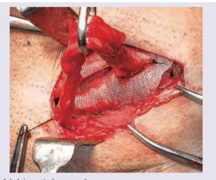
Which hernia repair procedure is shown in the image? (Recent NEET Pattern 2016-17)

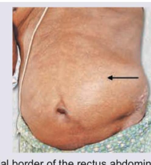
Hernia that is depicted in the image usually occurs at:

Explanation: ***Lateral thoracotomy*** - **Lateral thoracotomy** is associated with the **highest risk of postoperative pulmonary complications** among common surgical incisions, with complication rates ranging from **15-70%** depending on the procedure. - This incision **directly violates the chest wall** with rib resection or spreading, causing severe postoperative pain that significantly impairs respiratory mechanics. - The procedure disrupts **intercostal muscles**, damages **intercostal nerves**, and violates the **pleura**, leading to immediate risks like **pneumothorax**, **hemothorax**, and **pleural effusion**. - Severe pain leads to **splinting**, **shallow breathing**, **impaired cough**, and **reduced lung expansion**, markedly increasing the risk of **atelectasis**, **pneumonia**, and **respiratory failure**. - The **ipsilateral lung** is particularly affected with reduced functional residual capacity and impaired secretion clearance. *Vertical laparotomy* - **Upper abdominal vertical incisions** are indeed associated with high pulmonary complication rates (**30-50%**), second only to thoracotomy. - Pain leads to **diaphragmatic splinting** and impaired respiratory mechanics, increasing risk of **atelectasis** and **pneumonia**. - However, the chest wall itself remains intact, making complications generally less severe than with thoracotomy. *Median sternotomy* - While a major thoracic procedure, **median sternotomy** has relatively **lower pulmonary complication rates** compared to lateral thoracotomy. - The sternal split preserves **intercostal muscles** and **nerve integrity**, resulting in less severe pain and better preserved respiratory mechanics. - Postoperative pain management is generally more effective than with lateral thoracotomy. *Horizontal laparotomy* - **Transverse abdominal incisions** (e.g., Pfannenstiel, transverse supraumbilical) cause significantly less pain than vertical incisions. - These incisions follow **natural tissue planes**, cause less muscle disruption, and allow better respiratory mechanics. - Lower pain levels facilitate **effective coughing**, **deep breathing**, and **early mobilization**, reducing pulmonary complication risk.
Explanation: ***CO2 Exhalation*** - Measuring **CO2 exhalation** (capnography) is the most reliable method to confirm endotracheal tube placement, as CO2 is present in the trachea but not in the esophagus. - A persistent **waveform on the capnograph** indicates proper tracheal intubation. *Direct laryngoscopy* - While helpful for initial visualization during intubation, **direct laryngoscopy** cannot confirm continuous tracheal placement after the tube is advanced. - It only confirms the tube passing through the vocal cords, not its final position in the trachea versus esophagus. *Auscultation* - **Auscultation** can be misleading because stomach sounds can be transmitted to the chest, and breath sounds can be heard in the epigastrium even with esophageal intubation. - It relies on subjective interpretation and is less definitive than capnography. *Chest wall movement* - Observing **chest wall movement** is not a definitive sign, as the chest can still rise with esophageal intubation due to air entering the stomach. - This method is unreliable and can be mistaken for proper ventilation, leading to dangerous delays in correcting tube misplacement.
Explanation: ***Isthmus*** - The **isthmic portion** of the fallopian tube is the most common and preferred site for laparoscopic ring application (e.g., Falope ring or Yoon ring) in female sterilization. - This segment is chosen because it is relatively **straight**, has a **narrow lumen**, and possesses a **thick muscular wall**, making it ideal for occlusion and effective contraception. *Fimbrial* - The **fimbrial end** is the most distal part of the fallopian tube, characterized by finger-like projections that capture the ovum. - Ligation at this site is less common due to its **delicate structure** and proximity to the ovary, increasing the risk of **ovarian damage** or incomplete occlusion. *Cornual* - The **cornual portion** is the segment of the fallopian tube that passes through the muscular wall of the uterus. - This site is generally avoided for ring application due to the **risk of uterine perforation** and increased **bleeding** from the uterine arteries within the myometrium. *Ampullary* - The **ampullary portion** is the widest and longest part of the fallopian tube, where fertilization typically occurs. - Its **dilated lumen** and **tortuous nature** make it less suitable for secure and effective ring placement, as the ring may not fully occlude the tube.
Explanation: ***Arrest hemorrhage and control contamination.*** * The overarching goal of a **damage control laparotomy** is to rapidly address immediate life threats, primarily **hemorrhage** and **bowel contamination**, in severely injured, unstable patients. * This approach prioritizes patient survival by performing essential steps quickly, deferring definitive repairs until the patient is physiologically stable. *Control contamination* * While **controlling contamination** is a critical component of damage control laparotomy, it is not the sole primary aim. * Uncontrolled bleeding, even without contamination, can rapidly lead to death in a trauma patient. *Prevent coagulopathy* * Preventing **coagulopathy** is an important consideration during damage control, but it is a consequence of uncontrolled hemorrhage and hypothermia, rather than a primary surgical aim in the initial damage control phase. * The surgical steps in damage control directly address the sources of bleeding and contamination. *Arrest hemorrhage* * **Arresting hemorrhage** is indeed a primary aim, but it is often accompanied by the need to control contamination from injured hollow organs. * Many abdominal trauma cases involve both significant bleeding and potential contamination.
Explanation: ***Cystoscopy*** - **Cystoscopy** with or without ureteric catheterization can be used as an adjunct in some complex pelvic surgeries, but it is **not considered a primary or routine preventive measure** during most surgeries where ureteric injury risk exists. - While **intraoperative cystoscopy** may help identify ureters or detect injury post-operatively, it is more of a **diagnostic/confirmatory tool** rather than a direct anatomical protective measure during the surgical dissection itself. - Compared to the other listed options, cystoscopy is the **least direct method** of preventing mechanical ureteric injury during the actual surgical dissection and clamping phases. - The other three options represent **direct anatomical protective techniques** employed during surgery. *Ureter should not be dissected off the peritoneum for a long distance* - This is a crucial **surgical principle** to prevent ureteric injury. - Extensive dissection of the ureter from the peritoneum compromises its **blood supply** from adventitial vessels. - Maintaining peritoneal attachments preserves **vascularity** and reduces risk of **ischemic injury** and subsequent necrosis. *Bladder should be pushed downwards and outwards while the clamps are placed near the angles of vagina* - This is an important **anatomical displacement technique** in pelvic surgery. - The ureters course near the **lateral vaginal fornices** (approximately 2 cm lateral to the cervix). - Repositioning the bladder helps displace the ureters away from surgical **clamps, sutures, and electrocautery** applied to vaginal angles. - This maneuver provides a **safety margin** during cardinal ligament and uterosacral ligament procedures. *Direct visualization during surgery* - **Direct visualization** is the gold standard for ureteric protection during surgery. - Allows the surgeon to **identify anatomical location** and confirm ureter position before clamping or ligating. - Essential in complex pelvic procedures with **distorted anatomy** (endometriosis, adhesions, malignancy). - May involve identification of the ureter at the **pelvic brim** and tracing it through the surgical field.
Explanation: ***X-ray*** - An **X-ray**, particularly an erect chest X-ray or an erect abdominal X-ray, is the **most sensitive and readily available investigation** to detect **free air under the diaphragm** (pneumoperitoneum). - This free air, indicating a perforated viscus, appears as a **Crescent-shaped lucency** shadowing the diaphragm. *USG* - While ultrasound can sometimes detect free air, it is **less sensitive and specific** than X-ray for this purpose, especially in early or small perforations. - Its utility is more in detecting other intra-abdominal pathologies like fluid collections or organomegaly. *Laparoscopy* - **Laparoscopy** is a **surgical procedure** that allows direct visualization of the peritoneal cavity. - While it can definitively identify free air and its source, it is an **invasive procedure** and not the primary diagnostic investigation for suspected pneumoperitoneum. *Laparotomy* - **Laparotomy** is an **open surgical procedure** involving a large incision to access the abdominal cavity. - It is used for definitive diagnosis and treatment, but it is **highly invasive** and not a diagnostic investigation in the initial workup for air in the peritoneal cavity.
Explanation: ***Ilioinguinal nerve*** - Damage to the ilioinguinal nerve during abdominal surgery, especially an appendectomy, can lead to muscle weakness in the anterior abdominal wall. - This weakness predisposes the patient to the formation of an **inguinal hernia**, which manifests as an inguinal swelling. *Spermatic cord* - Trauma to the spermatic cord could lead to **testicular atrophy**, pain, or issues with fertility due to vascular or ductal damage. - It is not directly associated with the development of an inguinal hernia as a primary consequence of isolated trauma during non-hernia repairs. *Genital branch of genitofemoral nerve* - Injury to the genital branch of the genitofemoral nerve primarily affects the **cremasteric reflex** and sensation in the scrotum/inner thigh. - While it can cause sensory disturbances, it does not directly lead to weakness of the abdominal wall sufficient to cause an inguinal hernia. *Pampiniform plexus* - The pampiniform venous plexus is involved in regulating testicular temperature. Injury primarily causes a **hydrocele** or **varicocele** due to impaired venous drainage. - It would not cause an inguinal hernia, which involves protrusion of abdominal contents through a weakened abdominal wall.
Explanation: ***Spleen*** - The **spleen** is the most commonly injured organ in **blunt abdominal trauma** (40-55% of cases) due to its superficial location in the left upper quadrant and its relatively fragile, highly vascular parenchyma. - Its anatomical position, without significant muscular or bony protection anteriorly, makes it vulnerable to compressive and shearing forces during blunt impacts. - Typically presents with left upper quadrant pain, left shoulder pain (Kehr's sign), and signs of hypovolemic shock. *Liver* - While the **liver** is the second most commonly injured organ in blunt abdominal trauma (35-45% of cases), it is less frequently ruptured than the spleen. - Its larger size and more protected position by the rib cage offer some degree of shielding compared to the spleen. - Presents with right upper quadrant pain and peritoneal signs. *Kidney* - **Kidney injuries** are less common than splenic or hepatic injuries in blunt abdominal trauma, requiring significant force due to their retroperitoneal location and protection by the back muscles and lower ribs. - Renal trauma is usually associated with flank pain and hematuria. - Protected retroperitoneal position makes injury less frequent. *Adrenals* - **Adrenal gland injuries** are extremely rare in blunt abdominal trauma, typically occurring only with severe, high-energy impact and often in conjunction with other significant organ damage. - Their small size and deep retroperitoneal location make them highly protected.
Explanation: ***Lichtenstein repair*** - The image clearly displays a **mesh patch** being used to reinforce the posterior wall of the inguinal canal, which is the hallmark of a **tension-free Lichtenstein repair**. - This technique is widely considered the **gold standard** for **inguinal hernia repair** due to its low recurrence rates and reduced postoperative pain. *Bassini herniorrhaphy* - **Bassini's repair** is a **tissue-based repair** that involves suturing the conjoined tendon and transversalis fascia to the inguinal ligament. - This method does **not use mesh** and is associated with higher tension and recurrence rates compared to mesh-based repairs. *Shouldice repair* - The **Shouldice repair** is another **tissue-based repair** from Canada, renowned for its strong, multilayered closure of the posterior wall of the inguinal canal. - It involves **four layers of suture repair** of the transversalis fascia and conjoined tendon, without the use of synthetic mesh as seen in the image. *Lord's procedure* - **Lord's procedure** is a historical method for **inguinal hernia repair** that primarily involved placing a small, tightly rolled mesh plug into the internal ring. - It is **not commonly used today** and does not involve the broad, flat mesh placement depicted in the image to reinforce the entire posterior wall.
Explanation: ***Lateral border of the rectus abdominis*** - The image depicts a **Spigelian hernia**, which is a rare type of ventral hernia that occurs through the **Spigelian aponeurosis**. - This aponeurosis is located at the **semilunar line**, which is the curved tendinous intersection found at the lateral border of the rectus abdominis muscle. *Medial border of the rectus abdominis* - Hernias at the medial border of the rectus abdominis are typically **umbilical or epigastric hernias**, which present differently and are not depicted here. - These are located closer to the midline, unlike the more lateral protrusion shown. *Medial border of transverse abdominis* - The transverse abdominis muscle generally lies deeper and its medial border is not a common site for a hernia like the one shown. - Hernias in this region would not typically present as a bulge along the semilunar line. *Lateral border of transverse abdominis* - The lateral border of the transverse abdominis is situated more posteriorly and superiorly, often near the flank or lumbar region. - Hernias in this area are typically **lumbar hernias**, which are distinct from the anterior bulge seen in the image.
Get full access to all questions, explanations, and performance tracking.
Start For Free