
Hepatobiliary Surgery — MCQs

On this page
The 'Push, Pringle, Plug and Pack' technique is used for managing which organ's bleeding?
What is the best treatment for the given type of cholecystitis?
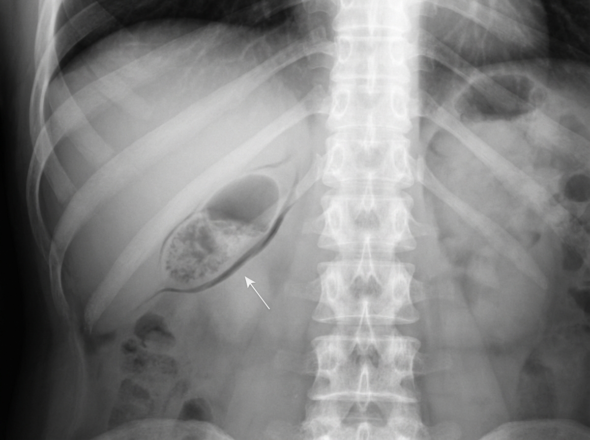
In gallbladder mucocele, where is the stone usually impacted?
Reynolds' pentad of fever, jaundice, right upper quadrant pain, septic shock, and altered mental status is typical of which of the following conditions?
A 30-year-old lady is found to have gallstones. She is asymptomatic and has never had any jaundice or dyspeptic symptoms in the past. What is the best course of management for her?
A patient presents with pain in the upper abdomen, melena, and jaundice three weeks after percutaneous transhepatic cholangiography. What is the most appropriate investigation for this patient?
Gall bladder stone formation is influenced by all except?
What is the most common surgical cause of obstructive jaundice?
Bile duct strictures are seen in all except?
Rigler's triad consists of all except?
Practice by Chapter
Liver Anatomy and Physiology
Practice Questions
Benign Liver Lesions
Practice Questions
Liver Abscess
Practice Questions
Hepatocellular Carcinoma
Practice Questions
Metastatic Liver Disease
Practice Questions
Cirrhosis and Portal Hypertension
Practice Questions
Liver Trauma
Practice Questions
Cholelithiasis and Cholecystitis
Practice Questions
Choledocholithiasis
Practice Questions
Biliary Tract Tumors
Practice Questions
ERCP and Its Complications
Practice Questions
Liver Transplantation Basics
Practice Questions
Want unlimited practice?
Get full access to all questions, explanations, and performance tracking.
Scan to download app