
Hepatobiliary Surgery — MCQs

On this page
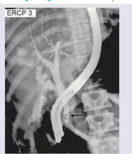
The following image shows: (DNB Pattern 2018)
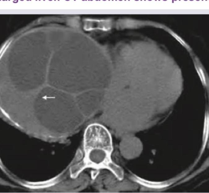
A 35-year-old shepherd presents with painless enlarged liver. CT abdomen shows presence of:
A Sengstaken-Blakemore tube is used for the management of :
Which of the following is NOT included in Grade II acute cholecystitis as per 'Tokyo Consensus Guidelines for Severity'? 1. Elevated white cell count (> 18000/mm3) 2. Renal dysfunction 3. Duration > 72 hours 4. Marked local inflammation
A 50 year old diabetic patient with asymptomatic gallstone (> 3 cm) will be best treated by
A young patient develops high grade fever with chills and rigors, mild jaundice and acute pain in the upper abdomen following cholecystectomy. On examination she was jaundiced, toxic, haemodynamically stable and having vague fullness upper abdomen. What is the most probable diagnosis ?
Which one of the following is not a common feature of bile duct stone ?
A retained stone in CBD (common bile duct) diagnosed by T-tube cholangiogram is best treated by :
A 25-year-old patient has 5 x 5 cm amoebic abscess in right lobe of liver. He is febrile and has pain in right hypochondrium. His primary management would include:
The scolicidal agents used in the surgery of a hydatid cyst include all of the following except
Practice by Chapter
Liver Anatomy and Physiology
Practice Questions
Benign Liver Lesions
Practice Questions
Liver Abscess
Practice Questions
Hepatocellular Carcinoma
Practice Questions
Metastatic Liver Disease
Practice Questions
Cirrhosis and Portal Hypertension
Practice Questions
Liver Trauma
Practice Questions
Cholelithiasis and Cholecystitis
Practice Questions
Choledocholithiasis
Practice Questions
Biliary Tract Tumors
Practice Questions
ERCP and Its Complications
Practice Questions
Liver Transplantation Basics
Practice Questions
Want unlimited practice?
Get full access to all questions, explanations, and performance tracking.
Scan to download app