
Hepatobiliary Surgery — MCQs

On this page
Sphincterectomy of the sphincter of Oddi is performed at which position?
A patient with gallstone disease underwent laparoscopic cholecystectomy. The pathology report revealed Stage 1A adenocarcinoma. What is the recommended treatment approach?
Which of the following is NOT an indication for cholecystectomy?
Spontaneous rupture of the liver occurs in which of the following conditions?
A 48-year-old man presents with severe abdominal pain, right hypochondrium tenderness, and a WBC count of 12,000. A HIDA scan fails to visualize the gallbladder after 4 hours, establishing a diagnosis of acute cholecystitis. Within what timeframe should cholecystectomy be performed following hospital admission?
What is the approximate association of choledocholithiasis in patients with cholelithiasis?
Which of the following is NOT a risk factor for malignant change in an asymptomatic patient with a gallbladder polyp identified on ultrasound?
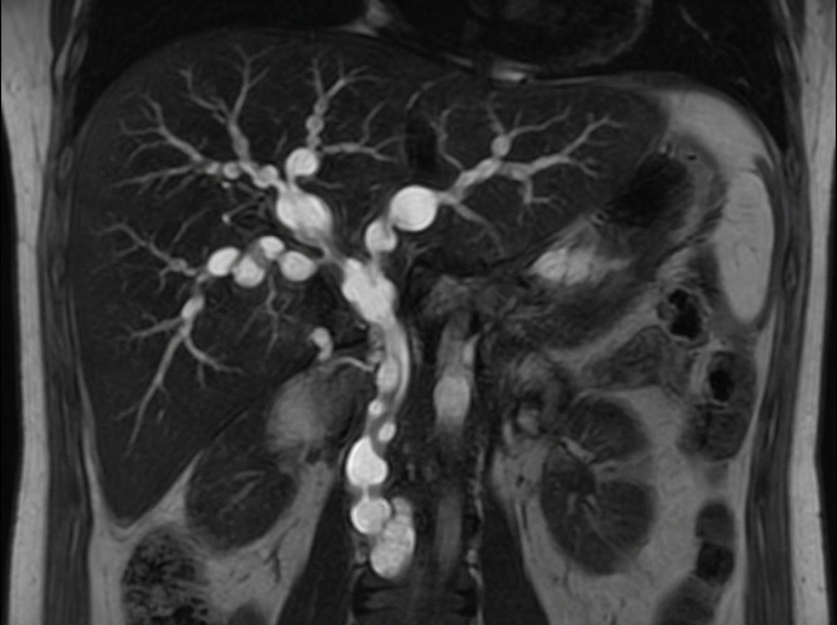
A 45-year-old male presented with recurrent attacks of cholangitis. MRCP and ERCP findings are suggestive of this condition. What is the treatment of choice for this condition?
A 65-year-old woman presents with right upper quadrant (RUQ) pain radiating to the right shoulder, along with nausea and vomiting. Physical examination reveals RUQ tenderness and a positive Murphy's sign, leading to a diagnosis of acute cholecystitis. What is the most likely associated finding?
Amoebic liver abscess ruptures most commonly into which cavity?
Practice by Chapter
Liver Anatomy and Physiology
Practice Questions
Benign Liver Lesions
Practice Questions
Liver Abscess
Practice Questions
Hepatocellular Carcinoma
Practice Questions
Metastatic Liver Disease
Practice Questions
Cirrhosis and Portal Hypertension
Practice Questions
Liver Trauma
Practice Questions
Cholelithiasis and Cholecystitis
Practice Questions
Choledocholithiasis
Practice Questions
Biliary Tract Tumors
Practice Questions
ERCP and Its Complications
Practice Questions
Liver Transplantation Basics
Practice Questions
Want unlimited practice?
Get full access to all questions, explanations, and performance tracking.
Scan to download app