
Head and Neck Surgery — MCQs

On this page
Facial nerve injury occurs during parotid surgery. What is the best management?
The 'Starch iodine test' is useful to diagnose:
A 24-year-old computer technician presents with a progressive increase in the size of his left jaw. Following radiographic and biopsy confirmation of ameloblastoma, what is the recommended next step in management?
Which ganglion needs to be avoided during cervical sympathectomy to prevent Horner syndrome?
Which of the following is not done in the treatment of ankylosis?
The tooth most commonly involved in chronic focal sclerosing osteomyelitis is?
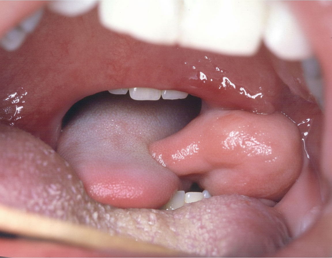
What is the most common organism that causes the following condition?
Sistrunk operation is the preferred treatment for which of the following conditions?
What is the standard treatment for verrucous carcinoma of the lip?
Excessive blood loss during mandibulectomy can be prevented by all of the following except?
Practice by Chapter
Salivary Gland Diseases
Practice Questions
Thyroid Gland Disorders
Practice Questions
Parathyroid Gland Disorders
Practice Questions
Neck Masses Evaluation
Practice Questions
Oral Cavity Lesions
Practice Questions
Laryngeal Disorders
Practice Questions
Head and Neck Cancer
Practice Questions
Reconstructive Techniques in Head and Neck Surgery
Practice Questions
Surgical Management of Sleep Apnea
Practice Questions
Airway Management in Head and Neck Surgery
Practice Questions
Surgical Approaches to the Neck
Practice Questions
Neck Dissection Techniques
Practice Questions
Want unlimited practice?
Get full access to all questions, explanations, and performance tracking.
Scan to download app