Which surgical landmarks are used for locating the facial nerve?
A 23-year-old male patient presents with midline swelling in the neck. The swelling moves with deglutition and protrusion of the tongue. What is the likely diagnosis?
What is Pott’s puffy tumor?
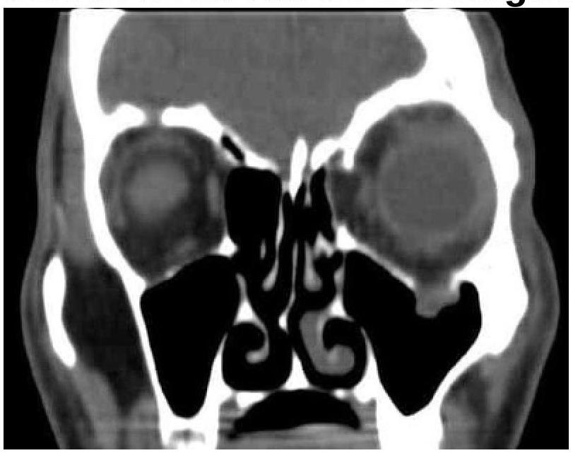
A patient presented with a history of diplopia and restricted eye movements. What is the most likely diagnosis based on the clinical and CT images?

In which condition is surgical intervention involving partial or full closure of the nasal cavity typically performed?
A middle-aged man presents with a swelling in the neck that has been present since childhood. The swelling has a bag or worm-like appearance and features a central black spot. Based on this description, what is the most likely diagnosis?
Where will be the placement location for Auditory Brainstem Implant?
What is the most common cause of a parotid mass that presents with mixed consistency?
Supraomohyoid dissection is a type of?
Which of the following neck dissections is considered the most conservative?
Explanation: ***All of the options*** - The **tympano-mastoid suture**, the **tragal pointer**, and the **posterior belly of the digastric** muscle are all commonly used surgical landmarks for identifying and preserving the facial nerve during parotidectomy or other procedures in the vicinity. - Using multiple landmarks provides a more reliable and precise localization of the facial nerve, reducing the risk of iatrogenic injury. *Tympano - mastoid suture* - This suture line is an important external landmark that helps guide the dissection towards the **stylomastoid foramen**, where the facial nerve exits the skull. - The facial nerve typically lies deep and anterior to this suture, providing a valuable starting point for identification. *Tragal pointer* - The **tragal pointer** (cartilaginous projection of the tragus) points directly towards the main trunk of the facial nerve as it enters the parotid gland. - It is a highly reliable superficial landmark, particularly useful in locating the superior aspect of the facial nerve trunk. *Posterior belly of digastric* - The **posterior belly of the digastric muscle** serves as a deep landmark, as the facial nerve typically courses immediately superior and lateral to it. - Dissection along the superior border of this muscle can lead directly to the facial nerve trunk, especially when dissecting inferiorly.
Explanation: ***Thyroglossal cyst*** - A **thyroglossal cyst** is a congenital anomaly that arises from the persistent **thyroglossal duct**, a remnant of the thyroid's embryologic descent. - Its classic diagnostic feature is its movement with **deglutition** (due to attachment to the hyoid bone, which moves during swallowing) and **protrusion of the tongue** (as the thyroglossal duct is connected to the base of the tongue). *Brachial cyst* - A **brachial cyst** is a congenital neck mass that typically presents as a lateral neck swelling, often located along the anterior border of the **sternocleidomastoid muscle**. - Unlike a thyroglossal cyst, it does not typically move with **deglutition** or **tongue protrusion**. *Plunging ranula* - A **plunging ranula** is a type of mucocele that arises from the **sublingual gland** and extends below the mylohyoid muscle into the neck. - It presents as a cervical mass but is typically located in the floor of the mouth or submandibular region and does not move with **deglutition** or **tongue protrusion**. *Dermoid cyst* - A **dermoid cyst** is a congenital cyst that can occur anywhere on the body, including the head and neck, often presenting as a painless mass. - It arises from sequestered embryonic ectoderm and mesoderm, containing skin appendages, but it does not move with **deglutition** or **tongue protrusion**.
Explanation: ***Subperiosteal abscess of frontal bone*** - **Pott's puffy tumor** is a rare complication resulting from untreated or inadequately treated **frontal sinusitis**, leading to osteomyelitis of the frontal bone. - The infection spreads through the venous system or direct erosion, forming a **subperiosteal abscess** on the outer table of the frontal bone, often presenting as a tender, doughy swelling on the forehead. *Subperiosteal abscess of ethmoid bone* - While abscesses can occur in conjunction with ethmoid sinusitis, this condition does not specifically correspond to the definition of **Pott's puffy tumor**, which is characteristic of frontal bone involvement. - **Ethmoid sinusitis** is more commonly associated with orbital complications rather than a forehead swelling. *Mucocele of frontal bone* - A **mucocele** is an expansile, cystic lesion filled with mucus, typically resulting from obstruction of a sinus ostium. It is not an infectious process involving an abscess. - While it can cause swelling or bone erosion, it does not involve the acute inflammatory and infectious features of a **subperiosteal abscess**. *Mucocele of ethmoid bone* - Similar to a frontal mucocele, an ethmoid mucocele is a **non-infectious cystic lesion** filled with mucus due to obstruction of the ethmoid sinus drainage. - It does not involve a collection of pus or an active infectious process characteristic of **Pott's puffy tumor**.
Explanation: ***Blowout fracture*** - The CT image shows a **fracture of the orbital floor**, with associated **herniation of orbital contents** (fat and inferior rectus muscle) into the maxillary sinus. - This type of fracture commonly causes **diplopia** (due to muscle entrapment) and **restricted eye movements**. *Le Fort fracture* - Le Fort fractures involve the **midface and maxilla** as a whole, typically causing widespread facial instability. - They are classified into three types (I, II, III), none of which are exclusively characterized by an isolated orbital floor fracture with muscle entrapment. *Maxillary fracture* - While an orbital floor fracture involves the maxilla, a "maxillary fracture" is a broad term and does not specifically describe the characteristic features of **orbital content prolapse** and their resulting symptoms. - Isolated maxillary fractures might not cause diplopia or restricted eye movements unless they directly involve the orbit. *Zygomatic fracture* - A zygomatic fracture (either isolated or as part of a zygomaticomaxillary complex fracture) primarily affects the **cheekbone**. - While it can involve the orbital rim, it typically presents with **facial asymmetry**, **numbness** in the infraorbital nerve distribution, and sometimes **trismus**, rather than isolated muscle entrapment causing diplopia, as seen in the image.
Explanation: ***Atrophic rhinitis*** - Surgical intervention, specifically **Young's operation** (partial or full closure of the nostril), is performed to create a moist environment and promote mucosal healing in severe cases. - This condition involves **progressive atrophy of nasal mucosa** and turbinates, leading to dryness, crusting, and a foul odor, which surgery aims to alleviate. *Allergic rhinitis* - This is an inflammatory condition triggered by **allergens**, typically managed with antihistamines, nasal corticosteroids, and allergen avoidance. - Surgical intervention is generally not indicated, as it does not address the underlying allergic immune response. *Vasomotor rhinitis* - Characterized by **non-allergic, non-infectious rhinitis** due to autonomic nervous system dysfunction, often triggered by irritants or temperature changes. - Management focuses on symptom relief with nasal sprays (anticholinergics or corticosteroids) and trigger avoidance, not surgical closure. *Occupational rhinitis* - This type is caused by **exposure to irritants or sensitizers** in the workplace, leading to nasal inflammation. - Treatment involves identifying and avoiding the offending agent, alongside symptomatic relief, with no role for surgical closure of the nasal cavity.
Explanation: ***Plexiform neurofibroma*** - The description of a swelling since childhood with a **bag-of-worms appearance** and a **central black spot** is highly characteristic of a plexiform neurofibroma, a benign tumor of peripheral nerves. - These lesions are often associated with **neurofibromatosis type 1 (NF1)** and can grow quite large, causing cosmetic and functional concerns. - The **central black spot** represents pigmentation often seen in neurofibromas, particularly in NF1. *Cirsoid aneurysm* - A cirsoid aneurysm is a **congenital arteriovenous malformation** characterized by a tortuous mass of dilated blood vessels. - While it can manifest as a swelling, it typically presents with a **palpable thrill and audible bruit**, and its appearance is not usually described as having a central black spot. *Varicocele* - A varicocele is an **abnormal enlargement of the pampiniform plexus veins** in the scrotum, often described as a **bag of worms**, but it occurs exclusively in the scrotum. - It would not be found in the neck and does not feature a central black spot. *Lymphangioma* - A lymphangioma is a **benign malformation of the lymphatic system** that can present as a soft, compressible mass, often in the neck. - While present since childhood, its appearance is typically **cystic or multicystic** and does not usually have the distinct bag-of-worms texture or a central black spot described.
Explanation: ***Recess of 4th ventricle*** - An **Auditory Brainstem Implant (ABI)** is placed on the **cochlear nucleus** in the brainstem, which is anatomically located near the **lateral recess of the fourth ventricle**. - The implant stimulates these nuclei directly, bypassing the damaged auditory nerve in patients who cannot benefit from cochlear implants. *Scala tympani* - The **scala tympani** is the location for electrode placement in a **cochlear implant**, not an auditory brainstem implant. - Cochlear implants stimulate the intact auditory nerve within the cochlea. *IAC* - The **internal auditory canal (IAC)** houses the auditory and facial nerves, but it is not the target site for an ABI. - The ABI is designed for patients whose auditory nerve (which passes through the IAC) is non-functional. *Back of ear* - The "back of the ear" is the general area where the **external processor of a cochlear implant** or a **bone-anchored hearing aid** is typically worn, not the surgical placement site for an ABI. - The ABI's internal component is surgically placed within the cranium.
Explanation: ***Pleomorphic adenoma*** - The presence of a **mixed consistency** parotid mass, often noted as a **firm yet compressible** lump, is characteristic of pleomorphic adenoma. - This tumor's **benign nature** allows for gradual growth and is the most common parotid gland tumor, often being mobile and well-defined on examination [1]. *All* - The term "all" is too vague and does not specify a particular pathology or characteristics relevant to a **mixed consistency** mass. - It does not represent any known entity, making it an unsuitable answer in the context of **specific diagnosis**. *Sebaceous cyst* - Sebaceous cysts are typically **smooth, soft, and fluctuant**, lacking the mixed consistency seen in pleomorphic adenomas. - They are also usually associated with **skin** and hair follicles, not commonly arising in the parotid gland region. *Dermoid cyst* - Dermoid cysts usually present as **soft, cystic lesions** filled with **keratin**, which does not align with the mixed consistency nature of pleomorphic adenomas. - They can occur in various locations but are not the typical presentation for a parotid mass. **References:** [1] Kumar V, Abbas AK, et al.. Robbins and Cotran Pathologic Basis of Disease. 9th ed. Head and Neck, pp. 751-753.
Explanation: ***Selective neck dissection*** - **Supraomohyoid dissection** specifically refers to a type of selective neck dissection, characterized by the removal of lymph node levels **I, II, and III**. - This procedure is commonly performed for early-stage oral cavity cancers due to their typical lymphatic spread patterns. *Modified radical neck dissection* - This dissection preserves one or more **non-lymphatic structures** (e.g., sternocleidomastoid muscle, internal jugular vein, spinal accessory nerve) that are typically removed in a radical neck dissection. - It involves a broader range of lymph node levels (typically **I-V**) compared to a supraomohyoid dissection. *Radical neck dissection* - This is a more extensive procedure involving the removal of all lymph node groups (levels **I-V**), along with the **sternocleidomastoid muscle**, **internal jugular vein**, and **spinal accessory nerve**. - It is reserved for advanced neck disease due to its significant morbidity. *Posterolateral dissection* - **Posterolateral neck dissection** is a term not commonly used within the standard classification of neck dissections (radical, modified radical, selective). - Lymphatic dissection is typically categorized based on anatomical levels rather than a general directional term like posterolateral.
Explanation: ***Supraomohyoid neck dissection*** - This dissection is highly **selective**, removing only lymph nodes from **levels I, II, and III**, which are the most superficial and anterior groups in the neck. - It preserves the **internal jugular vein**, spinal accessory nerve, and sternocleidomastoid muscle, minimizing functional and cosmetic morbidity. *Radical neck dissection* - This is the **most extensive** neck dissection, involving the removal of all lymph node levels (I-V), the **internal jugular vein**, the **spinal accessory nerve**, and the **sternocleidomastoid muscle**. - It is reserved for advanced cancers with extensive nodal involvement due to its significant associated morbidity and functional deficits. *Modified radical neck dissection* - This dissection removes lymph nodes in levels I-V but **spares at least one non-lymphatic structure**, such as the spinal accessory nerve, internal jugular vein, or sternocleidomastoid muscle. - While less radical than a full radical neck dissection, it is still more extensive than a supraomohyoid dissection as it targets a broader range of lymph node levels. *All options are conservative.* - This statement is incorrect because **radical neck dissection** is by definition the most extensive and least conservative surgical approach to neck nodal disease. - The different types of neck dissections represent a spectrum of extensiveness, with supraomohyoid being the most selective and conservative.
Salivary Gland Diseases
Practice Questions
Thyroid Gland Disorders
Practice Questions
Parathyroid Gland Disorders
Practice Questions
Neck Masses Evaluation
Practice Questions
Oral Cavity Lesions
Practice Questions
Laryngeal Disorders
Practice Questions
Head and Neck Cancer
Practice Questions
Reconstructive Techniques in Head and Neck Surgery
Practice Questions
Surgical Management of Sleep Apnea
Practice Questions
Airway Management in Head and Neck Surgery
Practice Questions
Surgical Approaches to the Neck
Practice Questions
Neck Dissection Techniques
Practice Questions
Get full access to all questions, explanations, and performance tracking.
Start For Free