Which type of collagen is most abundant in hyaline cartilage?
Which of the following structures in the spermatic cord is typically preserved (not divided) during vasectomy surgery?
For a midline incision in the abdomen, length of suture required is:
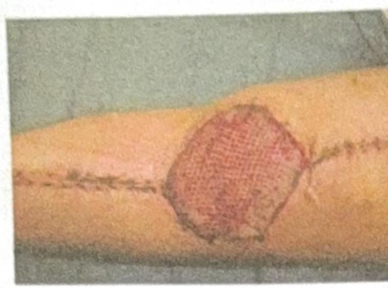
What type of graft or dressing is used to cover the post-burn wound shown in the image?

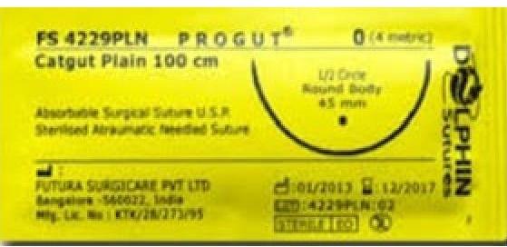
Which of the following statements is true about the suture material shown in the image?

A patient has a lacerated, untidy wound of the leg and attended the casualty department after 2 hours. His wound should be:
Vacuum assisted closure is contraindicated in which of the following conditions -
Which of the following layers are cut during fasciotomy ?
Which one of the following regarding absorbable meshes is NOT true?
What is the typical absorption duration of Polydioxanone sutures?
Explanation: ***Type II*** - **Type II collagen** is the predominant type found in **hyaline cartilage**, providing tensile strength and elasticity [1]. - It is crucial for the **structural integrity** and functionality of cartilage in articular surfaces [1]. *Type I* - Predominantly found in **bone**, tendons, and skin, contributing to tensile strength but not a major component of hyaline cartilage [2]. - It forms the structure of **fibrocartilage**, such as in the **intervertebral discs**. *Type IV* - Mainly located in **basement membranes** and plays a role in filtration and structural support of epithelial cells, not in hyaline cartilage. - It is critical in the formation of structures like **glomeruli** in kidneys, differing from cartilage's needs. *Type III* - Found in **reticular fibers** and supporting tissues throughout the body, important for organ structure but not prominent in hyaline cartilage. - Often associated with **vascular structures** and is not involved in the composition of cartilage.
Explanation: ***Testicular artery*** - The goal of a vasectomy is to interrupt sperm transport, not the blood supply to the testis. The **testicular artery** is the most critical structure to preserve as it provides the primary blood supply to the testis. - Preserving the **testicular artery** ensures continued blood flow to the testis, preventing ischemia and maintaining both spermatogenesis (though sperm won't exit) and endocrine function (testosterone production). - Surgeons carefully isolate and preserve the testicular artery while dividing only the vas deferens. *Vas deferens* - The **vas deferens** is the target structure that is deliberately divided and ligated during vasectomy. - Cutting the **vas deferens** interrupts the pathway for sperm transport from the epididymis to the ejaculatory duct, achieving permanent contraception. - This is the only structure within the spermatic cord that is intentionally divided during the procedure. *Autonomic nerves* - While **autonomic nerves** (sympathetic postganglionic fibers) are present in the spermatic cord and innervate the vas deferens, they may be inadvertently damaged during the vasectomy procedure. - The primary function of these **autonomic nerves** related to the vas deferens is smooth muscle contraction for sperm transport, which becomes irrelevant once the vas deferens is divided. - These nerves are not actively preserved as their division doesn't significantly impact testicular function. *Testicular vein* - The **testicular vein** (pampiniform plexus) drains blood from the testis and is also typically preserved during vasectomy, along with the testicular artery. - However, the **testicular artery** is considered more critical as arterial blood supply is essential for tissue viability, whereas venous drainage has collateral pathways through cremasteric and deferential veins. - Both vessels are preserved, but the arterial supply takes priority in surgical technique.
Explanation: ***4 times the length of incision*** - The standard recommendation for interrupted abdominal fascial closure is to use a **suture-to-wound length ratio** of approximately **4:1**. - This ratio ensures sufficient material for adequate fascial apposition, overlapping bites, and knots, which are crucial for preventing wound dehiscence. *3 times the length of incision* - A 3:1 suture-to-wound ratio might be insufficient for secure fascial closure, potentially leading to increased tension on the suture lines and a **higher risk of dehiscence**. - This ratio could be considered for very specific continuous closure techniques, but it's generally not recommended for standard interrupted closures. *2 times the length of incision* - A 2:1 ratio is generally considered **inadequate** for most fascial closures, especially in the abdomen. - This ratio would likely result in insufficient suture material, leading to very large bites and an insecure closure, significantly increasing the risk of **wound dehiscence** and **herniation**. *5 times the length of incision* - While it ensures enough material, a 5:1 ratio suggests using **excessive suture material** which might extend operating time. - Using significantly more suture than necessary offers no proven benefit in terms of wound security and can sometimes introduce more foreign material into the wound.
Explanation: ***Split thickness skin graft*** - The image shows a **meshed pattern** on the skin graft, which is characteristic of a **split-thickness skin graft** that has been expanded to cover a larger area. - This type of graft consists of the epidermis and a portion of the dermis, making it more flexible and able to **"take" more reliably** on various wound beds, commonly used for burn wounds. *Full thickness skin graft* - A **full-thickness skin graft** includes the entire epidermis and dermis and typically does not have a meshed appearance. - They are used for smaller defects where cosmesis is a priority, but have a **lower take rate** than split-thickness grafts, making them less suitable for large burn wounds. *VAC dressing* - A **VAC (Vacuum-Assisted Closure) dressing** is a system that applies negative pressure to a wound to promote healing and is not a skin graft itself. - It involves a foam or gauze dressing sealed with an adhesive film, connected to a vacuum pump, which is not what is depicted in the image. *Normal saline dressing* - A **normal saline dressing** is a simple wet-to-dry or wet-to-wet dressing for wound care, involving gauze soaked in normal saline. - This is a basic wound management technique and does not involve grafting or have the characteristic meshed appearance seen in the image.
Explanation: ***Degraded by enzymatic degradation*** - Catgut sutures are **natural absorbable sutures** made primarily from purified collagen. - Their absorption in the body occurs through **enzymatic degradation** and phagocytosis, which is a key characteristic distinguishing them from synthetic absorbable sutures that undergo hydrolysis. *Made of cat submucosa* - While historically referred to as "catgut," these sutures are not actually made from cat intestines. - They are typically derived from the **submucosa of sheep or goat intestines**. *Not degraded* - Catgut sutures are explicitly classified as **absorbable surgical sutures**, as stated on the packaging. - Absorbable sutures by definition are designed to be broken down and **resorbed by the body** over time. *Made of rabbit submucosa* - Catgut sutures are not typically made from rabbit submucosa. - The primary sources for natural absorbable sutures like catgut are the **intestines of sheep or goats**.
Explanation: ***Debrided and sutured secondarily*** - An **untidy wound** indicates contamination, irregular edges, and devitalized tissue, which significantly increases the **risk of wound infection**. - The standard management involves **thorough debridement** to remove all contaminated and non-viable tissue, followed by **delayed primary closure** (suturing after 3-5 days once the wound shows healthy granulation) or **healing by secondary intention**. - This approach is especially important for **lower extremity wounds**, which have a higher infection risk due to relatively poorer blood supply compared to facial wounds. - Even though the patient presented within 2 hours (well within the "golden period"), the **untidy nature** of the wound makes **immediate primary closure risky** and secondary closure the safer, preferred option. *Debrided and sutured immediately* - While **debridement is essential** for untidy wounds, **immediate primary closure** after debridement is generally reserved for **tidy wounds** with minimal contamination. - For untidy wounds, immediate closure increases the risk of **trapping bacteria and devitalized tissue**, leading to **wound infection**, abscess formation, or dehiscence despite being within the golden period. - Primary closure may be considered in select cases with minimal contamination and excellent debridement, but this is not the standard teaching for untidy wounds. *Sutured immediately* - **Immediate suturing without debridement** of an untidy wound would be dangerous, as it would trap contaminants, foreign material, and devitalized tissue. - This approach would significantly increase the risk of **serious wound infection**, including **gas gangrene** or necrotizing fasciitis in contaminated wounds. - Proper wound preparation is mandatory before any closure is considered. *Cleaned and dressed* - Simple **cleaning and dressing** is insufficient for an untidy wound as it does not address the devitalized tissue that requires **surgical debridement**. - While this avoids the risk of premature closure, it fails to provide adequate treatment for a wound that needs formal surgical debridement to remove non-viable tissue and reduce bacterial load. - This approach might be acceptable only as a temporary measure if surgical debridement cannot be performed immediately.
Explanation: ***Large amount of necrotic tissue with eschar*** - The presence of a large amount of **necrotic tissue** and **eschar** is a contraindication for VAC therapy because it prevents effective contact between the foam and viable tissue, impairing wound healing. - Eschar acts as a physical barrier, trapping bacteria and hindering the proper function of negative pressure by preventing uniform pressure distribution and fluid removal from the wound bed. *Chronic osteomyelitis* - While chronic osteomyelitis can be challenging, VAC therapy can sometimes be used as an **adjunctive treatment** after surgical debridement to manage the wound and promote granulation tissue formation. - It helps in controlling infection and closing the wound by removing exudates, reducing edema, and improving blood flow. *Abdominal wound* - VAC therapy is commonly used for **abdominal wounds**, especially after damage control surgery or in cases of open abdomen management. - It facilitates closure by promoting granulation, reducing edema, and protecting the abdominal contents. *Surgical wound dehiscence* - **Surgical wound dehiscence** is a common indication for VAC therapy, as it helps to manage the open wound, promote granulation tissue, and prepare the wound for eventual secondary closure or grafting. - VAC therapy reduces surgical site infections, removes exudates, and enhances tissue perfusion, leading to better wound healing outcomes.
Explanation: ***Skin+subcutaneous tissue+Superficial fascia+deep fascia*** - A **fasciotomy** is a surgical procedure to relieve **compartment syndrome** by releasing the **deep fascia** that constricts muscle compartments. - To access and incise the deep fascia, all overlying layers must be cut: **skin**, **subcutaneous tissue** (also called superficial fascia or hypodermis), and finally the **deep fascia** itself. - Note: "Superficial fascia" and "subcutaneous tissue" refer to the same anatomical layer, but both terms are listed here to reflect common clinical terminology. *Skin* - Cutting only the skin does not provide access to the deep fascia and cannot relieve compartment syndrome. - The skin is merely the outermost protective layer. *Skin+subcutaneous tissue* - While both these layers must be incised, stopping here leaves the **deep fascia** intact. - The deep fascia is the primary constricting structure in compartment syndrome and must be released. *Skin+subcutaneous tissue+Superficial fascia* - This option is anatomically redundant since superficial fascia and subcutaneous tissue are the same layer. - More importantly, this still does not include division of the **deep fascia**, which is essential for decompression in a true fasciotomy.
Explanation: ***They show very good results as collagen deposition is maximum*** - Absorbable meshes are **resorbed by the body** over time, leading to less collagen deposition compared to non-absorbable meshes, which provide a permanent scaffold for tissue integration. - While they can be useful in certain situations, the statement implies **superior results due to maximum collagen deposition**, which is contradictory to their nature and purpose in situations where permanent reinforcement is needed. *They are made of polyglycolic acid fibre* - Many absorbable meshes, such as **Dexon** and **Vicryl**, are indeed made from synthetic polymers like **polyglycolic acid (PGA)** or polylactic acid (PLA). - These materials are designed to be **hydrolyzed and absorbed** by the body. *They are used to buttress sutured repair* - Absorbable meshes can be used to **reinforce a primary suture line** in contaminated fields or when there is concern for tissue breakdown. - They provide **temporary support** while the native tissue heals. *They are used in temporary abdominal wall closure* - In cases of **abdominal compartment syndrome** or severe contamination, absorbable meshes may be used for **temporary closure** of the abdominal wall. - This allows for staged repair and reduces the risk of infection often associated with permanent meshes in these scenarios.
Explanation: ***Correct: 6 months*** - **Polydioxanone (PDS) sutures** are known for their **prolonged absorption time**, typically ranging from 180 to 210 days, or approximately 6 months. - This characteristic makes PDS sutures suitable for tissues requiring **extended support** during the healing process. - PDS retains approximately **50% tensile strength at 4 weeks** and **25% at 6 weeks**, with complete absorption occurring over 6-7 months. *Incorrect: 2 weeks* - An absorption duration of 2 weeks is characteristic of **rapidly absorbing sutures**, such as **chromic gut** or **fast-absorbing synthetic sutures**, which are used for tissues that heal quickly or require minimal support. - PDS sutures offer much longer tensile strength retention and absorption than this brief period. *Incorrect: 4 weeks* - A 4-week absorption time is considerably shorter than that of PDS sutures. This duration might be seen with some **intermediate-absorbing sutures**, but not with the long-lasting PDS. - Sutures absorbed in this timeframe would not provide sufficient support for tissues where PDS is typically indicated. *Incorrect: 6 weeks* - While longer than 2 or 4 weeks, 6 weeks (approximately 42 days) is still much shorter than the typical absorption profile of PDS sutures. - Sutures like **Vicryl Rapide** might fall into this absorption range, but PDS is designed for applications needing several months of support.
Get full access to all questions, explanations, and performance tracking.
Start For Free