A 35-year-old surgeon develops a tremor in her dominant hand. Neurological evaluation suggests early Parkinson's disease. She performs complex microsurgical procedures and is concerned about patient safety. Her department chair is unaware of the diagnosis. Evaluate the ethical and professional approach to this situation.
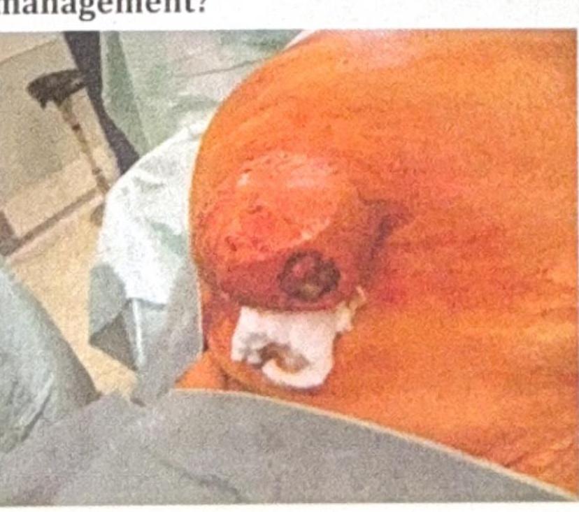
A patient presents with an umbilical mass, which was previously reducible but has now become irreducible with discharge coming out, as shown in the image. What is the most appropriate management?

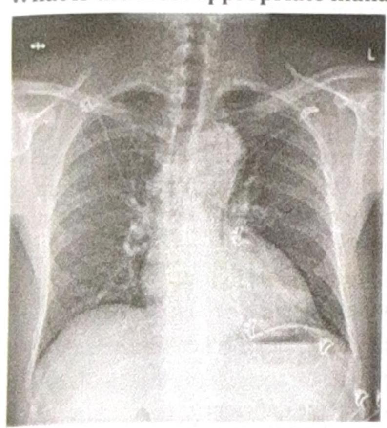
A patient presents with constant chest pain, and the radiological finding is as shown in the image. What is the most appropriate management?

A jeep driver presents with pain in the gluteal region along with swelling and pus discharge for the past 6 months. What is the most likely diagnosis?
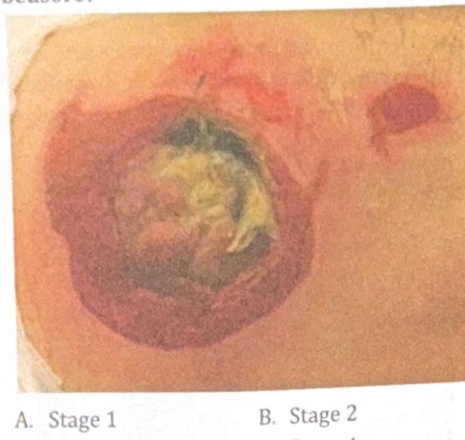
A 78-year-old immobile patient presents with a pressure ulcer on the sacral area, as shown in the image below. How would you grade this bedsore?

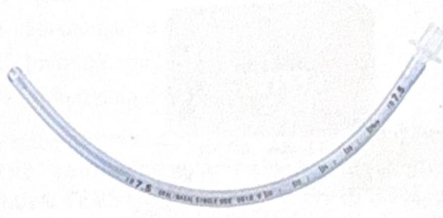
Identify the instrument shown in the image:

In which condition is gas under the diaphragm most commonly seen on imaging?
Which of the following is the most commonly performed repair for a direct inguinal hernia?
Identify the given bone marrow biopsy instrument

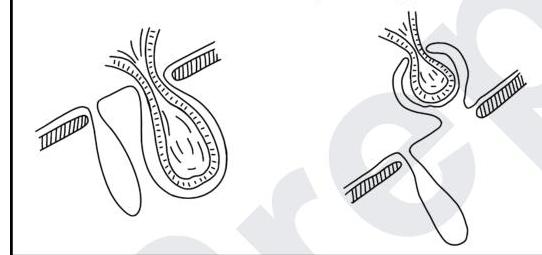
A surgeon examined the case of hernia. Forcefully reduces the sac in abdominal cavity, without actually pushing back the contents. Identify type of hernia with the image given.

Explanation: ***Disclosure to department with gradual transition plan*** - This option balances the **surgeon's well-being**, **patient safety**, and **professional responsibility**. - **Early disclosure** allows for confidential discussion, workplace accommodations, and a planned transition while minimizing risk to patients. - This approach aligns with **medical ethics principles** including beneficence, non-maleficence, and professional integrity. *Transition to non-operative subspecialty* - While a potential long-term solution, this is a **premature and abrupt decision** without exploring other possibilities or accommodations. - Doing so immediately may cause **unnecessary career disruption** and financial strain without fully assessing the impact of early-stage Parkinson's. *Immediately cease all surgical practice* - This is an **overly drastic measure** at the early stage of Parkinson's disease, especially before evaluating treatment options or functional impairment. - It could lead to significant **personal and professional hardship** without adequate assessment of the actual risk. *Continue practice without disclosure until symptoms worsen* - This approach **prioritizes self-interest over patient safety** and professional ethics. - Withholding critical information about a condition that affects motor skills in a surgeon performing microsurgery is a **breach of ethical conduct** and could lead to serious harm to patients. - This violates the fundamental principle of **informed consent** and the duty to prevent harm.
Explanation: ***Umbilical excision with mesh hernioplasty*** - The presence of an **irreducible umbilical mass with discharge** indicates a complicated umbilical hernia, likely with **incarceration, strangulation, or infection**. - Management requires **excision of compromised tissue** (umbilicus and surrounding necrotic/infected skin) followed by **hernia defect repair**. - **Mesh hernioplasty** provides strong, durable reinforcement and prevents recurrence. - **Note:** In heavily contaminated fields, primary tissue repair or biologic mesh may be preferred over synthetic mesh, or staged repair may be considered. However, if contamination is minimal after debridement, mesh repair can be performed in the same setting. *Umbilical excision* - While **excision of the compromised umbilical skin and necrotic tissue** is necessary due to the discharge (suggesting infection or necrosis), **excision alone does not address the underlying hernia defect**. - Simply excising the umbilicus without repairing the hernia would lead to **persistent hernia or recurrence**. *Mesh hernioplasty* - A mesh hernioplasty alone is appropriate for **uncomplicated, reducible umbilical hernias** to reinforce the abdominal wall. - However, it **does not account for the irreducibility and skin changes/discharge**, which necessitate **excision of potentially infected or necrotic tissue** first. - Placing mesh without addressing the compromised tissue would risk ongoing infection and mesh complications. *Conservative* - **Conservative management** is reserved for **asymptomatic, reducible umbilical hernias** in adults (especially if small) or for infants where spontaneous closure can occur. - An **irreducible mass with discharge** signifies an **acute surgical emergency** (incarceration, strangulation, or infection) requiring **urgent surgical intervention**, not observation.
Explanation: ***Surgical management*** - The chest X-ray shows a **widened mediastinum** and abnormal aortic contour, highly suggestive of **aortic dissection involving the ascending aorta (Type A)**. - **Type A aortic dissection** (involving the ascending aorta) is a **surgical emergency** requiring immediate operative repair to prevent life-threatening complications such as cardiac tamponade, acute aortic regurgitation, or rupture. - The constant chest pain with these radiological findings indicates urgent surgical intervention is the definitive management. *Vasodilator* - Vasodilators **alone** should never be used in aortic dissection as they can increase aortic wall shear stress and propagate the dissection. - They must always be preceded by beta-blockade to prevent reflex tachycardia. - Vasodilators do not address the structural defect requiring surgical correction in Type A dissection. *Beta blocker* - Beta-blockers are essential for **initial medical stabilization** to reduce heart rate (target <60 bpm) and blood pressure, thereby decreasing aortic wall stress (dP/dt). - However, in **Type A dissection**, beta-blockers alone do not address the structural defect and are used as a bridge to emergency surgery, not as definitive treatment. - For Type B dissections (descending aorta), medical management with beta-blockers may be definitive in uncomplicated cases. *Beta blocker plus vasodilator* - This combination represents optimal **medical management** for blood pressure and heart rate control in aortic dissection. - In **Type B (descending) aortic dissections**, this is often the definitive treatment for uncomplicated cases. - However, in **Type A dissections** (as indicated by the widened mediastinum suggesting ascending aortic involvement), this serves only as initial stabilization before **mandatory surgical repair**, not as definitive therapy. - Surgery cannot be delayed in Type A dissection due to high mortality risk (1-2% per hour).
Explanation: ***Pilonidal sinus*** - This is the classic presentation of **pilonidal sinus disease**, historically known as **"Jeep disease"** due to its high incidence in military personnel during WWII who sat for prolonged periods in jeeps - The **occupational clue "jeep driver"** is pathognomonic for pilonidal sinus, as prolonged sitting causes friction and pressure in the **sacrococcygeal/natal cleft region** - Presents with **chronic pain, swelling, and intermittent pus discharge** in the gluteal region, typically over weeks to months - Risk factors include: prolonged sitting, friction, deep natal cleft, obesity, and excessive body hair - The **chronic 6-month duration** with ongoing discharge is characteristic of pilonidal sinus with secondary infection, not an acute abscess *Gluteal abscess* - While this can cause pain, swelling, and pus discharge, it typically presents **acutely** (days to weeks, not 6 months) - Does not have the specific **occupational association with prolonged sitting** (jeep driver) - Would be expected to either resolve with drainage/antibiotics or progress to sepsis, not persist chronically for 6 months - Lacks the classic "Jeep disease" eponym *Fistula in ano* - This involves an abnormal tract between the **anal canal and perianal skin** - Discharge would be localized **perianally**, close to the anus, not in the broader gluteal/sacrococcygeal region - Does not have the jeep driver occupational association *Fissure in ano* - A **tear in the anal canal lining** causing severe pain during defecation with **bright red bleeding** - Does not present with swelling or chronic pus discharge - Pain is characteristically related to bowel movements, not constant
Explanation: *Stage 1* - A Stage 1 pressure ulcer is characterized by **non-blanchable erythema** of intact skin, typically over a bony prominence. - There is no break in the skin, which is not what is visible in the image. *Stage 2* - A Stage 2 pressure ulcer involves **partial-thickness skin loss** presenting as a shallow open ulcer with a red-pink wound bed, without slough or bruising. - The depth and tissue involvement shown in the image are far more extensive than a Stage 2 ulcer. *Stage 3* - A Stage 3 pressure ulcer involves **full-thickness tissue loss** where subcutaneous fat may be visible, but bone, tendon, or muscle are not exposed. - The visible structures in the image suggest involvement beyond the subcutaneous fat layer, distinguishing it from a Stage 3 ulcer. ***Stage 4*** - The image shows **full-thickness tissue loss** with visible **bone, tendon, or muscle**, indicating a Stage 4 pressure ulcer. - There is also evidence of **slough and eschar** in the wound bed, which is consistent with severe tissue damage extending beyond the subcutaneous fat. - This is the **correct answer** as the exposed deeper structures are pathognomonic for Stage 4.
Explanation: ***Uncuffed endotracheal (ET) tube*** - This image displays a transparent, flexible tube with a distinct connector at one end and no inflated cuff near the distal tip, which is characteristic of an **uncuffed endotracheal tube**. - Uncuffed ET tubes are commonly used in **pediatric patients** where a cuff could potentially damage the narrower, cone-shaped trachea. *Nasogastric tube* - A nasogastric tube typically has a much **smaller diameter** and a distinctly different tip design, often with multiple side ports for fluid aspiration or administration. - It does not feature the large, universal connector seen on endotracheal tubes. *Oropharyngeal tube* - An oropharyngeal (Guedel) airway is a **rigid, curved device** inserted into the mouth to maintain an open airway, and it looks distinctly different from the flexible tube shown. - It does not extend into the trachea like an ET tube. *Tracheostomy tube* - A tracheostomy tube is typically shorter, often with a curved neck flange for securement to the neck, and frequently made with an outer and inner cannula, which are absent in the image. - While it helps maintain an airway, its design is specific for insertion directly into a tracheostomy stoma, unlike the longer tube for oral/nasal intubation.
Explanation: ***Correct: Secondary*** - **Gas under the diaphragm** is a classic sign of a **perforated viscus**, which is a severe form of **secondary peritonitis**. - Secondary peritonitis typically arises from the **rupture or perforation of an abdominal organ**, allowing gas and contents to leak into the peritoneal cavity. *Incorrect: Tertiary* - **Tertiary peritonitis** refers to persistent or recurrent peritonitis after seemingly adequate surgical and antibiotic treatment for primary or secondary peritonitis. - It is associated with **immunocompromised states** or **low-virulence organisms** and does not typically present with free gas under the diaphragm. *Incorrect: Retained stone* - A **retained stone** (e.g., gallstone, renal stone) can cause obstruction or inflammation but does not directly lead to **gas under the diaphragm**. - While it might indirectly lead to perforation if neglected, it's not the most common direct cause of **free peritoneal gas**. *Incorrect: Primary* - **Primary peritonitis** (also known as spontaneous bacterial peritonitis) occurs without an apparent source of contamination within the abdominal cavity, often in patients with ascites and **liver cirrhosis**. - It is characterized by bacterial infection of the ascitic fluid but does not involve a perforated viscus or **free gas under the diaphragm**.
Explanation: ***Lichtenstein repair*** - The **Lichtenstein repair** is a **tension-free mesh repair** and is the most commonly performed technique for direct inguinal hernias due to its low recurrence rates. - This technique involves placing a **synthetic mesh** over the deficient posterior wall of the inguinal canal, reinforcing the weakened area. *Bassini's repair* - **Bassini's repair** is a tissue-based repair that involves suturing the conjoined tendon to the inguinal ligament. - It creates tension on the repair, which historically led to **higher recurrence rates** compared to mesh repairs. *Herniotomy* - **Herniotomy** is the surgical removal of the **hernia sac** without repairing the defect in the abdominal wall. - This procedure is typically reserved for **pediatric patients** with indirect inguinal hernias due to the good intrinsic muscle tone and relatively small defect, but it is not the primary repair for direct inguinal hernias in adults. *All of the options* - While all listed procedures are methods for hernia management, only the **Lichtenstein repair** is considered the most commonly performed repair for direct inguinal hernias in current practice. - The other options are less commonly performed or reserved for specific patient populations.
Explanation: ***Jamshidi needle*** - The image displays a **Jamshidi needle**, characterized by its **tapered, beveled tip** designed to facilitate entry into the bone and procure an intact core of bone marrow. - This needle is widely considered the **gold standard** for bone marrow biopsy due to its effectiveness in obtaining high-quality trephine samples. *Salah needle* - A Salah needle is primarily used for **bone marrow aspiration**, not typically for a trephine biopsy, and it has a different design meant for aspirating liquid marrow. - It features a **shorter, sturdier design** with a sharp bevel, optimized for safely extracting marrow fluid. *Tru-cut needle* - The Tru-cut needle is primarily designed for obtaining **soft tissue biopsies** (e.g., liver, kidney, prostate) and is not typically used for bone marrow biopsies. - Its mechanism involves an inner cutting stylet and an outer cutting cannula, which is unsuitable for penetrating dense bone and retrieving a bone core. *Vim Silverman needle* - The Vim Silverman needle is also designed for **soft tissue biopsies**, similar to the Tru-cut, and not specifically for bone marrow. - It utilizes a split needle design to capture tissue, which is not appropriate for obtaining a solid bone marrow core.
Explanation: ***Reduction en masse*** - **Reduction en masse** is a dangerous complication that occurs during attempted hernia reduction where the entire hernia sac, along with its incarcerated contents, is pushed back into the abdominal cavity. - The key feature is that **the contents remain trapped within the sac** after reduction, creating a false sense of successful reduction. - The scenario explicitly describes this: "forcefully reduces the sac... without actually pushing back the contents" - this is the textbook definition of reduction en masse. - This complication is dangerous because the incarcerated/strangulated bowel remains undetected inside the abdomen, potentially leading to **peritonitis and bowel necrosis**. - The hernia defect appears reduced externally, but the obstruction persists internally. *Incarcerated hernia* - An **incarcerated hernia** is the state where hernia contents are trapped and cannot be reduced back into the abdominal cavity. - This represents the **pre-existing condition** before the forceful reduction attempt was made. - While incarceration may have been present initially, the question asks about the outcome after the surgeon "forcefully reduces the sac" - this action creates a reduction en masse. *Sliding hernia* - A **sliding hernia** involves a retroperitoneal organ (colon, bladder, ovary) forming part of the hernia sac wall itself. - This is a structural variant unrelated to the reduction complication described in the scenario. *Maydl's hernia* - **Maydl's hernia** (W-hernia or retrograde strangulation) involves a loop of bowel where both ends remain in the abdomen while the intermediate segment is trapped in the hernia sac. - The strangulated segment is the intra-abdominal portion, not the part in the sac. - This is a specific type of hernia content configuration, not related to the reduction complication described.
Wound Healing and Care
Practice Questions
Surgical Infections
Practice Questions
Fluid and Electrolyte Management
Practice Questions
Nutrition in Surgical Patients
Practice Questions
Hemostasis and Blood Transfusion
Practice Questions
Surgical Instruments and Equipment
Practice Questions
Sutures and Stapling Devices
Practice Questions
Minimal Access Surgery Principles
Practice Questions
Surgical Complications
Practice Questions
Anesthesia Principles for Surgeons
Practice Questions
Surgical Oncology Principles
Practice Questions
Evidence-Based Surgery
Practice Questions
Get full access to all questions, explanations, and performance tracking.
Start For Free