Which retractor is shown in the image?

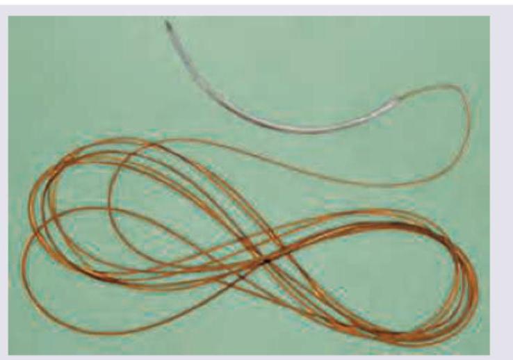
A surgical suture material is shown in the image below. Which of the following statements is TRUE about this suture?

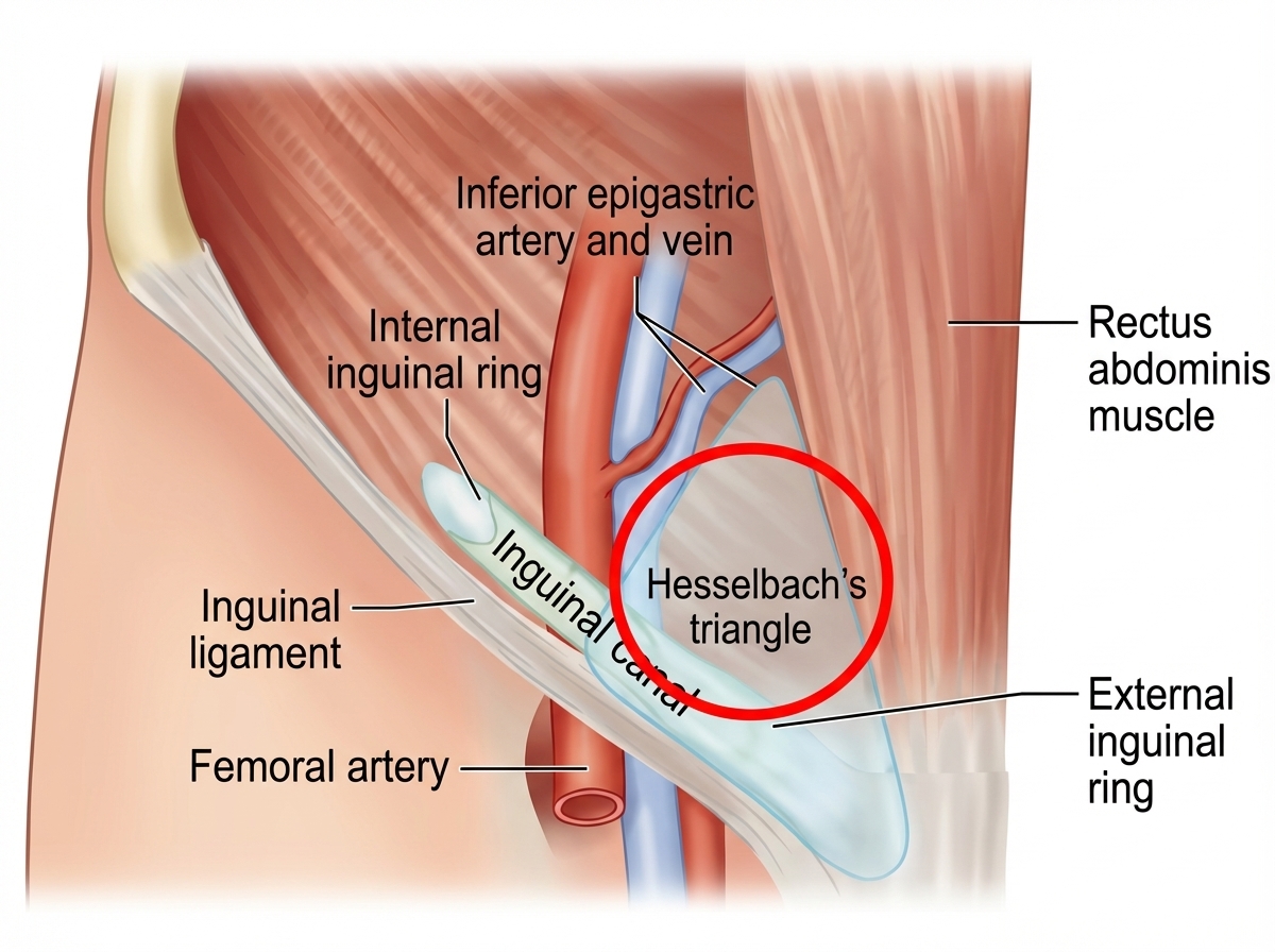
The patient has developed a hernia at the site highlighted by red circle. What is this hernia called as per NYHUS classification? (NEET Pattern 2018)

Identify the instrument shown in the image, which is a lumbar puncture needle with a sharp cutting edge and a hub indicating CSF.

A 60-year-old male presents to the emergency department on the second day of symptom onset with progressive deterioration of consciousness due to large right MCA territory infarct. Which of the following is the most appropriate management? (Image: img-179.jpeg)

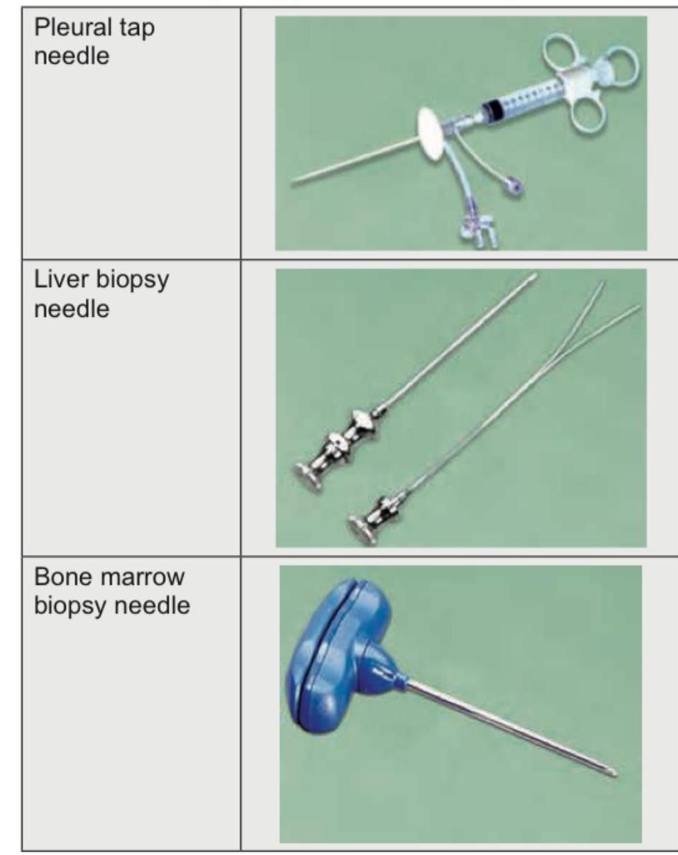
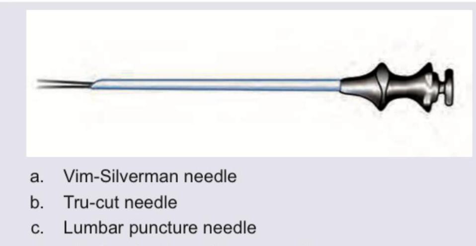
Identify the instrument shown in the figure below.

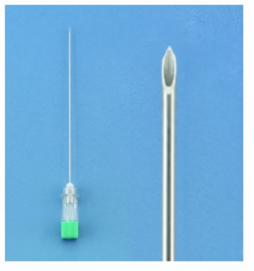
Which needle is shown below?

Which of the following are the techniques commonly used to close the raw area after excision of a pilonidal sinus in order to avoid a midline wound? I. Limberg procedure II. Y-V plasty III. Z-plasty IV. Karydakis procedure Select the correct answer using the code given below :
Which of the following statements are correct regarding sutures in surgery? I. Barbed sutures have the advantage of eliminating the need for knots. II. Vertical mattress sutures help in eversion of wound edges. III. Aberdeen knot is used for continuous suturing. IV. Silk is preferred for subcuticular suturing. Select the answer using the code given below :
A surgeon is about to start a laparoscopic procedure on a patient. The floor nurse asks the surgeon about the identity of the patient, site of the procedure to be performed and any anticipated critical events during the surgery. These questions are a part of the
Explanation: ***Deaver retractor*** - The Deaver retractor is easily identifiable by its distinct **flat, C-shaped, curved blade** at one end and a **long handle with a fenestrated grip**. - It is commonly used in deep abdominal and thoracic procedures to **retract organs and tissues**, providing excellent exposure. *Morris retractor* - The Morris retractor typically features a **curved blade** that is less pronounced than a Deaver and often includes a **fenestrated handle**. - However, its blade is generally **flatter and broader** compared to the distinctive C-shape of the Deaver. *Langenbeck retractor* - A Langenbeck retractor is characterized by a **small, L-shaped blade** and a handle, designed for retracting small incisions and delicate tissues. - Its blade is significantly smaller and has a **sharper, angled bend** compared to the broad, C-shaped blade shown. *Doyen retractor* - The Doyen retractor, also known as a Doyen rib raspatory, is typically used to **retract ribs** and protect lung tissue during thoracic surgeries. - It has a distinctive **hooked end** or a spoon-like blade, which is quite different from the broad, curved blade in the image.
Explanation: ***Absorbed by phagocytosis and enzymatic degradation*** - The image shows a **braided absorbable suture**, which undergoes absorption through **tissue reaction** involving enzymatic breakdown and phagocytosis - This is the characteristic mechanism of absorption for most **absorbable sutures** (both natural like catgut and synthetic like Vicryl, Monocryl, PDS) - The body's macrophages phagocytose suture particles while proteolytic enzymes break down the material components, allowing complete removal from tissues *Derived from catgut* - While **catgut sutures** (derived from sheep/bovine intestinal submucosa) are absorbable, modern synthetic absorbable sutures like **polyglactin (Vicryl)** or **polyglycolic acid (Dexon)** are more commonly used - The image appearance and braided structure suggest a synthetic absorbable suture rather than catgut - Catgut is less commonly used today due to higher tissue reactivity and unpredictable absorption *Non-absorbable* - **Non-absorbable sutures** (silk, nylon, polypropylene) remain permanently in tissues or require removal - The mechanism of absorption by phagocytosis and enzymatic degradation specifically defines **absorbable sutures**, making this option incorrect - Non-absorbable sutures maintain tensile strength indefinitely *Absorbed over 3 months* - Absorption time varies significantly by suture type: **rapidly absorbing gut (5-7 days)**, **Vicryl (56-70 days)**, **PDS (180-210 days)** - While some sutures absorb around 90 days, this is not a universal characteristic - The statement is too specific and not applicable to all absorbable sutures shown
Explanation: ***Nyhus Type IIIB*** - The image shows a hernia occurring within **Hesselbach's triangle**, which is the anatomical site for a **direct inguinal hernia**. A direct inguinal hernia is classified as **Nyhus Type III**, as it involves a defect in the posterior wall of the inguinal canal, medial to the inferior epigastric vessels. - The "B" in Nyhus Type IIIB specifically denotes a **direct inguinal (III) hernia with a weakness in the posterior inguinal wall**. While the question asks for the specific Nyhus classification, options like Nyhus Type I, Type II, and Type IIIA, as well as a femoral hernia (Nyhus Type IV), are inconsistent with the highlighted location. *Nyhus Type I* - This classification refers to an **indirect inguinal hernia** where the internal ring is normal, typically seen in children. - It would originate lateral to the inferior epigastric artery, unlike the highlighted region. *Nyhus Type II* - This type describes an **indirect inguinal hernia** with an enlarged internal ring but an intact posterior inguinal wall. - This type of hernia would still originate lateral to the inferior epigastric vessels, not within Hesselbach's triangle. *Nyhus Type IIIA* - Nyhus Type IIIA specifically classifies an **indirect inguinal hernia** with an enlarged internal ring and a completely destructed transversalis fascia. - While it's also a Type III (indirect), Type IIIA refers to an indirect hernia, which emerges lateral to the inferior epigastric vessels, whereas a direct hernia (within Hesselbach's triangle) emerges medial to these vessels. *Nyhus Type IV* - This classification is for a **femoral hernia**, which occurs inferior to the inguinal ligament and through the femoral canal. - The highlighted area is clearly superior to the inguinal ligament and within the inguinal region, not the femoral region.
Explanation: ***Quincke's needle*** - This image shows a **Quincke's needle**, which is a type of spinal needle commonly used for lumbar punctures. - It features a **sharp, beveled cutting edge** designed to cut through tissue layers, and the hub allows for CSF flow immediately upon entry into the subarachnoid space. - Quincke needles are **cutting needles** with a traditional beveled tip. *Whitacre needle* - This is a **pencil-point, atraumatic needle** with a rounded tip and side opening. - Designed to separate rather than cut dural fibers, reducing post-dural puncture headache risk. *Sprotte needle* - Another **pencil-point, atraumatic needle** similar to Whitacre but with a larger side opening. - Also designed to minimize post-dural puncture headache. *Tuohy needle* - A **curved-tip needle** primarily used for **epidural anesthesia**, not lumbar puncture. - Features a Huber tip that helps guide the epidural catheter.
Explanation: ***Decompressive surgery*** - A large **MCA territory infarct** causes significant brain edema, leading to increased **intracranial pressure** and progressive deterioration of consciousness, especially on day 2. - **Decompressive craniectomy** is often necessary in such cases to reduce pressure, prevent herniation, and improve outcomes by removing a portion of the skull. *Anticoagulation* - Anticoagulation is primarily used for preventing future thrombotic events due to conditions like **atrial fibrillation**, or for managing an evolving stroke if there's no major hemorrhage risk. - In the setting of a **large ischemic stroke** with significant edema and risk of hemorrhagic transformation, anticoagulation can increase the risk of bleeding into the infarct. *Dual antiplatelet therapy* - **Dual antiplatelet therapy** (DAPT) is typically used to prevent recurrent ischemic events following a **minor stroke** or **transient ischemic attack (TIA)**. - It is not indicated for managing acutely deteriorating consciousness due to brain edema in a **large established infarct**. *Mechanical thrombectomy* - **Mechanical thrombectomy** is an acute intervention performed within a narrow time window (typically up to 24 hours in select patients, but ideally much earlier) to remove the clot and **restore blood flow**. - On the second day of symptom onset, with a fully developed **large infarct** and progressive neurological deterioration due to edema, the brain tissue is likely already irreversibly damaged, making thrombectomy ineffective and potentially harmful due to reperfusion injury.
Explanation: ***Quincke's needle*** - A **Quincke's needle** is a spinal needle with a **sharp, beveled cutting tip** designed for lumbar punctures and cerebrospinal fluid access. - The cutting edge creates a clean penetration through the **dura mater** but may cause **post-dural puncture headache** due to larger dural tears compared to pencil-point needles. *Abraham's needle* - An **Abraham's needle** is a specialized **pleural biopsy needle** with a cutting mechanism designed to obtain pleural tissue samples. - Features a **notched cutting edge** and **outer cannula system** that differs significantly from the simple beveled design of spinal needles. *Liver biopsy needle* - **Liver biopsy needles** (such as Menghini or Jamshidi types) have **blunt or slightly beveled tips** designed to core tissue samples. - They typically feature **depth markings** and a **larger bore** for tissue acquisition, unlike the thin profile of spinal needles. *Bone marrow aspiration needle* - **Bone marrow needles** have a **trocar-style tip** designed to penetrate cortical bone and access marrow cavity. - Feature **depth guards**, **T-shaped handles**, and **larger gauge** construction for bone penetration, distinct from the delicate design of spinal needles.
Explanation: ***Vim-Silverman needle*** - The image displays a needle with two prongs at the tip, which is characteristic of the **Vim-Silverman needle** used for **biopsy**. - This design allows for the capture of a tissue core between the prongs for microscopic examination. *Tru-cut needle* - A Tru-cut needle is also a biopsy needle, but it typically has a **single, cutting cannula** with a notch in its inner stylet. - Its design is different from the dual-pronged tip shown in the image. *Lumbar puncture needle* - A **lumbar puncture needle** (spinal needle) is used to access the subarachnoid space for cerebrospinal fluid (CSF) collection or drug administration. - It usually has a single, sharp bevel and a stylet, but lacks the biopsy collecting mechanism seen in the image. *Abraham's pleural biopsy needle* - **Abraham's pleural biopsy needle** is specifically designed for pleural biopsies and typically has a hook or a cutting edge that can be closed to obtain a tissue sample. - Its structure differs significantly from the bifurcated tip shown in the image.
Explanation: ***I, II and IV*** - The **Limberg procedure** (rhomboid flap) and **Karydakis flap** are the most widely established plastic surgical techniques used to close the defect after pilonidal sinus excision. - These techniques aim to **flatten the natal cleft** and move the scar away from the midline, reducing tension and recurrence rates. - **Y-V plasty** is less commonly used specifically for pilonidal sinus compared to Limberg and Karydakis procedures, though it can be employed for tissue advancement in selected cases. - This combination excludes Z-plasty, which is not a primary technique for pilonidal sinus closure. *II, III and IV* - While **Karydakis procedure** is indeed a standard technique, this option incorrectly includes **Z-plasty**, which is generally used for **scar revision** or releasing contractures rather than primary closure of large excisional defects. - It also excludes the **Limberg flap**, which is one of the most commonly used techniques worldwide for pilonidal sinus surgery. *I, III and IV* - This option incorrectly includes **Z-plasty** while excluding **Y-V plasty**. - Z-plasty is not a primary technique for closing pilonidal sinus defects as it doesn't provide adequate tissue coverage for large excisions. - The **Limberg and Karydakis procedures** are the mainstay techniques from this list. *I, II and III* - This combination incorrectly includes **Z-plasty** as a primary technique for closing the excisional defect. - It omits the **Karydakis procedure**, which is one of the most widely recognized and effective flaps for pilonidal disease with excellent outcomes. - The Karydakis flap specifically focuses on **modifying the natal cleft contour** and displacing the incision laterally for better healing and lower recurrence rates.
Explanation: ***I, II and III*** - **I. Barbed sutures** have unidirectional or bidirectional barbs that grip tissue, negating the need for traditional knots to secure the suture line. This property can significantly **reduce operating time** and the volume of foreign material left in the wound. - **II. Vertical mattress sutures** are designed to achieve precise wound edge approximation and eversion, which are crucial for optimal healing and cosmesis, particularly in areas under tension or for thick skin. - **III. The Aberdeen knot** is a slip knot technique specifically designed to secure the end of a **continuous suture line** efficiently and reliably. It provides a flat, secure knot that minimizes bulk and is less prone to loosening. *I, II and IV* - While statements I and II are correct, statement IV is incorrect. **Silk is a braided, non-absorbable multifilament suture** that can cause significant tissue reaction. - It is generally not preferred for subcuticular suturing due to its increased risk of infection, visibility, and foreign body reaction compared to monofilament, absorbable sutures. *II, III and IV* - Statements II and III are correct, but statement IV is incorrect. **Silk is avoided for subcuticular closure** due to its inflammatory properties and potential for suture extrusion or sinus formation. - Subcuticular sutures typically use **absorbable monofilament sutures** (e.g., poliglecaprone 25 or polydioxanone) to minimize tissue reaction and achieve good cosmetic results. *I, III and IV* - Statements I and III are correct regarding barbed sutures and the Aberdeen knot, respectively. However, statement IV is incorrect because **silk suture is a non-absorbable, braided material that is highly reactive and not suitable for subcuticular placement**, where monofilament absorbable sutures are preferred for minimal tissue reaction and good cosmesis.
Explanation: **WHO surgical safety checklist** - The questions about patient identity, procedure site, and anticipated critical events are key components of the **"Sign In"** and **"Time Out"** sections of the **WHO Surgical Safety Checklist**. - This checklist is designed to improve **patient safety** by ensuring communication and adherence to essential steps before, during, and after surgery, thereby reducing surgical errors. *nurses safety checklist* - While nurses play a crucial role in patient safety, there isn't a universally recognized "nurses safety checklist" that specifically encompasses these exact comprehensive surgical verification steps. - The comprehensive framework described, with its specific questions, aligns more closely with the broader, interdisciplinary **WHO Surgical Safety Checklist**. *universal precautions checklist* - **Universal precautions** focus on preventing the transmission of bloodborne pathogens and other infectious agents by treating all bodily fluids as potentially infectious. - This checklist primarily addresses **infection control** measures and does not cover patient identification, surgical site verification, or critical event anticipation. *MCI patient safety checklist* - A "MCI patient safety checklist" is not a widely recognized or standardized medical safety protocol. - The scenario describes a standard, internationally adopted set of safety checks specifically for surgical procedures, which is the **WHO Surgical Safety Checklist**.
Wound Healing and Care
Practice Questions
Surgical Infections
Practice Questions
Fluid and Electrolyte Management
Practice Questions
Nutrition in Surgical Patients
Practice Questions
Hemostasis and Blood Transfusion
Practice Questions
Surgical Instruments and Equipment
Practice Questions
Sutures and Stapling Devices
Practice Questions
Minimal Access Surgery Principles
Practice Questions
Surgical Complications
Practice Questions
Anesthesia Principles for Surgeons
Practice Questions
Surgical Oncology Principles
Practice Questions
Evidence-Based Surgery
Practice Questions
Get full access to all questions, explanations, and performance tracking.
Start For Free