A 25-year-old male presents with inguinal swelling. He had surgery for acute abdomen 2 years ago but could not tell the reason behind it. Trauma to which structure during the surgery conducted 2 years ago would have resulted in this inguinal swelling?
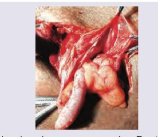
Contents of the inguinal hernia sac are displayed intraoperatively and appendix is seen at 7 o'clock. What is the diagnosis?

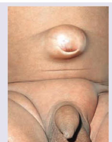
A 35-year-old patient presents with a painless swelling at the umbilicus that has been gradually increasing in size over the past 6 months. The swelling increases on coughing and straining. On examination, there is a reducible bulge at the umbilicus. What is the likely diagnosis?

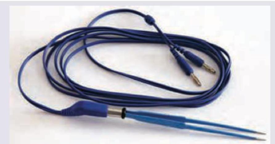
What is the functional capability of the instrument shown in the image?

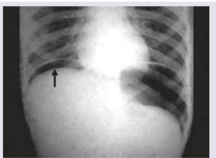
A 30-year-old patient presents with abdominal pain, fever, nausea, vomiting and respiratory distress. On admission BP=80/40 mm Hg and pulse rate is 120 BPM. The following CXR was performed. What is the immediate management? (AIIMS May 2016)

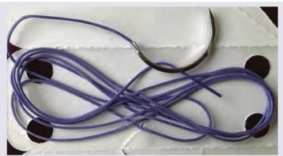
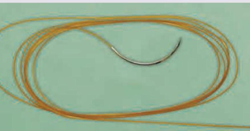
What type of suture is this?

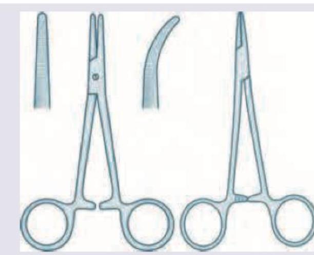
Identify these two surgical instruments.

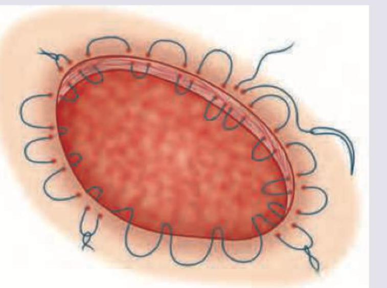
The following suture material gets absorbed in how many days?

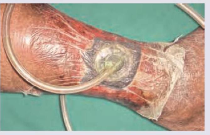
The method of ulcer healing shown below generates a pressure of: (Recent NEET Pattern 2016-17)

Which is correct about the suture material shown in the image?
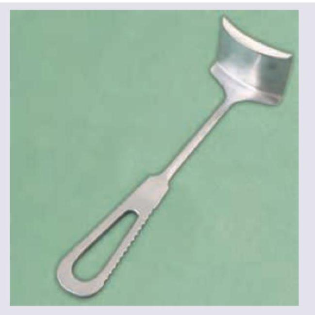
Explanation: ***Ilioinguinal nerve*** - Damage to the ilioinguinal nerve during abdominal surgery, especially an appendectomy, can lead to muscle weakness in the anterior abdominal wall. - This weakness predisposes the patient to the formation of an **inguinal hernia**, which manifests as an inguinal swelling. *Spermatic cord* - Trauma to the spermatic cord could lead to **testicular atrophy**, pain, or issues with fertility due to vascular or ductal damage. - It is not directly associated with the development of an inguinal hernia as a primary consequence of isolated trauma during non-hernia repairs. *Genital branch of genitofemoral nerve* - Injury to the genital branch of the genitofemoral nerve primarily affects the **cremasteric reflex** and sensation in the scrotum/inner thigh. - While it can cause sensory disturbances, it does not directly lead to weakness of the abdominal wall sufficient to cause an inguinal hernia. *Pampiniform plexus* - The pampiniform venous plexus is involved in regulating testicular temperature. Injury primarily causes a **hydrocele** or **varicocele** due to impaired venous drainage. - It would not cause an inguinal hernia, which involves protrusion of abdominal contents through a weakened abdominal wall.
Explanation: ***Amyand hernia*** - An **Amyand hernia** is a rare type of inguinal hernia that contains the **vermiform appendix** within the hernia sac. - The discovery of the appendix, as shown in the image, within an inguinal hernia sac definitively identifies this as an Amyand hernia. *Richter hernia* - A **Richter's hernia** occurs when only a portion of the circumference of the bowel wall becomes incarcerated in the hernia sac. - It does not specifically involve the appendix and is characterized by the potential for **strangulation without obstruction**. *Pantaloons hernia* - A **pantaloons hernia** (or saddlebag hernia) is a combined direct and indirect inguinal hernia, with the inferior epigastric vessels located between the two sacs. - It describes the anatomical configuration of the hernia sacs rather than the contents within them. *Maydl's hernia* - **Maydl's hernia** (or W-hernia) occurs when two loops of bowel are contained within the hernia sac, with a connecting loop lying within the abdominal cavity. - This type of hernia is particularly dangerous due to the high risk of **strangulation of the intervening loop** of bowel.
Explanation: ***Umbilical hernia*** - The image clearly displays a bulge at the **umbilicus**, which is characteristic of an umbilical hernia. - This type of hernia occurs when abdominal contents protrude through a weakness in the **umbilical ring**. - The clinical presentation of a **reducible swelling that increases with coughing** is pathognomonic of a hernia. - Umbilical hernias are common in adults and are associated with increased intra-abdominal pressure. *Epigastric hernia* - An epigastric hernia occurs in the **midline** between the **xiphoid process** and the umbilicus. - The location of the bulge in the image is specifically at the umbilicus, not superior to it. - These typically present as small, often tender nodules in the epigastric region. *Spigelian hernia* - A Spigelian hernia protrudes through the **Spigelian fascia**, which is lateral to the rectus abdominis muscle, typically at the **arcuate line**. - This location is **lateral**, distinct from the periumbilical region shown in the image. - These are rare and often difficult to palpate due to their interparietal nature. *Paraumbilical hernia* - A paraumbilical hernia occurs **adjacent to the umbilicus** through the linea alba, rather than directly through the umbilical ring. - While close in location, the image shows a hernia **directly at the umbilicus**, not beside it. - Paraumbilical hernias are more common in adults, while true umbilical hernias through the ring can occur in both children and adults.
Explanation: ***Used for both cutting and coagulation*** - The image displays a **bipolar electrosurgical forceps**, which is specifically designed to deliver **high-frequency electrical current** for both cutting and coagulating tissue. - The electrical energy is localized between the two tips of the forceps, allowing for precise tissue manipulation with minimal collateral damage and reducing the risk of current spread to other parts of the body. *Used for coagulation* - While this instrument is excellent for **coagulation**, its capabilities extend beyond just stopping bleeding. - It can also be used for **cutting tissue efficiently** by using a different electrical waveform or power setting. *Used for cutting* - This instrument is indeed used for **cutting tissue**, but it also has the critical function of **coagulation**. - Restricting its description to only cutting would be incomplete and overlook its dual utility in surgery. *Cannot be used in patient with artificial valves* - Bipolar electrosurgery is generally considered **safe for patients with pacemakers, ICDs, or artificial valves** because the current is confined between the two tips of the instrument. - This localized current flow **minimizes the risk of interference** with implanted medical devices, unlike monopolar electrosurgery which has a greater risk of current dispersion.
Explanation: ***IV fluids and antibiotics followed by laparotomy*** - The imaging shows **free air under the diaphragm** (pneumoperitoneum), confirming a perforated viscus. The patient is also in **septic shock** (low blood pressure, high heart rate, fever, abdominal pain), requiring immediate resuscitation with **IV fluids and broad-spectrum antibiotics** to stabilize before surgery. - While definitive management is surgical, **resuscitation** must precede laparotomy in an unstable patient to improve outcomes from septic shock. *Immediate laparotomy* - Performing an immediate laparotomy without prior resuscitation in a patient with **septic shock** (BP 80/40, HR 120) significantly increases surgical risk and mortality. - The patient's **hemodynamic instability** must be addressed first to withstand the stress of surgery. *Intravenous fluids* - While critical for initial resuscitation in septic shock, **IV fluids alone** are insufficient as the definitive management for a perforated viscus. - The source of sepsis (perforation) must be addressed surgically, and **antibiotics** are essential to combat the infection. *Intravenous potassium* - There is no indication in the clinical presentation or image for **intravenous potassium administration**. - This intervention is used for treating **hypokalemia**, which is not mentioned as a concern here, and does not address the underlying pathology.
Explanation: ***Purse string suture*** - The image shows a suture placed in a **circular pattern** around a wound opening, which can be **tightened like a drawstring** to close the defect. - This technique is characteristic of a **purse-string suture**, commonly used to close circular openings or to reduce the size of a defect for easier closure. *Pare's suture* - This is not a recognized or standard surgical suture technique. - Suture names are typically associated with their creators (e.g., Halsted) or their specific pattern/function (e.g., mattress, purse-string). *Halsted's suture* - Refers to a type of **interrupted mattress suture** or a principle of meticulous surgical technique, not a specific circular closing suture like the one pictured. - Halsted's sutures are typically used for strong wound closure and **eversion of wound edges**. *Vertical mattress interrupted suture* - This suture involves a "far-far, near-near" pattern, passing deep then superficial on each side of the wound, resulting in **strong closure** and **wound edge eversion**. - It consists of **individual, non-continuous stitches** (interrupted) and its appearance is distinct from the encircling pattern shown.
Explanation: **Artery forceps and needle holder** - The instrument on the left is an **artery forceps (hemostat)**, characterized by its serrated jaws and a locking mechanism, designed for clamping vessels to control bleeding. - The instrument on the right is a **needle holder**, which has short, robust jaws with a cross-hatch pattern for secure gripping of surgical needles, and a ratcheted handle to lock the needle in place. *Thumb forceps and sponge holder* - **Thumb forceps** are non-locking instruments held like a pen, used for grasping tissue, and are not depicted here. - A **sponge holder** (also known as Foerster sponge forceps) typically has large, fenestrated jaws to hold surgical sponges, which is different from either of the instruments shown. *Sponge holder and Allis forceps* - As mentioned, a **sponge holder** is distinct and not shown in the image. - **Allis forceps** have multiple fine teeth that interlock to hold tissue firmly but atraumatically, which differs from the jaw patterns seen in these instruments. *Needle holder and ovum forceps* - While a **needle holder** is correctly identified on the right, the instrument on the left is not an **ovum forceps**. - **Ovum forceps** (or ovum polyp forceps) are used in gynecology, often having wide, fenestrated, cup-shaped jaws for grasping tissue during uterine procedures.
Explanation: **90 days** - The image displays **Vicryl suture**, which is a **braided, absorbable suture** material made of polyglactin 910. - Vicryl typically retains tensile strength for about 2-3 weeks and is **completely absorbed** by hydrolysis in approximately **60 to 90 days**. *7 days* - Sutures that absorb in roughly 7 days, like **plain gut**, have a much **faster absorption profile** and are usually reserved for rapidly healing tissues. - Vicryl maintains its strength much longer and is used for tissues requiring more prolonged support, such as muscle repair. *21 days* - While Vicryl retains sufficient tensile strength for about **21 days (3 weeks)**, this refers to the period it provides adequate wound support, not its complete absorption time. - Complete absorption of Vicryl takes significantly longer than 21 days. *Non-absorbable* - **Non-absorbable sutures**, such as nylon or polypropylene, are designed to remain in the tissue permanently or until physically removed. - Vicryl's composition and mode of degradation (hydrolysis) clearly classify it as an **absorbable suture**.
Explanation: ***Minus 125 mm Hg*** - The image depicts **Negative Pressure Wound Therapy (NPWT)**, also known as vacuum-assisted closure (VAC), which applies subatmospheric pressure to promote wound healing. - The standard therapeutic pressure for NPWT is **-125 mmHg**, which aids in wound contraction, granulation tissue formation, removal of exudate, and increased blood flow to the wound bed. - This pressure setting is well-established in clinical practice and supported by surgical literature. *Minus 100 cm of water* - While this represents negative pressure, **mmHg is the standard unit** used for measuring and reporting NPWT pressure settings, not cm H₂O. - Converting -100 cm H₂O to mmHg yields approximately -73.5 mmHg, which is lower than the commonly effective therapeutic pressure range for NPWT. *Plus 100 cm of water* - This indicates **positive pressure**, whereas NPWT fundamentally relies on **negative (subatmospheric) pressure** to achieve its therapeutic effects. - Positive pressure would not facilitate wound fluid removal, tissue contraction, or granulation tissue formation as intended by NPWT. *Plus 125 cm of water* - This represents **positive pressure**, which contradicts the core mechanism of action of **Negative Pressure Wound Therapy**. - Positive pressure would push against the wound rather than creating the suction needed to draw out exudate and promote wound closure.
Explanation: ***Absorbed by phagocytosis and enzymatic degradation*** - This description is characteristic of **natural absorbable sutures**, such as **catgut** (which appears to be depicted by the golden, multi-filament material with a needle). Catgut is absorbed by the body's enzymatic processes and macrophage activity. - This type of absorption is a key feature distinguishing natural absorbable sutures from synthetic ones (which are absorbed by hydrolysis) and non-absorbable sutures. *It is less reactive* - Natural absorbable sutures like catgut are generally associated with a **higher inflammatory reaction** in tissues compared to synthetic absorbable sutures due to their proteinacious nature. - The body's immune system recognizes these natural proteins, leading to a more pronounced foreign body response. *Derived from cat gut mucosa* - **Catgut sutures** are indeed derived from the sub-mucosal layer of the sheep or goat **intestine**, not the mucosa itself. - The term "catgut" is historical and misleading, as it is not derived from cats. *Used for bowel anastomosis* - While absorbable sutures are used for bowel anastomosis, the use of **plain catgut** (which the image suggests due to its color and appearance) is generally not preferred for critical anastomoses due to its variable and rapid absorption time, which can compromise wound strength before healing is complete. - **Chromic catgut** or synthetic absorbable sutures (like Vicryl or PDS) with more predictable absorption profiles are typically favored for bowel anastomoses.
Wound Healing and Care
Practice Questions
Surgical Infections
Practice Questions
Fluid and Electrolyte Management
Practice Questions
Nutrition in Surgical Patients
Practice Questions
Hemostasis and Blood Transfusion
Practice Questions
Surgical Instruments and Equipment
Practice Questions
Sutures and Stapling Devices
Practice Questions
Minimal Access Surgery Principles
Practice Questions
Surgical Complications
Practice Questions
Anesthesia Principles for Surgeons
Practice Questions
Surgical Oncology Principles
Practice Questions
Evidence-Based Surgery
Practice Questions
Get full access to all questions, explanations, and performance tracking.
Start For Free