
General Surgery Principles — MCQs

On this page
Which of the following has an increased risk?
Malignant change in lipoma is most common in which anatomical location?
A 70-year-old, moderately obese woman presents with a large midline incisional hernia. She underwent colon resection for adenocarcinoma one year previously. Which of the following statements is truest regarding her condition?
What is the clinical significance of the triangle of safety in thoracic procedures?
What is the most appropriate treatment for a desmoid tumor diagnosed in the abdomen?
Bassini's repair is indicated for which type of hernia?
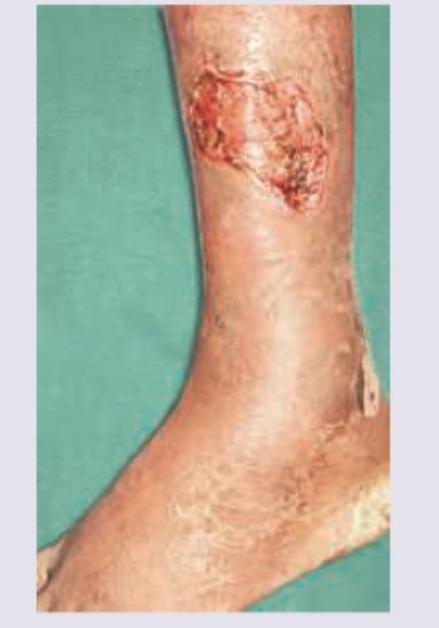
A 50-year-old patient developed a wound infection post-laparotomy for pyoperitoneum and was treated conservatively. Granulation tissue is now seen in the wound. What is the next step in management?
Which surgical blade number is most useful for performing a tracheotomy?
Which of the following is NOT a basic rule for wound closure?
What is the concentration of Potassium in Ringer's Lactate solution?
Practice by Chapter
Wound Healing and Care
Practice Questions
Surgical Infections
Practice Questions
Fluid and Electrolyte Management
Practice Questions
Nutrition in Surgical Patients
Practice Questions
Hemostasis and Blood Transfusion
Practice Questions
Surgical Instruments and Equipment
Practice Questions
Sutures and Stapling Devices
Practice Questions
Minimal Access Surgery Principles
Practice Questions
Surgical Complications
Practice Questions
Anesthesia Principles for Surgeons
Practice Questions
Surgical Oncology Principles
Practice Questions
Evidence-Based Surgery
Practice Questions
Want unlimited practice?
Get full access to all questions, explanations, and performance tracking.
Scan to download app