
General Surgery Principles — MCQs

On this page
Which of the following sutures has maximum tensile strength and minimum tissue reaction?
Most important factor in causation of ingrown toenail is?
Which of the following is typical of rectus sheath hematoma?
Which of the following statements about umbilical hernias is true?
Prolonged surgery time of vaginal hysterectomy may lead to damage to which of the following nerves?
For how many weeks do cortisol levels typically remain elevated following a hemorrhage?
Which type of healing occurs in an incisional wound with infection?
Which of the following types of gangrene is least likely to be associated with diabetes?
Which of the following is NOT a criterion for the viability of muscle?
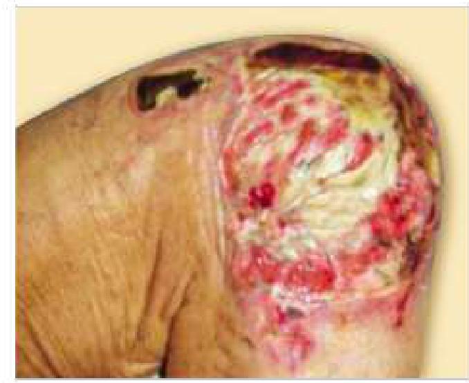
The image below shows a pressure sore. Which stage does this belong to?
Practice by Chapter
Wound Healing and Care
Practice Questions
Surgical Infections
Practice Questions
Fluid and Electrolyte Management
Practice Questions
Nutrition in Surgical Patients
Practice Questions
Hemostasis and Blood Transfusion
Practice Questions
Surgical Instruments and Equipment
Practice Questions
Sutures and Stapling Devices
Practice Questions
Minimal Access Surgery Principles
Practice Questions
Surgical Complications
Practice Questions
Anesthesia Principles for Surgeons
Practice Questions
Surgical Oncology Principles
Practice Questions
Evidence-Based Surgery
Practice Questions
Want unlimited practice?
Get full access to all questions, explanations, and performance tracking.
Scan to download app