
General Surgery Principles — MCQs

On this page
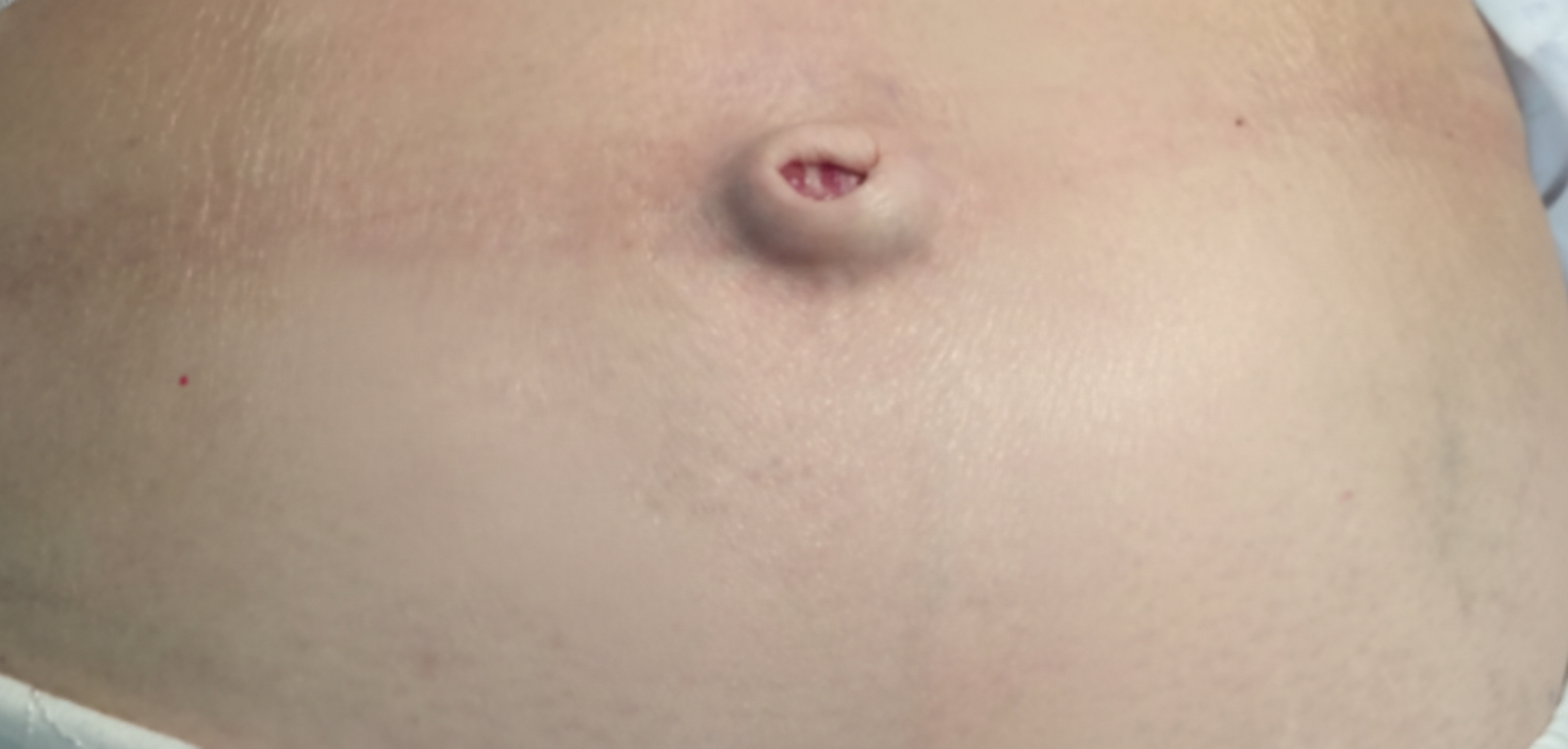
All of the following carcinomas are associated with this condition shown in the image except:
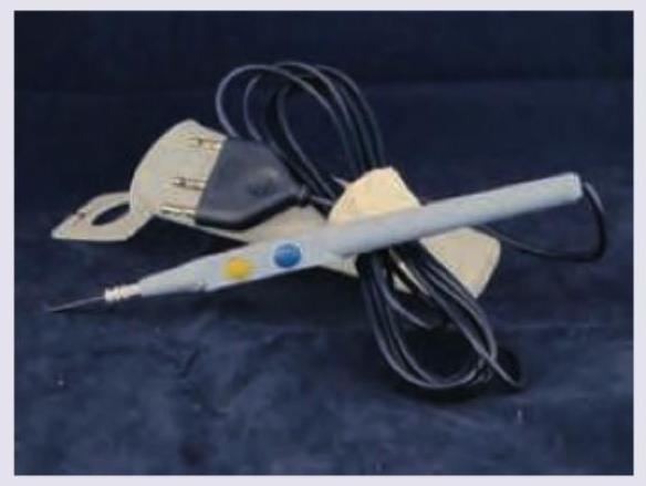
What is the functional capability of the instrument shown in the image?

A 60-year-old male came with bleeding per rectum and was diagnosed to have carcinoma colon. The patient underwent extended hemicolectomy. Identify the instrument the surgeon is using: (AIIMS Nov 2016)
What is the most likely diagnosis based on the image provided? The patient had a snake bite at this site 5 years ago.

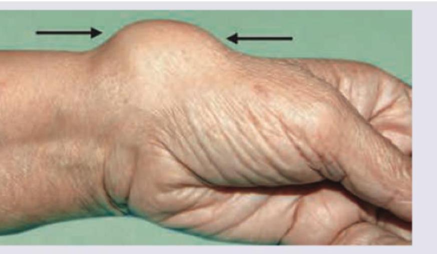
A 60-year-old male patient with a history of weight gain and polyuria presents with the following lesion. What is the most possible diagnosis?

What is shown in the image given below?
All are correct about the image shown below except:

All are true about the lesion shown below except:

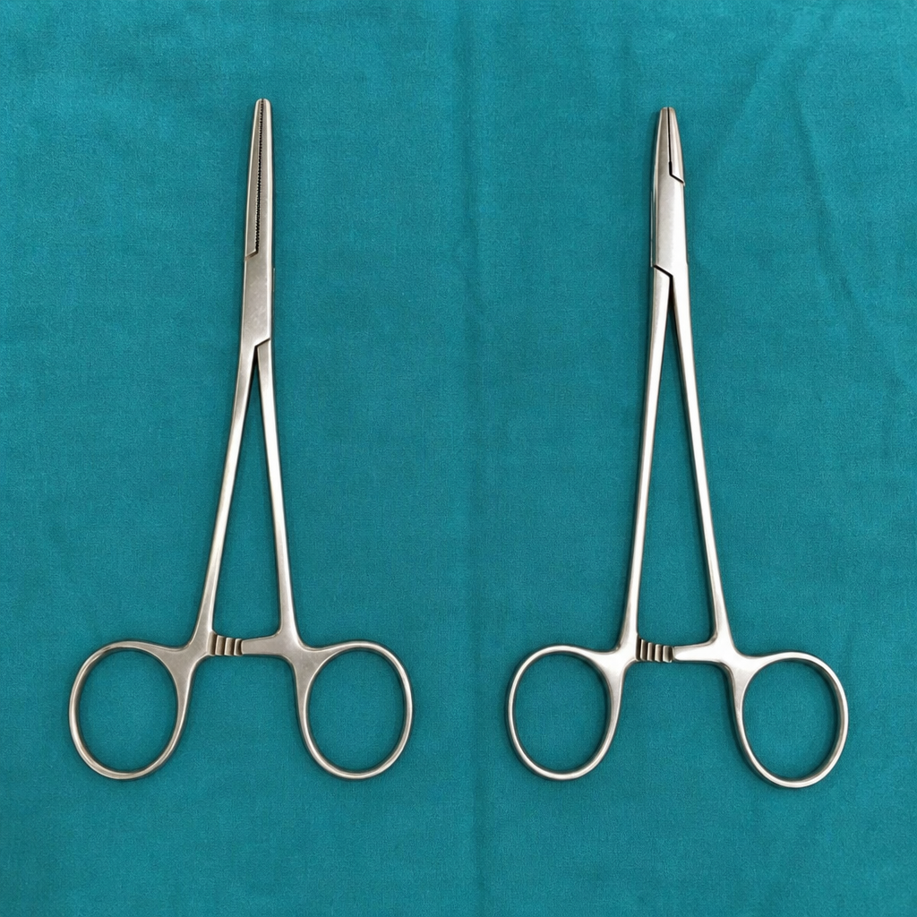
Identify these two surgical instruments.
All are true about the lesion shown below except:

Practice by Chapter
Wound Healing and Care
Practice Questions
Surgical Infections
Practice Questions
Fluid and Electrolyte Management
Practice Questions
Nutrition in Surgical Patients
Practice Questions
Hemostasis and Blood Transfusion
Practice Questions
Surgical Instruments and Equipment
Practice Questions
Sutures and Stapling Devices
Practice Questions
Minimal Access Surgery Principles
Practice Questions
Surgical Complications
Practice Questions
Anesthesia Principles for Surgeons
Practice Questions
Surgical Oncology Principles
Practice Questions
Evidence-Based Surgery
Practice Questions
Want unlimited practice?
Get full access to all questions, explanations, and performance tracking.
Scan to download app