
General Surgery Principles — MCQs

On this page
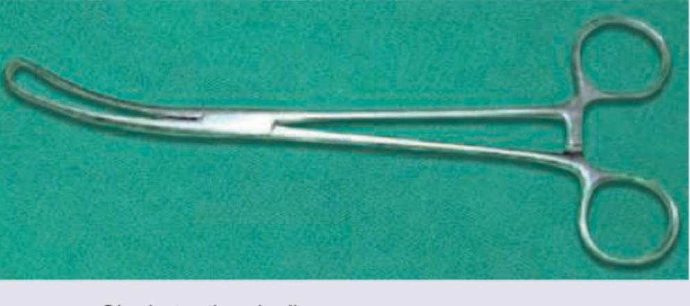
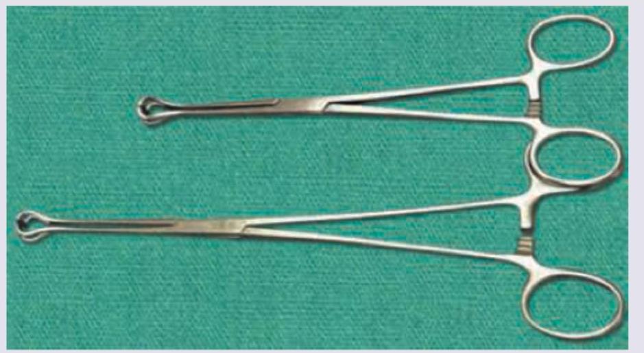
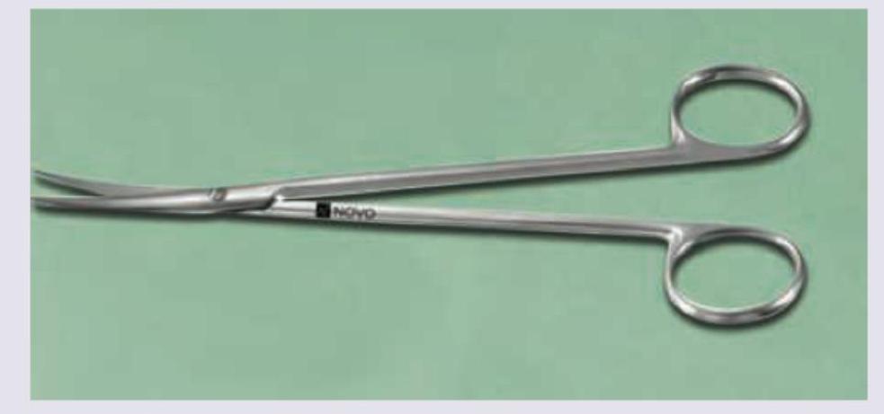
Identify the instrument:
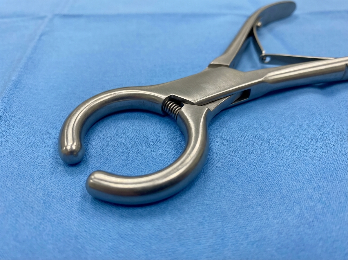
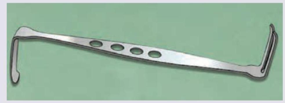
Identify the instrument:
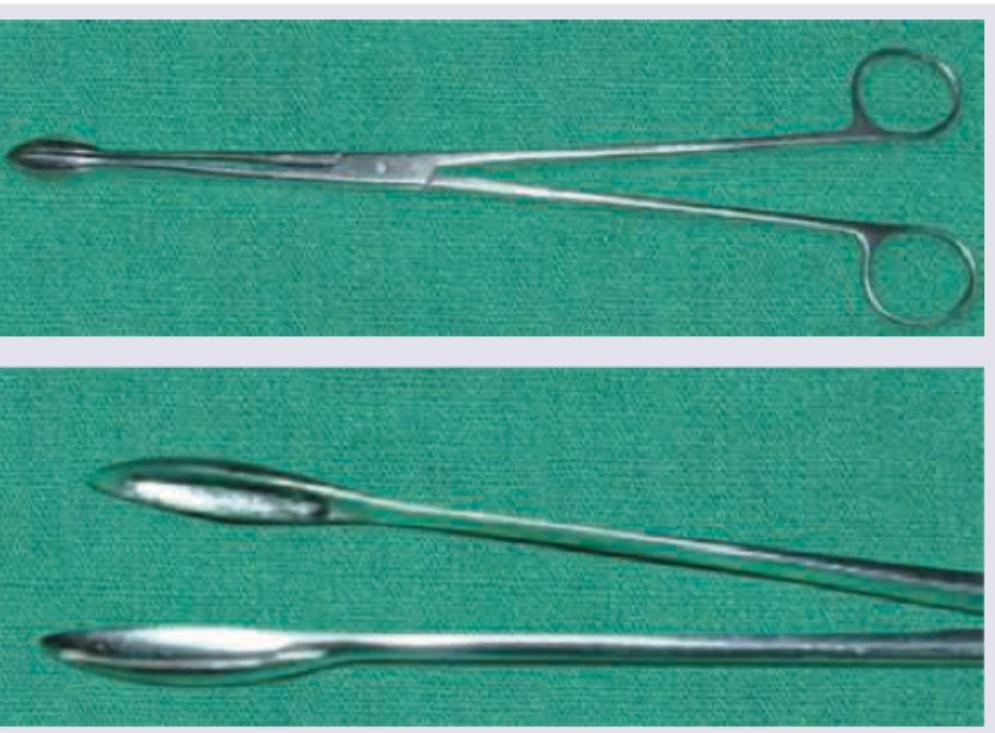
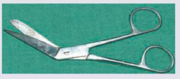
Identify the instrument:
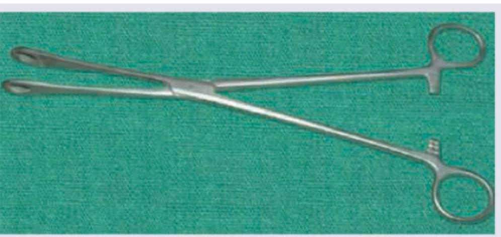
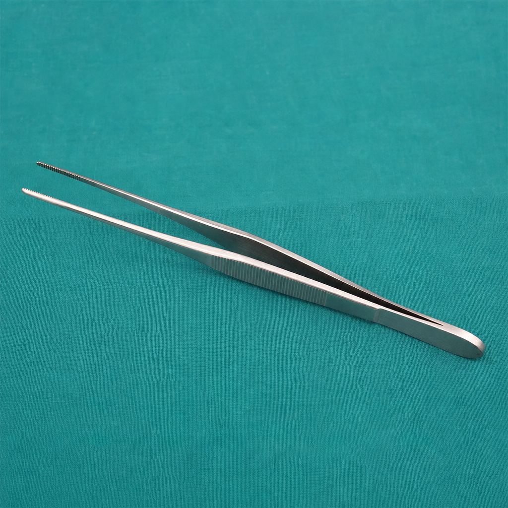
Identify the instrument:
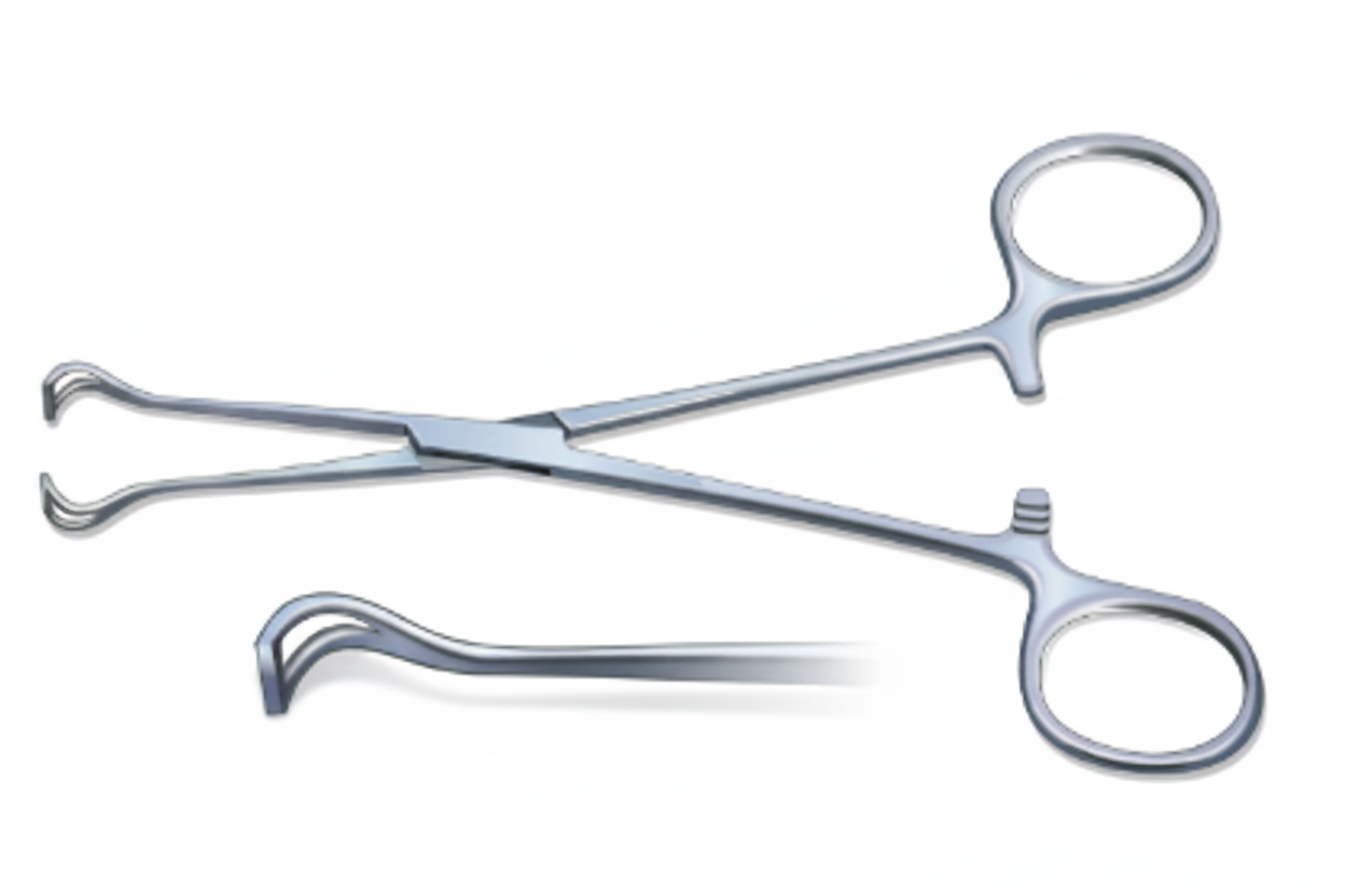
Identify the instrument shown:
Identify the instrument: (Recent Neet Pattern 2016-17)
Identify the instrument shown.
Identify the instrument in the image.
Identify the instrument in the image:
Identify the image given below:
Practice by Chapter
Wound Healing and Care
Practice Questions
Surgical Infections
Practice Questions
Fluid and Electrolyte Management
Practice Questions
Nutrition in Surgical Patients
Practice Questions
Hemostasis and Blood Transfusion
Practice Questions
Surgical Instruments and Equipment
Practice Questions
Sutures and Stapling Devices
Practice Questions
Minimal Access Surgery Principles
Practice Questions
Surgical Complications
Practice Questions
Anesthesia Principles for Surgeons
Practice Questions
Surgical Oncology Principles
Practice Questions
Evidence-Based Surgery
Practice Questions
Want unlimited practice?
Get full access to all questions, explanations, and performance tracking.
Scan to download app