
General Surgery Principles — MCQs

On this page
Arthrocentesis can be performed efficiently by:
In an elective laparoscopic cholecystectomy with no gross spillage, what is the recommended antibiotic prophylaxis?
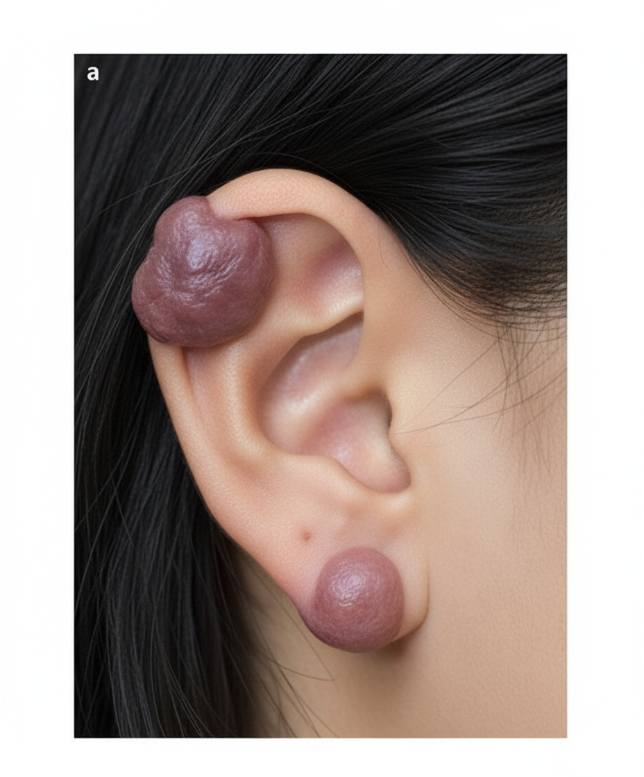
Which of the following is the best treatment for this condition?
Which of the following is the least likely complication after massive blood transfusion?
All are the features of rheumatoid arthritis except?
An ulcer with undermined edges is seen in which condition?
Schwann cells are derived from:
Identify the suturing technique shown in the image.
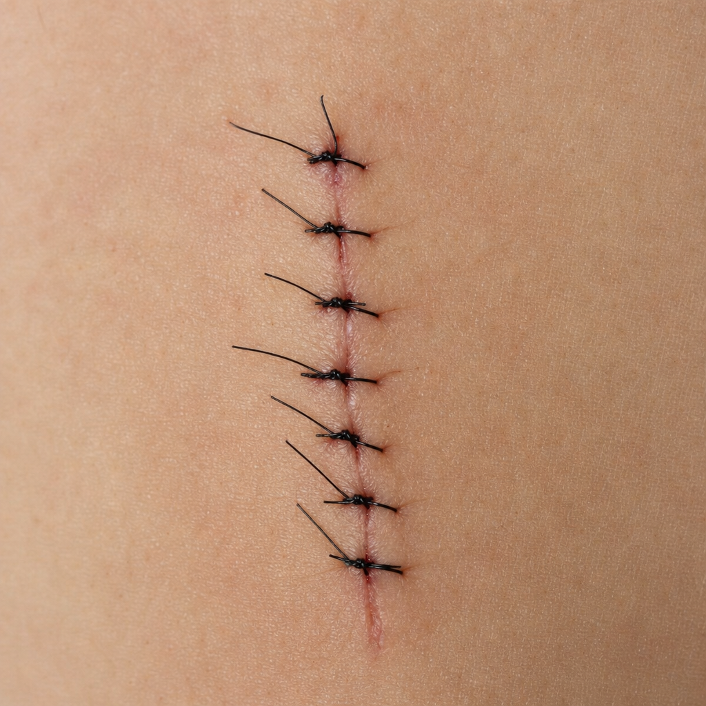
Identify the type of suture?
A 20-year-old male presents to the outpatient department with a swelling on his wrist. He reports fluctuation in size, mild numbness in the hand, and occasional pain. What is the most likely diagnosis?
Practice by Chapter
Wound Healing and Care
Practice Questions
Surgical Infections
Practice Questions
Fluid and Electrolyte Management
Practice Questions
Nutrition in Surgical Patients
Practice Questions
Hemostasis and Blood Transfusion
Practice Questions
Surgical Instruments and Equipment
Practice Questions
Sutures and Stapling Devices
Practice Questions
Minimal Access Surgery Principles
Practice Questions
Surgical Complications
Practice Questions
Anesthesia Principles for Surgeons
Practice Questions
Surgical Oncology Principles
Practice Questions
Evidence-Based Surgery
Practice Questions
Want unlimited practice?
Get full access to all questions, explanations, and performance tracking.
Scan to download app