
General Surgery Principles — MCQs

On this page
Which of the following statements about surgical wounds is incorrect?
Which of the following is a non-absorbable suture?
Splenectomy is contraindicated in which of the following hemolytic anemias?
In an adult patient with pleural effusion, what is the most appropriate site for thoracentesis?
From the surgery performed, what is the gold standard investigation used to diagnose this condition?
Which of the following muscles is not incised during a posterolateral thoracotomy?
What is the preservative used for this suture material?

In a patient with lung cancer, which of the following is a contraindication for surgical resection?
What is the most common tumour of the posterior mediastinum?
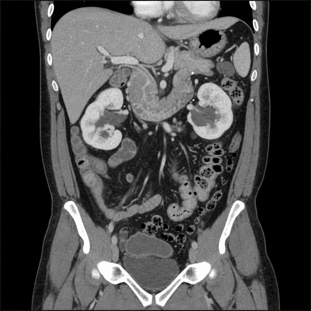
A 20-year-old female presents to the casualty with right iliac fossa pain, local guarding, and tenderness. Which of the following management interventions should NOT be done?
Practice by Chapter
Wound Healing and Care
Practice Questions
Surgical Infections
Practice Questions
Fluid and Electrolyte Management
Practice Questions
Nutrition in Surgical Patients
Practice Questions
Hemostasis and Blood Transfusion
Practice Questions
Surgical Instruments and Equipment
Practice Questions
Sutures and Stapling Devices
Practice Questions
Minimal Access Surgery Principles
Practice Questions
Surgical Complications
Practice Questions
Anesthesia Principles for Surgeons
Practice Questions
Surgical Oncology Principles
Practice Questions
Evidence-Based Surgery
Practice Questions
Want unlimited practice?
Get full access to all questions, explanations, and performance tracking.
Scan to download app