
General Surgery Principles — MCQs

On this page
Diabetic gangrene is due to:
Risk of Clostridium difficile infection increases with the use of which of the following medications?
What is the use of the instrument shown here?
When the development of a wound seroma is a potential problem after an appendectomy in an obese patient, what is the best effective method of wound management?
What is the tensile strength of a wound at different stages?
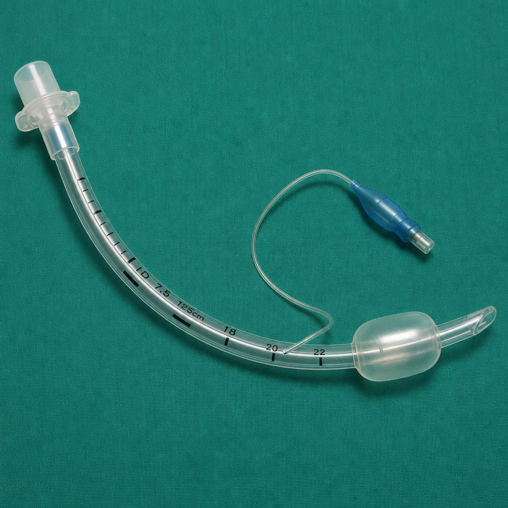
A 49-year-old man suffering from depression attempts suicide by jumping out of the window of his third-floor apartment. He requires multiple operations during a prolonged, complicated hospital stay. Endotracheal intubation is attempted in the ICU but is unsuccessful because of tracheal stenosis, which is attributed to which of the following?
What is true about abdominal compartment syndrome?
What is this instrument used for?
During splenectomy for ITP, at what point is platelet infusion typically given?
A patient underwent surgery two months ago using a midline incision. Now, the patient requires a second operation. What is the ideal incision to be used for this subsequent procedure?
Practice by Chapter
Wound Healing and Care
Practice Questions
Surgical Infections
Practice Questions
Fluid and Electrolyte Management
Practice Questions
Nutrition in Surgical Patients
Practice Questions
Hemostasis and Blood Transfusion
Practice Questions
Surgical Instruments and Equipment
Practice Questions
Sutures and Stapling Devices
Practice Questions
Minimal Access Surgery Principles
Practice Questions
Surgical Complications
Practice Questions
Anesthesia Principles for Surgeons
Practice Questions
Surgical Oncology Principles
Practice Questions
Evidence-Based Surgery
Practice Questions
Want unlimited practice?
Get full access to all questions, explanations, and performance tracking.
Scan to download app