
General Surgery Principles — MCQs

On this page
An elderly woman underwent a radical mastectomy with radiation to the axilla 20 years ago. For 25 years, she has had an open wound that has never healed. It is not a recurrence of breast cancer. What is the most likely diagnosis?
Which of the following is NOT an absolute indication for splenectomy?
To prevent pressure ulcers, which intervention should be included in the plan of care?
Which statement is NOT true regarding bone removal during impacted third molar extraction?
When using colloid infusion for the treatment of shock, what is the ratio at which blood loss is compensated?
What is the first step in performing a pneumonectomy for cancer of the bronchus?
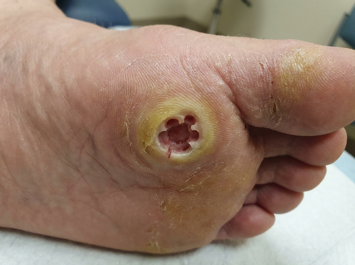
A 45-year-old male with a history of polyuria presents with a foot condition. What is the most likely diagnosis?
What is the primary treatment for a perforated peptic ulcer?
Indications for surgery in Bronchiectasis include all of the following except?
What is true about Marjolin's ulcer?
Practice by Chapter
Wound Healing and Care
Practice Questions
Surgical Infections
Practice Questions
Fluid and Electrolyte Management
Practice Questions
Nutrition in Surgical Patients
Practice Questions
Hemostasis and Blood Transfusion
Practice Questions
Surgical Instruments and Equipment
Practice Questions
Sutures and Stapling Devices
Practice Questions
Minimal Access Surgery Principles
Practice Questions
Surgical Complications
Practice Questions
Anesthesia Principles for Surgeons
Practice Questions
Surgical Oncology Principles
Practice Questions
Evidence-Based Surgery
Practice Questions
Want unlimited practice?
Get full access to all questions, explanations, and performance tracking.
Scan to download app