
General Surgery Principles — MCQs

On this page
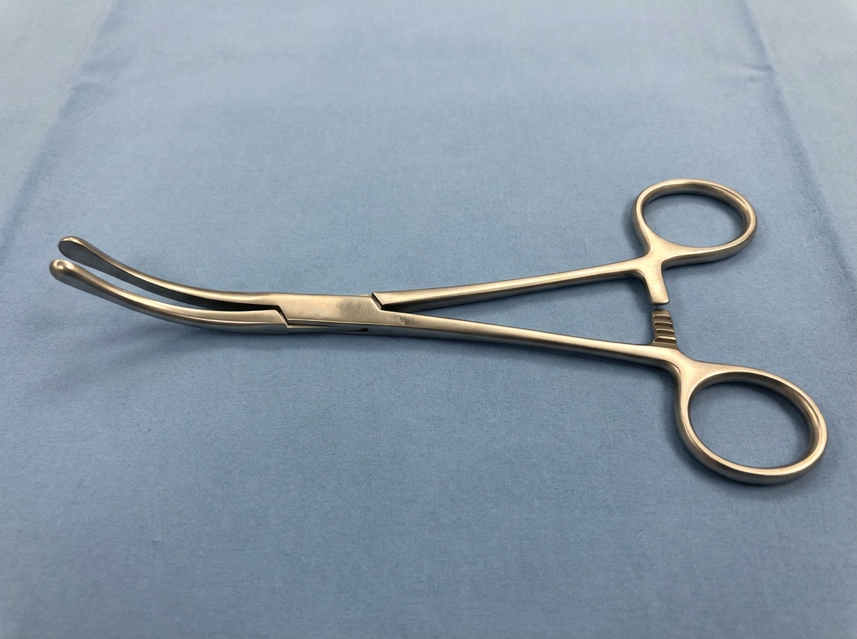
This instrument, historically used for opening sinus ostia in sinusitis, has a blunt tip. Surgeons currently use it to drain abscesses in which of the following regions?
A patient with Child's C cirrhosis has had repeated episodes of variceal bleeding and ascites. What is the treatment of choice?
What is the treatment of an uncomplicated hydatid cyst in the lung?
Surgical approaches for open thymectomy include all except?
Which suture material is least adherent to tissue and possesses plasticity?
A flexible bronchoscope is designed for all of the following functions EXCEPT?
Which of the following complications is likely to result after several units of blood have been transfused?
Reactionary hemorrhage occurs within which time frame?
Which of the following statements is true about hernias?
Cock’s peculiar tumour is described as:
Practice by Chapter
Wound Healing and Care
Practice Questions
Surgical Infections
Practice Questions
Fluid and Electrolyte Management
Practice Questions
Nutrition in Surgical Patients
Practice Questions
Hemostasis and Blood Transfusion
Practice Questions
Surgical Instruments and Equipment
Practice Questions
Sutures and Stapling Devices
Practice Questions
Minimal Access Surgery Principles
Practice Questions
Surgical Complications
Practice Questions
Anesthesia Principles for Surgeons
Practice Questions
Surgical Oncology Principles
Practice Questions
Evidence-Based Surgery
Practice Questions
Want unlimited practice?
Get full access to all questions, explanations, and performance tracking.
Scan to download app