
Gastrointestinal Surgery — MCQs

On this page
A 25 year old man presents with a 3-day history of pain in the right lower abdomen and vomiting. The patient's general condition is satisfactory, and clinical examination reveals a tender lump in the right iliac fossa. What is the most appropriate management in this case?
Which nerve is most commonly damaged during hernia repair?
In which type of hernia can the cecum be found as part of the hernia contents?
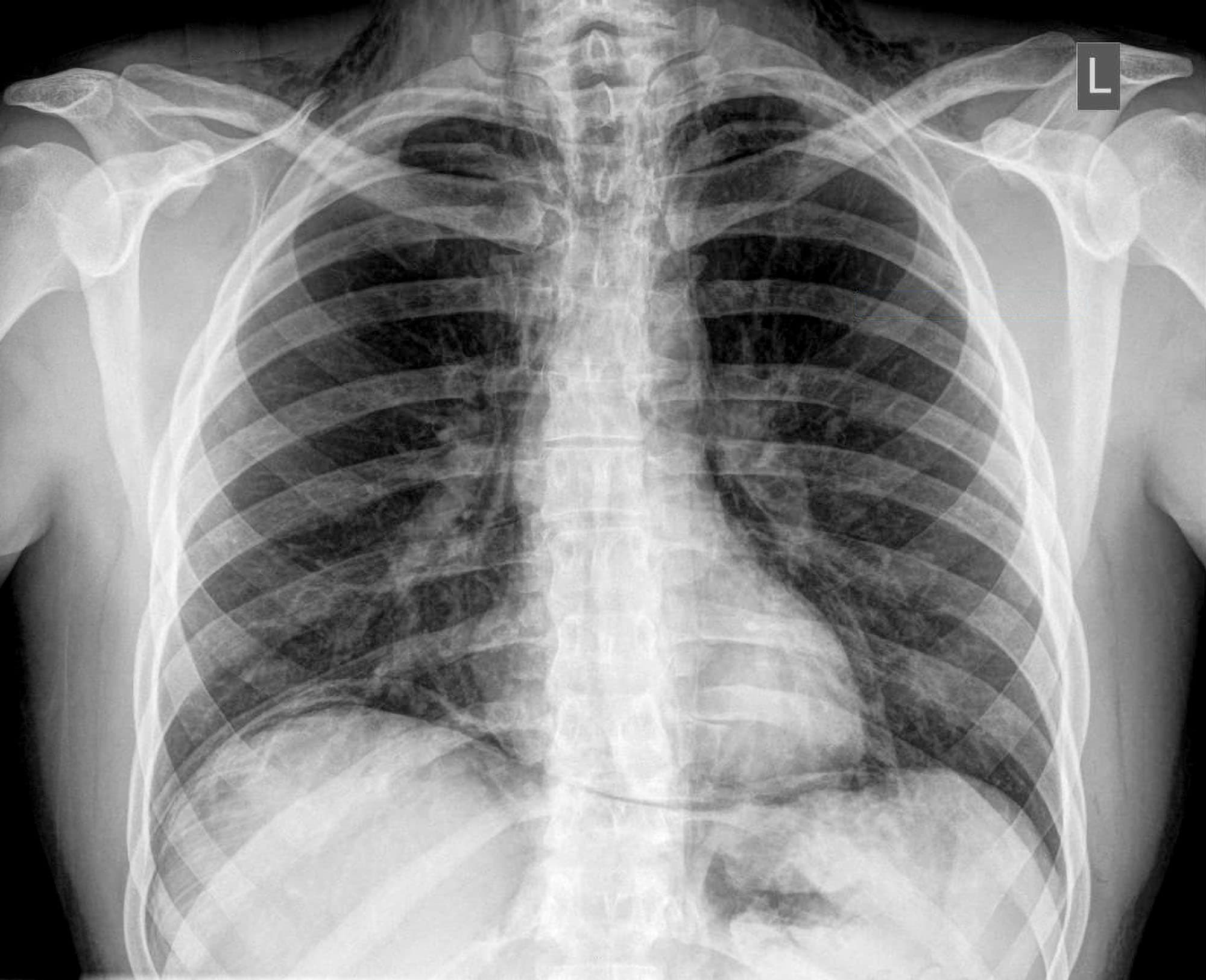
A patient after a heavy meal and episode of forceful vomiting presents with severe epigastric pain. On examination, there is tenderness and rigidity in the upper abdomen. X-ray shows pneumomediastinum. What is the most likely cause?
A 6-year-old boy experienced life-threatening shock. His CT scan showed a large amount of ascites, bowel wall thickening, and poor or absent enhancement of the strangulated bowel segment, showing gangrenous bowel on surgical exploration. Which of the following statements about anastomosis is true?
A patient presents with abdominal pain. On physical examination there was abdominal guarding and tenderness. A plain erect chest X-ray reveals air under diaphragm. Probable diagnosis is
Where does spontaneous esophageal rupture (Boerhaave syndrome) most commonly occur?
Which of the following characteristics is most accurate about Boerhaave syndrome?
What is the appropriate management for a patient with a carcinoid tumor of the appendix larger than 2 cm?
A 25 year old male is receiving conservative management for an appendicular mass since 3 days now presents with a rising pulse rate, tachycardia and fever. The mode of management must be -
Practice by Chapter
Esophageal Disorders
Practice Questions
Gastric Disorders
Practice Questions
Small Intestine Pathology
Practice Questions
Appendicitis
Practice Questions
Inflammatory Bowel Disease
Practice Questions
Intestinal Obstruction
Practice Questions
Gastrointestinal Bleeding
Practice Questions
Diverticular Disease
Practice Questions
Anorectal Disorders
Practice Questions
Colorectal Neoplasms
Practice Questions
Gastrointestinal Stomas
Practice Questions
Bariatric Surgery Principles
Practice Questions
Want unlimited practice?
Get full access to all questions, explanations, and performance tracking.
Scan to download app