
Gastrointestinal Surgery — MCQs

On this page
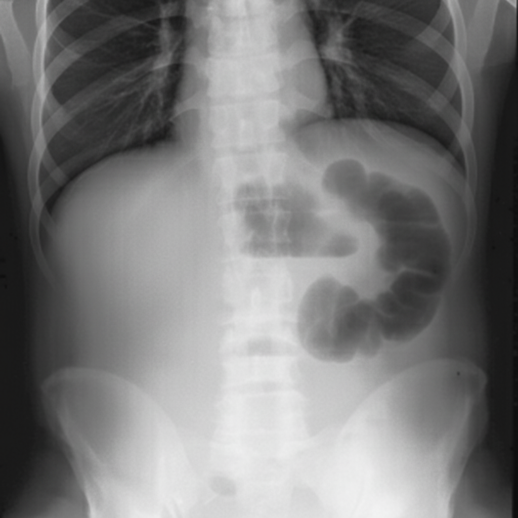
This condition is caused by:
Diffuse peritonitis in acute appendicitis is caused due to:
What is the most common cause of upper gastrointestinal bleeding?
Which one of the following is not performed in Lichtenstein tension-free hernioplasty?
All of the following benign conditions are associated with increased rates of gastric cancer except?
What is the most common early post-operative complication of ileostomy?
All are true about the Tillaux triad of a mesenteric cyst, except?
What is an uncommon cause of upper gastrointestinal bleeding?
All of the following are true for Meckel's diverticulum EXCEPT?
An epiplocele contains:
Practice by Chapter
Esophageal Disorders
Practice Questions
Gastric Disorders
Practice Questions
Small Intestine Pathology
Practice Questions
Appendicitis
Practice Questions
Inflammatory Bowel Disease
Practice Questions
Intestinal Obstruction
Practice Questions
Gastrointestinal Bleeding
Practice Questions
Diverticular Disease
Practice Questions
Anorectal Disorders
Practice Questions
Colorectal Neoplasms
Practice Questions
Gastrointestinal Stomas
Practice Questions
Bariatric Surgery Principles
Practice Questions
Want unlimited practice?
Get full access to all questions, explanations, and performance tracking.
Scan to download app