
Gastrointestinal Surgery — MCQs

On this page
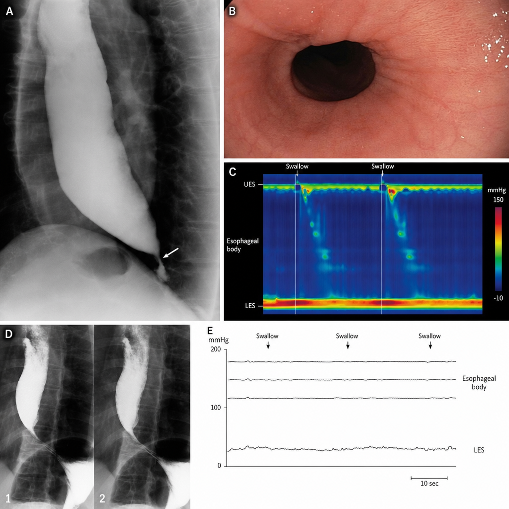
Regarding Achalasia Cardia, which of the following statements are true?
All of the following are indications for surgery in a case of duodenal ulcer except?
Which of the following statements about carcinoid of the appendix is NOT TRUE?
All are true about acute appendicitis except:
Malignant transformation is commonly seen in which type of ulcer?
Highest incidence of acute appendicitis occurs in which decade of life?
As per EHS classification, what is the designation for a primary, direct inguinal hernia with a 4 cm width?
All of the following are early complications arising after appendicectomy for acute appendicitis except?
What is the treatment of choice for a bleeding gastric ulcer?
A 64-year-old male presented with severe diarrhea, having more than 20 bowel movements per day, following an elective operation for duodenal ulcer disease. Medications have been ineffective. The exact details of his operation cannot be ascertained. What operation was most likely performed?
Practice by Chapter
Esophageal Disorders
Practice Questions
Gastric Disorders
Practice Questions
Small Intestine Pathology
Practice Questions
Appendicitis
Practice Questions
Inflammatory Bowel Disease
Practice Questions
Intestinal Obstruction
Practice Questions
Gastrointestinal Bleeding
Practice Questions
Diverticular Disease
Practice Questions
Anorectal Disorders
Practice Questions
Colorectal Neoplasms
Practice Questions
Gastrointestinal Stomas
Practice Questions
Bariatric Surgery Principles
Practice Questions
Want unlimited practice?
Get full access to all questions, explanations, and performance tracking.
Scan to download app