
Gastrointestinal Surgery — MCQs

On this page
What is the most common tumor of the appendix?
A patient presents with non-progressive dysphagia exclusively for solids. A barium study reveals proximal esophageal dilatation with distal constriction. What is the most likely diagnosis?
Which of the following is not a surgical treatment for GERD?
A 60-year-old male presents with an inguinal hernia of recent onset. Which of the following is true?
What are the similarities between fissure in ano and achalasia cardiae?
Which of the following is considered a true diverticulum?
What are the causes of recurrent hernia?
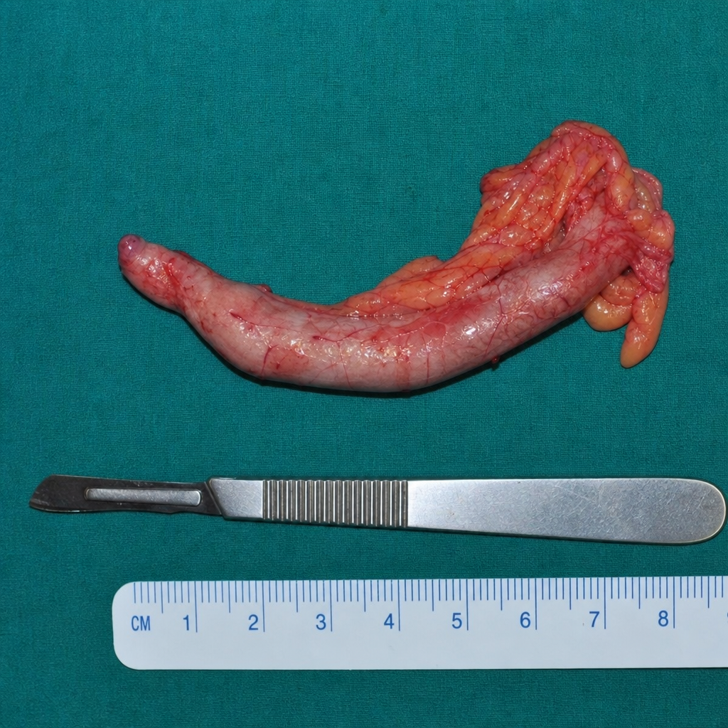
What scoring system is used to diagnose the condition for which the surgery below was performed?
Which of the following statements is NOT true regarding hyperplastic ileocecal tuberculosis?
Physiological gastrectomy is defined as:
Practice by Chapter
Esophageal Disorders
Practice Questions
Gastric Disorders
Practice Questions
Small Intestine Pathology
Practice Questions
Appendicitis
Practice Questions
Inflammatory Bowel Disease
Practice Questions
Intestinal Obstruction
Practice Questions
Gastrointestinal Bleeding
Practice Questions
Diverticular Disease
Practice Questions
Anorectal Disorders
Practice Questions
Colorectal Neoplasms
Practice Questions
Gastrointestinal Stomas
Practice Questions
Bariatric Surgery Principles
Practice Questions
Want unlimited practice?
Get full access to all questions, explanations, and performance tracking.
Scan to download app