
Gastrointestinal Surgery — MCQs

On this page
An elderly male presents with gradually increasing dysphagia more for solids, hoarseness of voice, and a palpable cervical node. What is the most likely diagnosis?
Which hernia often simulates a peptic ulcer?
All of the following are true about carcinoid crisis, EXCEPT:
A triad of vomiting, abdominal distension, and a "string of beads" sign on abdominal X-ray is typically suggestive of which condition?
Which of the following factors is responsible for causing diarrhea after vagotomy?
What percentage of patients with a perforated peptic ulcer show free gas under the diaphragm?
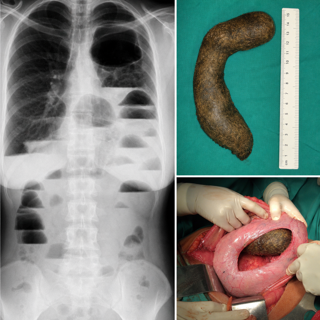
A 23-year-old woman with a history of an eating disorder complains of vomiting, nausea, and severe abdominal pain. Physical examination shows abdominal distension and constipation. An X-ray film of the abdomen reveals air-fluid levels and a hyperlucent shadow at the epigastric area. The material obstructing the gastrointestinal tract is removed surgically and shown. Which of the following is the most likely diagnosis?
Which of the following nutritional consequences is possible following peptic ulcer surgeries other than vagotomy?
Charcot's triad includes all EXCEPT:
A 35-year-old woman presents to the emergency department with abdominal pain and bilious vomiting, but no bowel distension. Abdominal X-ray shows no air-fluid levels. What is the most likely diagnosis?
Practice by Chapter
Esophageal Disorders
Practice Questions
Gastric Disorders
Practice Questions
Small Intestine Pathology
Practice Questions
Appendicitis
Practice Questions
Inflammatory Bowel Disease
Practice Questions
Intestinal Obstruction
Practice Questions
Gastrointestinal Bleeding
Practice Questions
Diverticular Disease
Practice Questions
Anorectal Disorders
Practice Questions
Colorectal Neoplasms
Practice Questions
Gastrointestinal Stomas
Practice Questions
Bariatric Surgery Principles
Practice Questions
Want unlimited practice?
Get full access to all questions, explanations, and performance tracking.
Scan to download app