Primary peritonitis is more common in females because:
In Duke's classification of cancer rectum, what does stage B2 denote?
A 63-year-old man undergoes a partial gastrectomy with Billroth II reconstruction for intractable peptic ulcer disease. He presents several months postoperatively with a megaloblastic anemia. Which of the following is the best treatment for this surgical complication?
Malignant transformation is commonly seen in which of the following conditions?
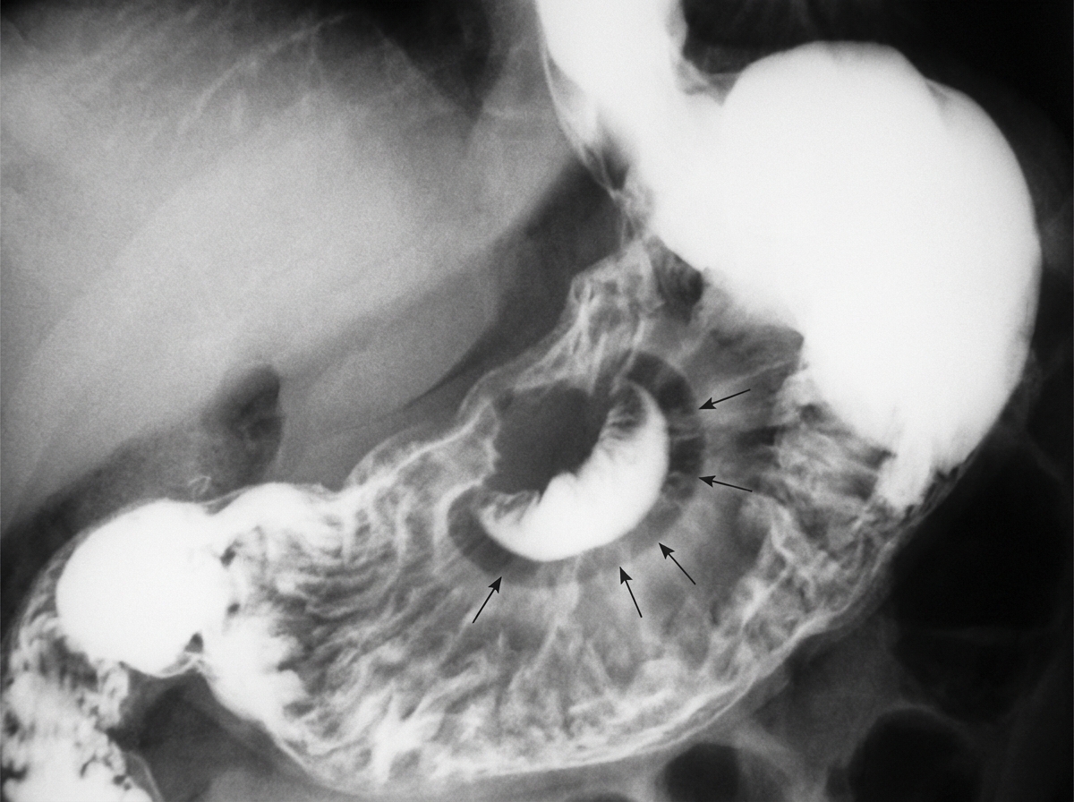
Which condition is diagnosed by the following sign?

Reflux esophagitis is common in which type of hiatus hernia?
A 24-year-old man presents with symptoms of acute intestinal obstruction. His temperature is 38°C (101°F), respirations are 25 per minute, and blood pressure is 120/80 mm Hg. Physical examination reveals a mass in the right lower abdominal quadrant. Laparoscopy shows numerous small bowel strictures and a fistula extending into a loop of small bowel. Which of the following is the most likely diagnosis?
Which of the following is false regarding cecal volvulus?
Which condition presents with a 'bird's beak' appearance?
Lung volume reduction surgery is used in the management of which of the following conditions?
Explanation: **Explanation:** **Primary peritonitis** (also known as Spontaneous Bacterial Peritonitis) is an infection of the peritoneal cavity without an evident intra-abdominal source of sepsis (like a perforated viscus). **Why Option A is Correct:** In females, the peritoneal cavity is technically "open" to the external environment. The **ostia of the Fallopian tubes** provide a direct anatomical communication between the vagina, uterus, and the peritoneal cavity. This allows for the **ascending migration of bacteria** from the female genital tract into the peritoneum. This unique anatomical feature explains why primary peritonitis has a higher incidence in females compared to males, where the peritoneal cavity is a completely closed sac. **Why Other Options are Incorrect:** * **Option B:** The peritoneum overlying the uterus is a normal anatomical relationship (forming the broad ligament and pouch of Douglas) but does not provide a pathway for infection. * **Option C:** Rupture of a functional ovarian cyst usually causes chemical irritation or hemoperitoneum, leading to "acute abdomen," but it is not a primary source of bacterial infection. **High-Yield Clinical Pearls for NEET-PG:** * **Most common organism (General):** *Escherichia coli* is the most common isolate overall. * **Most common organism (Post-Splenectomy):** *Streptococcus pneumoniae*. * **Diagnosis:** Established by paracentesis showing an **Absolute Neutrophil Count (ANC) > 250 cells/mm³**. * **Associated Conditions:** Most commonly seen in patients with **cirrhosis with ascites** or **Nephrotic syndrome** (especially in children). * **Management:** Unlike secondary peritonitis, primary peritonitis is managed **medically** (antibiotics like Cefotaxime) rather than surgically.
Explanation: **Explanation:** The **Dukes’ classification** (modified by Astler and Coller) is a classic staging system for colorectal cancer based on the depth of invasion and nodal involvement. 1. **Why Option C is Correct:** In the **Astler-Coller modification** of Dukes' classification, **Stage B2** specifically denotes a tumor that has extended **through the muscularis propria** and invaded the **perirectal fat (subserosa/serosa)**, but without lymph node involvement. This is a critical prognostic marker as it indicates a T3 lesion in the TNM system. 2. **Analysis of Incorrect Options:** * **Option A:** Growth extending into but not through the muscularis propria is classified as **Stage B1**. * **Option B:** Any regional lymph node involvement automatically moves the classification to **Stage C**. Specifically, C1 involves nodes but the primary tumor is limited to the wall, while C2 involves nodes with the primary tumor extending through the wall. * **Option C:** This represents **Stage D**, indicating systemic spread (e.g., liver or lung metastasis). **High-Yield Clinical Pearls for NEET-PG:** * **Dukes’ A:** Limited to mucosa/submucosa (T1). * **Dukes’ B1:** Extends into muscularis propria but not through it (T2, N0). * **Dukes’ B2:** Extends through muscularis propria (T3, N0). * **Dukes’ C:** Lymph node positive (Any T, N1/N2). * **Dukes’ D:** Distant metastasis (M1). * **Prognostic Note:** The most important prognostic factor in colorectal cancer is the **status of regional lymph nodes**. * **Modern Practice:** While Dukes' is historically significant, the **TNM staging system** is now the gold standard for clinical management.
Explanation: **Explanation:** The patient is presenting with **Vitamin B12 deficiency (megaloblastic anemia)** following a partial gastrectomy. This occurs because the resection of the stomach (specifically the body and fundus) leads to a loss of **parietal cells**, which are responsible for secreting **Intrinsic Factor (IF)**. Intrinsic factor is essential for the absorption of Vitamin B12 in the terminal ileum. **Why Option D is Correct:** In post-gastrectomy patients, the primary issue is the **absence of Intrinsic Factor**. Because B12 cannot be absorbed via the enteral route without IF, oral supplementation is generally ineffective. Therefore, **parenteral (Intravenous or Intramuscular) Vitamin B12** is the treatment of choice to bypass the gastrointestinal absorption barrier and replenish stores directly. **Why Other Options are Incorrect:** * **Option A:** Transfusion is reserved for symptomatic or life-threatening anemia (Hb <7 g/dL). It does not address the underlying nutritional deficiency. * **Option B:** While iron deficiency (microcytic anemia) is the *most common* anemia post-gastrectomy (due to bypass of the duodenum), the question specifically mentions **megaloblastic anemia**, which points to B12 or folate deficiency. * **Option C:** Oral B12 relies on intrinsic factor for active transport. While extremely high doses of oral B12 can sometimes work via passive diffusion, parenteral administration remains the gold standard for post-surgical deficiency. **High-Yield Clinical Pearls for NEET-PG:** * **Most common anemia post-gastrectomy:** Iron deficiency anemia (due to decreased acid and bypass of the duodenum). * **Megaloblastic anemia post-gastrectomy:** Usually due to Vitamin B12 deficiency (loss of IF) or Folate deficiency (poor intake). * **Billroth II specific complication:** Blind Loop Syndrome can also cause B12 deficiency due to bacterial overgrowth consuming the vitamin. * **Schilling Test:** Historically used to diagnose B12 malabsorption (now largely replaced by serology).
Explanation: **Explanation:** The risk of malignant transformation is a critical distinction between gastric and duodenal ulcer disease. **Why Gastric Ulcer is the Correct Answer:** Gastric ulcers, particularly those located on the **greater curvature** or the **prepyloric region**, carry a significant risk of being malignant at the time of presentation or undergoing malignant transformation (approximately 3–5%). Because it is clinically difficult to distinguish a benign chronic gastric ulcer from an early gastric carcinoma, the standard of care dictates that **all gastric ulcers must be biopsied** (usually 6–8 samples from the ulcer margin) and followed to complete healing to rule out malignancy. **Why the Other Options are Incorrect:** * **Chronic Duodenal Ulcer (B) & Postbulbar Ulcer (D):** Duodenal ulcers, whether in the first part (bulbar) or distal to it (postbulbar), are **virtually never malignant**. The duodenum is a highly resistant site for primary carcinomas. If a "duodenal ulcer" appears malignant, it is usually due to direct invasion from a neighboring primary (e.g., pancreatic or biliary cancer). * **Stomal Ulcer (A):** Also known as a marginal ulcer, these occur at the site of a gastrojejunostomy. While they are a known complication of gastric surgery (often due to ischemia or high acid output), they are typically inflammatory/peptic in nature and do not have a primary association with malignant transformation. **NEET-PG High-Yield Pearls:** * **Rule of Thumb:** "Gastric ulcers can be malignant; Duodenal ulcers are always benign." * **Biopsy Protocol:** Always biopsy the **margins** of a gastric ulcer. * **Risk Factors:** *H. pylori* infection and chronic atrophic gastritis are common precursors for both gastric ulcers and gastric adenocarcinoma. * **Location:** Ulcers on the **lesser curvature** are more common, but ulcers on the **greater curvature** have a higher suspicion for malignancy.
Explanation: ***Carcinoma of the stomach*** - The **leather bottle stomach** (linitis plastica) sign on **barium meal** is pathognomonic for diffuse gastric adenocarcinoma, showing a rigid, non-distensible stomach. - This appearance results from **signet ring cells** infiltrating the gastric wall, causing **fibrosis** and loss of normal gastric rugae and distensibility. *Peptic ulcer disease* - Barium meal typically shows **crater-like ulcers** with surrounding **mucosal edema** and inflammation, not the rigid leather bottle appearance. - The stomach maintains its **normal distensibility** and **peristaltic activity**, unlike the fixed appearance in linitis plastica. *Strawberry gallbladder* - This is a **cholesterolosis** condition affecting the **gallbladder wall** with characteristic **yellow cholesterol deposits** creating a strawberry-like appearance. - It is diagnosed by **ultrasound** or **cholecystography**, not by barium meal, and does not involve the stomach. *Patent vitellointestinal duct* - This congenital anomaly presents as a **Meckel's diverticulum** or **umbilical discharge** in infants, diagnosed by **technetium-99m scan**. - It involves the **small intestine** and umbilicus, with no characteristic barium meal findings in the stomach.
Explanation: **Explanation:** **1. Why Sliding Hiatus Hernia (Type I) is the correct answer:** In a sliding hiatus hernia, the gastroesophageal (GE) junction and the cardia of the stomach "slide" upward through the esophageal hiatus into the posterior mediastinum. This displacement disrupts the normal anatomy of the **Lower Esophageal Sphincter (LES)** and the **Angle of His** (the acute angle between the esophagus and the stomach fundus). The loss of the intra-abdominal segment of the esophagus and the widening of the hiatus compromise the anti-reflux mechanism, making **Gastroesophageal Reflux Disease (GERD)** and reflux esophagitis the hallmark clinical features of this type. **2. Why the other options are incorrect:** * **Paraesophageal Hiatus Hernia (Type II):** In this type, the GE junction remains in its normal anatomical position (fixed by the phrenoesophageal ligament), but the fundus of the stomach herniates alongside the esophagus. Because the GE junction is intact, the anti-reflux mechanism usually remains functional; therefore, reflux is rare. These patients are more at risk for **gastric volvulus, incarceration, or strangulation.** * **Both/None:** Since the pathophysiology of reflux is specifically tied to the displacement of the GE junction (unique to Type I), these options are incorrect. **Clinical Pearls for NEET-PG:** * **Most Common Type:** Sliding hiatus hernia is the most common (approx. 95% of cases). * **Cameron Ulcers:** Linear gastric erosions found within the hernial sac (due to mechanical trauma) which can cause chronic occult blood loss. * **Surgical Management:** Indications for surgery in sliding hernia are persistent symptoms of GERD despite medical therapy. In contrast, paraesophageal hernias are often operated on even if asymptomatic due to the risk of life-threatening complications like strangulation. * **Investigation of Choice:** Barium swallow is excellent for anatomy, but **Endoscopy** is used to grade esophagitis.
Explanation: ### Explanation **1. Why Crohn Disease is Correct:** The clinical presentation is classic for **Crohn Disease (CD)**. The patient is young (typical bimodal age distribution: 15–30 and 50–80 years) and presents with signs of **acute intestinal obstruction** and inflammation (fever, tachypnea). Key diagnostic features provided include: * **Transmural Inflammation:** Leads to the formation of **fistulas** (e.g., entero-enteric, as seen here) and **strictures** due to fibrosis. * **Skip Lesions:** "Numerous small bowel strictures" suggest the characteristic non-contiguous involvement. * **Right Lower Quadrant Mass:** Often represents "creeping fat," thickened bowel loops, or an inflammatory phlegmon/abscess involving the terminal ileum. **2. Why Other Options are Incorrect:** * **Adenocarcinoma:** While CD increases the risk of small bowel adenocarcinoma, it usually presents in older patients and typically as a single mass/stricture rather than multiple strictures and fistulas. * **Carcinoid Tumor:** These are the most common tumors of the appendix and ileum. While they can cause obstruction (via desmoplastic reaction), they do not typically cause fever or fistula formation. * **Pseudomembranous Colitis:** Caused by *C. difficile* toxin, it presents with profuse diarrhea and "volcano" lesions on colonoscopy. It involves the mucosa of the colon, not the small bowel, and does not cause strictures or fistulas. **3. NEET-PG High-Yield Pearls:** * **String Sign of Kantor:** Radiologic finding in CD due to terminal ileal strictures. * **Cobblestone Appearance:** Due to deep longitudinal and transverse ulcers. * **Histology:** Non-caseating granulomas (pathognomonic but seen in only ~50% of cases). * **Surgery:** Not curative; indicated only for complications (obstruction, fistula, perforation). The procedure of choice for multiple strictures is **Stricturoplasty** (to preserve bowel length).
Explanation: **Explanation:** **1. Why Option D is the correct (False) statement:** While **cecopexy** (fixing the cecum to the lateral abdominal wall) was historically practiced, it is **not the procedure of choice** due to high recurrence rates (up to 40%). The current gold standard for a viable cecum is **ileocolic resection** with primary anastomosis. If the patient is unstable or the bowel is gangrenous, resection with an ileostomy is preferred. **2. Analysis of other options:** * **Option A (True):** Since the cecum is the proximal part of the large bowel, its torsion often leads to a functional or mechanical blockage of the terminal ileum, presenting as **small bowel obstruction** (vomiting, central distension). * **Option B (True):** On X-ray, cecal volvulus typically shows a "coffee bean" appearance. Unlike sigmoid volvulus (which points to the RUQ), cecal volvulus usually displaces the dilated cecum to the **left upper quadrant (LUQ)** or epigastrium, with the convexity directed toward the **left**. * **Option C (True):** Endoscopic detorsion is successful in <20% of cases for cecal volvulus, compared to >80% for sigmoid volvulus. This is due to the technical difficulty of reaching the right colon and the high risk of perforation. **Clinical Pearls for NEET-PG:** * **Predisposing factor:** Incomplete fixation of the ascending colon to the retroperitoneum (**"mobile cecum"**). * **Radiological Signs:** "Coffee bean" sign, "Bird’s beak" sign on gastrografin enema. * **Axial Torsion vs. Cecal Bascule:** Axial torsion (true volvulus) is more common; Cecal bascule involves the cecum folding anteriorly over the ascending colon. * **Management Rule:** If viable → Resection/Anastomosis; If gangrenous → Resection/Stoma.
Explanation: **Explanation:** The **'bird’s beak' appearance** is a classic radiological sign seen in **Sigmoid Volvulus**. This occurs when the sigmoid colon twists on its mesenteric axis, causing a high-grade mechanical obstruction. On a contrast enema (Barium enema), the contrast tapers at the site of the twist, resembling a bird's beak or an ace of spades. On a plain X-ray, this condition typically presents as a 'coffee bean' sign. **Analysis of Options:** * **Volvulus (Correct):** Specifically, sigmoid volvulus. The torsion creates a funnel-shaped narrowing at the point of the twist. * **Testicular torsion:** This is a urological emergency. While it involves twisting, the diagnosis is clinical or via Doppler ultrasound (showing absent blood flow), not a 'bird's beak' sign. * **Meconium ileus:** Associated with Cystic Fibrosis, it shows a 'soap bubble' appearance (Neuhauser sign) on X-ray due to air mixing with thick meconium in the terminal ileum. * **Ileal atresia:** Presents with multiple air-fluid levels and a 'triple bubble' sign (if high ileal) or microcolon on contrast enema. **NEET-PG High-Yield Pearls:** * **Sigmoid Volvulus:** Most common site of volvulus; associated with a high-fiber diet and chronic constipation. * **Management:** Sigmoidoscopic detorsion is the initial treatment of choice if there is no gangrene. * **Differential:** Achalasia Cardia also shows a 'bird's beak' appearance on a barium swallow due to the failure of the Lower Esophageal Sphincter (LES) to relax. * **Cecal Volvulus:** Shows a 'fetal lamb' or 'comma' sign on X-ray.
Explanation: **Explanation:** **Lung Volume Reduction Surgery (LVRS)** is a surgical procedure designed to improve respiratory mechanics in patients with severe **Emphysema** (a component of COPD). **Why Emphysema is the Correct Answer:** In emphysema, the destruction of alveolar walls leads to permanent enlargement of air spaces and loss of elastic recoil. This causes **pathological hyperinflation**, where "dead space" air is trapped in the lungs, flattening the diaphragm and making breathing inefficient. LVRS involves the excision of the most diseased, non-functioning lung tissue (usually 20-30%). This reduces hyperinflation, allowing the remaining healthier lung tissue to expand and the diaphragm to return to a more natural, dome-shaped position, thereby improving the work of breathing and gas exchange. **Why Other Options are Incorrect:** * **Bronchial Asthma:** This is a reversible airway inflammation and bronchoconstriction. Management is medical (bronchodilators/steroids), not surgical. * **Interstitial Lung Disease (ILD):** These are restrictive lung diseases characterized by fibrosis and "shrunken" lungs. LVRS would be detrimental as it further reduces already diminished lung volumes. * **Chronic Bronchitis:** While part of COPD, it is characterized by airway mucus hypersecretion rather than the localized bullous destruction seen in emphysema. LVRS does not address the underlying pathology of the conducting airways. **High-Yield Clinical Pearls for NEET-PG:** * **NETT Trial:** The National Emphysema Treatment Trial established that LVRS is most beneficial for patients with **upper-lobe predominant emphysema** and low exercise capacity. * **Gold Standard:** Lung transplantation remains the definitive treatment for end-stage COPD, but LVRS serves as a palliative bridge. * **Contraindication:** A Carbon Monoxide Diffusing Capacity (DLCO) <20% is a high-risk predictor for mortality in LVRS.
Esophageal Disorders
Practice Questions
Gastric Disorders
Practice Questions
Small Intestine Pathology
Practice Questions
Appendicitis
Practice Questions
Inflammatory Bowel Disease
Practice Questions
Intestinal Obstruction
Practice Questions
Gastrointestinal Bleeding
Practice Questions
Diverticular Disease
Practice Questions
Anorectal Disorders
Practice Questions
Colorectal Neoplasms
Practice Questions
Gastrointestinal Stomas
Practice Questions
Bariatric Surgery Principles
Practice Questions
Get full access to all questions, explanations, and performance tracking.
Start For Free