Most common site of chronic gastric ulcer:
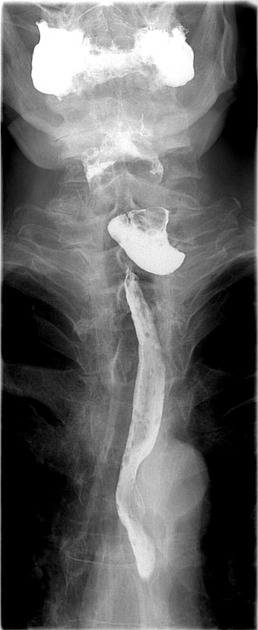
A 70 year old patient presented with history of fever, repeated aspiration and coughing in the night. On examination there is a swelling on left side of neck which produces gurgling sound on compression. Following is the barium swallow study of the patient. What is the most likely diagnosis?

A patient presents with abdominal pain, blood in stools and a palpable mass on examination. A Barium Study was performed, probable diagnosis is?
Which layer differentiates Boerhaave syndrome from Mallory-Weiss tear in terms of depth of involvement?
True about Mallory-Weiss tear is:
A 40-year-old male with right iliac fossa pain, fever. CT shows 4cm appendix with faecolith. Best management?
A 25-year-old patient presents with RLQ pain, fever, and vomiting. CT shows a ruptured appendix. What is the next step?
Which of the following findings on physical exam suggests a strangulated inguinal hernia?
What is the typical presentation of acute appendicitis?
What is the definitive treatment for gallstone-induced pancreatitis?
Explanation: ***Lesser curve near incisura*** - The **lesser curve** of the stomach, particularly the **incisura angularis** or the angular notch, is the most common site for gastric ulcers. - This area is susceptible to ulceration due to its anatomical location, which experiences significant **acid exposure** and **motility stress**. *Lesser curve near proximal stomach* - While the lesser curve is a common site, ulcers tend to be more prevalent in the **distal part** of the lesser curve, near the incisura, rather than the proximal stomach (cardia or fundus). - Gastric ulcers in the proximal stomach are less frequent compared to the **antrum** and **incisura region**. *Pylorus of stomach* - The **pylorus** is more commonly associated with duodenal ulcers but can occasionally be a site for gastric ulcers. - However, it is not the most common location for **chronic gastric ulcers** when compared to the lesser curve near the incisura. *Greater curvature* - Ulcers on the **greater curvature** are relatively rare and often raise suspicion for **malignancy**, necessitating careful biopsy and investigation. - The greater curvature is less exposed to the erosive effects of gastric acid and pepsin compared to the lesser curve, making ulcers there less common.
Explanation: ***Zenker's diverticulum*** - The patient's symptoms of **fever**, **repeated aspiration**, and **coughing at night** are classic for a Zenker's diverticulum, particularly in an older patient. - The presence of a **neck swelling** producing a **gurgling sound on compression** (Boyce's sign) is highly indicative of a Zenker's diverticulum, which is essentially a pharyngeal pouch. The barium swallow image likely shows contrast pooling in such a pouch. *Plummer Vinson syndrome* - Characterized by **dysphagia**, **iron-deficiency anemia**, and **esophageal webs**. - While it causes dysphagia, it does not typically present with a gurgling neck swelling or significant aspiration as described. *Dysphagia Lusoria* - This is a rare condition caused by an **aberrant right subclavian artery** compressing the esophagus. - It primarily causes dysphagia due to extrinsic compression, without the associated neck swelling, gurgling sound, or significant aspiration risk from food pooling within a diverticulum. *Laryngocoele* - A laryngocele is an **abnormal sac** or pouch that arises from the **laryngeal ventricle** and may extend externally, presenting as a neck swelling. - While it can cause a neck swelling, it is **air-filled**, not fluid or food-filled, and therefore would not typically produce a gurgling sound on compression or be clearly visible on a barium swallow as a contrast-filled pouch like in the image provided.
Explanation: ***Intussusception*** - This condition is characterized by a "telescoping" of one segment of the intestine into another, which can lead to **abdominal pain**, **rectal bleeding** (often described as "currant jelly" stools), and a **palpable sausage-shaped mass** on examination. - A barium study (specifically a **barium enema**) is often diagnostic and can also be therapeutic for intussusception, revealing a **coiled spring appearance** or an obstruction. *Volvulus* - Volvulus involves the **twisting of a loop of bowel** around its mesentery, often presenting with sudden onset, severe **abdominal pain**, vomiting, and constipation. - While it can cause an obstruction and pain, a palpable mass and bloody stools are less common initial findings compared to intussusception. *Meckel's Diverticulum* - Meckel's diverticulum is a **congenital outpouching** of the small intestine that can be asymptomatic or cause complications like **gastrointestinal bleeding** (due to ectopic gastric mucosa), obstruction, or diverticulitis. - While it can cause painless rectal bleeding, a palpable mass and acute, intermittent abdominal pain are not typical primary presentations for an uncomplicated Meckel’s diverticulum. *Diverticulitis* - Diverticulitis is the **inflammation of diverticula** (small pouches in the colon), typically presenting with **left lower quadrant abdominal pain**, fever, and changes in bowel habits. - While it can cause bleeding, a palpable mass is less common unless there's an abscess, and the clinical picture does not align as strongly with the "currant jelly stool" and classic palpable mass of intussusception.
Explanation: **Adventitia** - **Boerhaave syndrome** involves a **complete rupture** of the esophagus, extending through all layers, including the adventitia. - In contrast, a **Mallory-Weiss tear** is a **partial-thickness tear** that does not extend beyond the muscularis mucosae or submucosa and thus does not involve the adventitia. - The adventitia is the **key differentiating layer** - its involvement indicates full-thickness perforation (Boerhaave) versus partial-thickness tear (Mallory-Weiss). *Muscularis* - **Mallory-Weiss tears** can involve the **muscularis mucosae** or extend into the submucosa, but they do not typically penetrate the entire muscularis propria. - **Boerhaave syndrome** always involves the muscularis propria, as it is a full-thickness rupture. *Submucosa* - **Mallory-Weiss tears** can extend into the **submucosa**, which is the deepest layer they typically affect. - **Boerhaave syndrome** always involves the submucosa as part of its full-thickness esophageal perforation. *Mucosa* - Both **Boerhaave syndrome** and **Mallory-Weiss tears** involve the **mucosa** as it is the innermost layer of the esophagus. - However, the depth of involvement beyond the mucosa is what differentiates the two conditions.
Explanation: ***Occurs at GE junction*** - A **Mallory-Weiss tear** is a longitudinal mucosal laceration located in the **gastroesophageal junction** or upper stomach. - It is typically caused by a sudden increase in intra-abdominal pressure, such as from **retching** or **vomiting**. *Always needs surgery* - The vast majority of Mallory-Weiss tears **resolve spontaneously** and do not require surgical intervention. - Management usually involves supportive care and, if bleeding persists, endoscopic hemostasis. *Involves all layers* - Mallory-Weiss tears are **mucosal or submucosal lacerations** and do not typically penetrate through all layers of the esophageal or gastric wall. - Tears that extend through all layers are known as **Boerhaave syndrome**, which is a more severe condition and a medical emergency. *Common in elderly* - Mallory-Weiss tears can occur in any age group, but they are **more common in middle-aged adults**, often associated with conditions like alcoholism. - While not exclusive to younger populations, calling it "common in elderly" is not a primary characteristic.
Explanation: ***Immediate appendectomy*** - The presence of **right iliac fossa pain, fever**, and a **4cm appendix with a faecolith** on CT scan strongly indicates acute appendicitis, which requires urgent surgical intervention. - A faecolith suggests **luminal obstruction**, increasing the risk of perforation and complications if not treated promptly. *Conservative treatment* - While some cases of uncomplicated appendicitis can be managed conservatively with antibiotics, this patient's presentation with a **faecolith and inflamed appendix (4cm)** suggests a higher risk of progression and complications. - Delaying surgery could lead to **abscess formation** or **perforation**, increasing morbidity. *Interval appendectomy* - This approach is typically considered for patients who initially present with a **well-contained appendiceal mass or abscess** that is managed non-operatively in the acute phase. - The current presentation is one of **acute appendicitis** requiring immediate attention, not deferred surgery after initial conservative management. *Percutaneous drainage* - **Percutaneous drainage** is primarily indicated for patients with a **well-defined appendiceal abscess** large enough to be drained. - This patient's CT shows an inflamed appendix with a faecolith, but not explicitly a drained abscess, making immediate appendectomy the most appropriate first-line treatment for the acute inflammation.
Explanation: ***Open appendectomy*** - For a **ruptured appendix** with generalized peritonitis, **open appendectomy** is the traditional gold standard and most appropriate approach. - Open surgery allows for **thorough peritoneal lavage**, better visualization of the entire abdominal cavity, and effective drainage of contaminated fluid. - In the setting of **perforation with peritoneal contamination**, open approach ensures complete source control and reduces risk of missed abscesses or inadequate irrigation. *Laparoscopic appendectomy* - While laparoscopic appendectomy can be used in **selected cases** of perforated appendicitis, it is not the first-line approach for a ruptured appendix with generalized peritonitis. - Laparoscopic approach may be limited in cases with **extensive contamination** and may not allow adequate peritoneal toilet. - It is more appropriate for **uncomplicated appendicitis** or **early/localized perforation** in experienced hands. *Percutaneous drainage* - This is typically reserved for patients with a **well-defined appendiceal abscess** presenting late (>5 days after symptom onset) where a phlegmon or organized abscess has formed. - Used as part of **interval appendectomy** approach: drain abscess, treat with antibiotics, then perform appendectomy 6-8 weeks later. - Not appropriate for **acute rupture** with active peritonitis requiring immediate surgical source control. *Conservative treatment* - **Antibiotics alone** might be considered for **uncomplicated appendicitis** in select cases or when surgery is contraindicated. - A **ruptured appendix** is a surgical emergency requiring operative intervention to prevent sepsis, abscess formation, and other life-threatening complications. - Conservative management is contraindicated in the presence of perforation and peritonitis.
Explanation: ***Non-reducible mass*** - A **non-reducible (irreducible) mass** is the **primary clinical finding** that differentiates a strangulated or incarcerated hernia from a simple reducible hernia. - When herniated contents cannot be returned to the abdominal cavity, it indicates **bowel or tissue entrapment** within the hernia sac. - This is the **earliest and most consistent sign** suggesting progression from a simple hernia to one at risk of or already experiencing strangulation. - **Non-reducibility is the hallmark** that prompts urgent surgical evaluation to prevent or treat strangulation. *Tender mass* - **Tenderness** indicates inflammation or ischemia and is an important additional finding in strangulation. - However, tenderness can also occur with simple incarceration or localized inflammation without strangulation. - Tenderness **combined with** non-reducibility strengthens the diagnosis, but non-reducibility is the more fundamental finding. *Cyanotic skin over mass* - **Cyanotic or dusky skin** is a **very late sign** indicating advanced tissue ischemia and necrosis. - While it definitively confirms strangulation, by this stage significant tissue damage has already occurred. - This is **not the primary finding** that initially "suggests" strangulation—the diagnosis should be made much earlier based on non-reducibility and tenderness. *Bowel sounds over mass* - The presence of **bowel sounds over the hernia** suggests viable bowel with intact peristalsis. - This typically indicates an **uncomplicated or recently incarcerated hernia** without established strangulation. - **Absence of bowel sounds** would be more concerning for strangulation, but presence suggests viability.
Explanation: ***Periumbilical pain shifting to RLQ*** - This classic migratory pattern of pain, starting diffusely in the **periumbilical area** and localizing to the **right lower quadrant (RLQ)**, is highly characteristic of acute appendicitis. - The initial visceral pain from the inflamed appendix is referred to the umbilical region, followed by somatic pain as the inflammation irritates the parietal peritoneum in the RLQ. *Pain in the epigastrium* - While initial pain in acute appendicitis can be somewhat vague or generalized, it typically begins around the **umbilicus**, not the epigastrium. - Epigastric pain is more commonly associated with conditions like **gastritis**, **peptic ulcer disease**, or early **pancreatitis**. *Diffuse abdominal pain* - While initial pain can be somewhat generalized, it almost always localizes to the **right lower quadrant** as appendicitis progresses, making diffuse pain less typical for the entire course. - Diffuse abdominal pain could suggest conditions like **gastroenteritis**, **peritonitis** from a perforated viscus, or **ischemic bowel**. *Flank pain* - **Flank pain** primarily suggests conditions affecting the kidneys or retroperitoneal structures. - This type of pain is characteristic of **pyelonephritis**, **kidney stones**, or muscle strains in the back, not acute appendicitis.
Explanation: ***Cholecystectomy*** * **Cholecystectomy** is the definitive treatment for gallstone-induced pancreatitis because it removes the source of the obstructing gallstones (the gallbladder). * Typically, this procedure is performed once the acute inflammatory process has settled, to prevent recurrent episodes of pancreatitis. *Fasting* * **Fasting** is a supportive measure used to rest the pancreas during an acute pancreatitis attack, but it does not remove the underlying cause of gallstones. * While fasting helps alleviate pain and reduce pancreatic enzyme secretion, it is not a definitive long-term treatment. *ERCP* * **ERCP (Endoscopic Retrograde Cholangiopancreatography)** is primarily used for the *removal of obstructing common bile duct stones* in cases of gallstone pancreatitis, especially if there's evidence of cholangitis or persistent biliary obstruction. * ERCP can remove immediate obstruction but does not prevent future stone formation in the gallbladder, nor does it address the gallbladder itself as the source. *Pancreatic resection* * **Pancreatic resection** is a major surgical procedure reserved for severe complications of pancreatitis, such as necrotizing pancreatitis, or for pancreatic tumors. * It is **not** indicated for routine gallstone-induced pancreatitis and carries significant morbidity and mortality, making it inappropriate for this context.
Esophageal Disorders
Practice Questions
Gastric Disorders
Practice Questions
Small Intestine Pathology
Practice Questions
Appendicitis
Practice Questions
Inflammatory Bowel Disease
Practice Questions
Intestinal Obstruction
Practice Questions
Gastrointestinal Bleeding
Practice Questions
Diverticular Disease
Practice Questions
Anorectal Disorders
Practice Questions
Colorectal Neoplasms
Practice Questions
Gastrointestinal Stomas
Practice Questions
Bariatric Surgery Principles
Practice Questions
Get full access to all questions, explanations, and performance tracking.
Start For Free