Which one of the following parts of intussusception is most susceptible to ischaemia and perforation?
What is the treatment of choice in a patient with Crohn’s disease, where inflamed appendix was found on exploration?
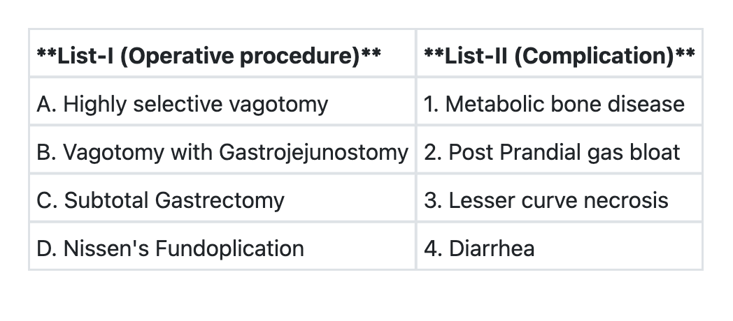
Match List-I with List-II and select the correct answer using the code given below the Lists: **List-I (Procedure)** A. Highly selective vagotomy B. Vagotomy with gastrojejunostomy C. Subtotal gastrectomy D. Nissen's fundoplication **List-II (Complication)** 1. Metabolic bone disease 2. Post-prandial gas bloat 3. Lesser curve necrosis 4. Diarrhea **Code:**

In a patient of gastric outlet obstruction nutritional support is best delivered by:
Mousseau-Barbin Tube (M.B.Tube) is used for:
The most common cause of intestinal obstruction is:
In gallstone ileus, obstruction most frequently occurs at:
A 48 year old male with the history of chronic duodenal ulcer presented in surgical emergency with the complaints of sudden severe pain in the abdomen. At presentation: Pulse = 120/m, BP = 90/60 mm of Hg Abdomen: Tenderness (+), Rigidity (+), Guarding (+) Respiratory Rate: 20/m X-ray: Gas under right dome of diaphragm The probable diagnosis is:
Which of the following is NOT a tissue repair surgery for inguinal hernia repair?
Valentino's syndrome is:
Explanation: ***Apex*** - The **apex** is the **leading edge** (distal tip) of the intussusceptum that protrudes furthest into the intussuscipiens. - It is the **most distal point** from its blood supply and experiences the **greatest degree of vascular compromise**. - The apex suffers from **pressure necrosis** due to compression against the intussuscipiens and maximal venous congestion. - This makes it the **most susceptible site for ischemia, necrosis, and perforation** in intussusception. - Clinically, when perforation occurs, it is **most commonly at the apex**. *Neck* - The **neck** is the constricted point where the intussusceptum enters the intussuscipiens. - While the neck does compress the **mesentery and blood vessels**, causing venous outflow obstruction that affects the entire intussusceptum, it is not itself the most susceptible site for perforation. - The neck causes the ischemia, but the apex suffers the most from it. *Intussuscipiens* - The **intussuscipiens** is the **outer receiving segment** that engulfs the intussusceptum. - Its blood supply remains relatively intact as it is not invaginated. - It is **not susceptible** to ischemia in the same way as the invaginated segment. *Intussusceptum* - The **intussusceptum** refers to the **entire invaginated inner segment**. - While the whole intussusceptum can become ischemic, the question asks for the **specific part** most susceptible. - Within the intussusceptum, the **apex is the most vulnerable point** for ischemia and perforation.
Explanation: ***Ileo-colic resection and anastomosis*** - This is the treatment of choice when an inflamed appendix is found during exploration in a patient with Crohn's disease, as the disease typically affects the **terminal ileum** and **right colon**. - The inflamed appendix is often a manifestation of Crohn's disease involving the **cecal base** and surrounding bowel. - **Ileo-colic resection** ensures removal of the diseased segment, including the inflamed appendix and involved bowel, thereby preventing future complications such as **fistulas** (risk up to 65% with simple appendectomy) and **strictures**. - If the cecal base is involved with Crohn's disease, simple appendectomy is contraindicated due to poor healing and high fistula risk. *Appendectomy* - Performing a simple appendectomy in the context of Crohn's disease carries a high risk of **fistula formation** and **poor wound healing** due to the underlying inflammatory process. - When the disease involves the **base of the appendix** and surrounding **cecum** (which is common), appendectomy alone is insufficient and dangerous. - Appendectomy may only be considered safe if the cecal base is completely **normal and uninvolved**, which is uncommon in this clinical scenario. *Closing the abdomen and starting medical treatment* - While medical treatment is crucial for managing Crohn's disease, an **inflamed appendix** found during exploration suggests an acute process that requires **surgical intervention**. - Delaying surgery by closing the abdomen could lead to complications such as **perforation** and **peritonitis**, especially if inflammation is severe. - Medical therapy alone is insufficient for acute complications requiring exploration. *Right hemicolectomy* - Right hemicolectomy is a more extensive resection than necessary for most cases of ileocecal Crohn's disease with appendiceal involvement. - **Ileo-colic resection** (removing terminal ileum, cecum, and ascending colon up to the hepatic flexure) is adequate and preferred as it is less extensive while addressing the pathology. - Right hemicolectomy would be reserved for more extensive colonic involvement beyond the typical ileocecal distribution.
Explanation: ***A→2 B→4 C→1 D→3*** - **Highly selective vagotomy** (HSV) aims to reduce gastric acid secretion by denervating only the parietal cell mass, sparing the antrum and pylorus. A potential complication due to altered gastric motility and emptying can be **post-prandial gas bloat** or fullness. - **Vagotomy with gastrojejunostomy** involves severing the vagus nerve, which can lead to altered gastrointestinal motility and malabsorption. **Diarrhea** is a common complication due to accelerated transit time and bacterial overgrowth. - **Subtotal gastrectomy** involves the removal of a significant portion of the stomach. This procedure can lead to malabsorption of nutrients, including calcium and vitamin D, resulting in **metabolic bone disease**. - **Nissen's Fundoplication** is a procedure to treat gastroesophageal reflux disease (GERD) by wrapping the gastric fundus around the lower esophageal sphincter. **Lesser curve necrosis** is a rare but severe complication that can occur due to devascularization during the procedure. *A→2 B→1 C→4 D→3* - This option incorrectly associates vagotomy with gastrojejunostomy with metabolic bone disease and subtotal gastrectomy with diarrhea. While diarrhea can occur after gastrectomy, metabolic bone disease is a more specific and significant long-term complication of subtotal gastrectomy due to malabsorption. - Furthermore, this option suggests that metabolic bone disease is a complication of vagotomy with gastrojejunostomy, which is not a primary or common complication of this procedure. *A→3 B→1 C→4 D→2* - This option incorrectly links highly selective vagotomy with lesser curve necrosis and vagotomy with gastrojejunostomy with metabolic bone disease. Lesser curve necrosis is a specific complication linked to Nissen's fundoplication, not HSV. - It also misassociates subtotal gastrectomy with diarrhea as the primary unique complication, and Nissen's fundoplication with post-prandial gas bloat, which is more typical of vagotomy. *A→3 B→4 C→1 D→2* - This option incorrectly pairs highly selective vagotomy with lesser curve necrosis, similar to one of the previous incorrect options. Lesser curve necrosis is a known specific complication of Nissen's fundoplication, not vagotomy. - It also incorrectly links Nissen's fundoplication with post-prandial gas bloat, which is a symptom more commonly associated with procedures that affect gastric emptying, such as vagotomy, rather than fundoplication.
Explanation: ***Jejunostomy*** - In **gastric outlet obstruction**, the stomach cannot empty properly, making gastric feeding routes (like Ryles tube or gastrostomy) ineffective. - A **jejunostomy** allows direct delivery of **enteral nutrition** into the jejunum, bypassing the obstructed stomach and duodenum. *Enteral nutrition by Ryles tube* - A **Ryles tube** delivers nutrition into the stomach, which is obstructed in this condition, leading to **stasis** and **vomiting**. - This method would be ineffective and potentially dangerous due to the inability of gastric contents to pass beyond the obstruction. *Gastrostomy* - A **gastrostomy** involves placing a tube directly into the stomach, which is still part of the obstructed system. - Feeding via gastrostomy would lead to accumulation of feed in the stomach, mimicking the issues with oral feeding or a Ryles tube. *Parenteral nutrition* - **Parenteral nutrition** is a viable option for nutritional support but is generally considered a second-line therapy after **enteral routes** fail or are contraindicated. - **Enteral feeding**, when possible (as with jejunostomy), is preferred due to lower cost, reduced risk of infection, and better maintenance of gut integrity.
Explanation: ***Advanced cancer oesophagus*** - The **Mousseau-Barbin tube** is a type of **endoscopic stent** used for palliative management of **dysphagia** caused by advanced **oesophageal cancer**. - It provides a lumen through obstructed oesophageal segments, allowing patients to swallow food and liquids more easily. *Advanced cancer oropharynx* - While dysphagia can be a symptom of oropharyngeal cancer, the Mousseau-Barbin tube is specifically designed for placement within the **oesophagus**. - Management for advanced oropharyngeal cancer often involves other interventions like **radiotherapy**, **chemotherapy**, or **surgical resection**. *All of these* - This option is incorrect because the Mousseau-Barbin tube has a specific application for the **oesophagus**. - It is not routinely used for cancers of the oropharynx or stomach due to differences in anatomical location and disease progression. *Advanced cancer stomach* - Advanced stomach cancer, particularly in the distal stomach, would not typically benefit from an oesophageal stent. - Gastric outlet obstruction can occur, but specific **gastric stents** or **surgical bypasses** are used for this.
Explanation: ***Bands and adhesions*** - **Post-surgical adhesions** are the most common cause of small bowel obstruction, often forming after abdominal surgeries due to tissue healing. - These fibrous bands can **constrict or kink** the bowel, leading to a mechanical blockage. *Inflammatory abdominal conditions* - Conditions like **Crohn's disease** or **diverticulitis** can cause obstruction, but they are less frequent than adhesions as a primary cause. - Obstruction due to inflammation often involves **strictures** or inflammation-induced narrowing of the lumen. *Obstructed hernia* - **Hernias** (inguinal, femoral, umbilical, incisional) can become obstructed or strangulated, causing acute obstruction. - While a significant cause, the overall incidence is lower than that of adhesions, especially looking at all cases of intestinal obstruction. *Gastrointestinal malignancy* - **Colorectal cancer** is a common cause of large bowel obstruction, and other GI malignancies can cause small bowel obstruction. - Malignancy-related obstructions typically involve **tumor growth** causing luminal narrowing, but adhesions remain the leading cause overall.
Explanation: ***Terminal ileum*** - In gallstone ileus, the **terminal ileum** (particularly at the **ileocecal valve region**) is the most common site of obstruction, accounting for **60-70%** of cases. - This occurs because the terminal ileum is the **narrowest portion of the small bowel**, creating a natural anatomical bottleneck where large gallstones become impacted. - The **ileocecal valve** represents the point of transition from small to large bowel, and its relatively fixed position and narrow caliber make it the classic site of obstruction. *Proximal ileum* - While gallstones can cause obstruction in the proximal or mid-ileum, this is **less frequent** than terminal ileum obstruction. - The proximal ileum has a relatively wider lumen compared to the terminal ileum, allowing larger stones to pass through more easily. *Jejunum* - The **jejunum** has the widest lumen of the small bowel, making obstruction at this site uncommon. - Gallstones typically pass through the jejunum without causing impaction. *Duodenum* - Duodenal obstruction by a gallstone is called **Bouveret's syndrome** and represents a rare variant (1-4% of gallstone ileus cases). - This occurs when a large stone impacts in the duodenal bulb or pylorus after eroding through a cholecystoduodenal fistula.
Explanation: ***Perforation Peritonitis*** - The patient's history of **chronic duodenal ulcer**, sudden severe abdominal pain, signs of **peritonitis** (**tenderness, rigidity, guarding**), and especially the X-ray finding of **gas under the right dome of the diaphragm** (indicating **pneumoperitoneum**) are all classic for a perforated viscus. - The **tachycardia** (Pulse = 120/m) and **hypotension** (BP = 90/60 mm Hg) further suggest a systemic inflammatory response syndrome (SIRS) or even **septic shock** due to peritonitis. *Acute appendicitis* - This typically presents with peri-umbilical pain migrating to the right lower quadrant, with localized tenderness and guarding, not diffuse peritonitis. - **Gas under the diaphragm** is not a feature of uncomplicated appendicitis but occurs with perforation of a hollow viscus. *Acute Pancreatitis* - While it can cause severe abdominal pain and signs of peritonitis, the pain often radiates to the back and is associated with elevated pancreatic enzymes. - **Gas under the diaphragm** is not a typical finding in acute pancreatitis unless there's a complication like colonic perforation. *Acute Myocardial infarction* - Though an MI can present with epigastric pain, it would not typically cause **diffuse abdominal tenderness, rigidity, guarding**, or **pneumoperitoneum**. - The primary symptoms would generally involve chest discomfort, and diagnostic tests would show cardiac enzyme elevation and EKG changes.
Explanation: ***Stoppa's repair*** - Stoppa's repair is a type of **giant prosthetic reinforcement of the visceral sac (GPRVS)**, which involves placing a large sheet of **synthetic mesh** in the preperitoneal space to buttress the entire myopectineal orifice. - This technique is primarily a **mesh repair** and thus not considered a pure tissue repair method. *Bassini's repair* - This is a classic **tissue repair** method where the conjoint tendon is sutured to the inguinal ligament, reinforcing the posterior wall of the inguinal canal. - It involves using the patient's own tissues without the implantation of synthetic mesh. *Shouldice repair* - Considered a gold standard among **tissue repairs**, it involves a multi-layered reconstruction of the posterior wall of the inguinal canal by approximating the transversalis fascia, conjoint tendon, and iliopubic tract. - The Shouldice repair also avoids the use of mesh. *Desarda repair* - This is a newer **tissue repair** method that utilizes a strip of the external oblique aponeurosis to create a new posterior wall for the inguinal canal. - It is promoted as a tension-free repair that does not use foreign mesh materials.
Explanation: ***Pain in right iliac fossa in perforated peptic ulcer*** - **Valentino's syndrome** (also known as **Valentino's sign**) specifically describes the clinical presentation of **right iliac fossa (RIF) pain** in patients with a **perforated peptic ulcer**. - This occurs when gastric or duodenal contents from the perforation track down along the **right paracolic gutter** due to gravity and peritoneal fluid flow, accumulating in the RIF and causing **localized peritonitis**. - This can **mimic acute appendicitis** clinically, making it an important differential diagnosis. - Named after Rudolph Valentino, the famous actor who died from complications of a perforated gastric ulcer. *Pain over left shoulder in left hypochondriac collection* - This describes **Kehr's sign**, which is referred pain to the left shoulder due to **diaphragmatic irritation** from blood or fluid in the left upper quadrant (e.g., splenic rupture, subphrenic abscess). - Caused by irritation of the phrenic nerve (C3-C5), which also supplies sensation to the shoulder. - This is **not** Valentino's syndrome. *Pain on per-vaginal examination in pelvic abscess* - Cervical excitation pain or adnexal tenderness on vaginal examination suggests **pelvic pathology** such as pelvic inflammatory disease, ectopic pregnancy, or pelvic abscess. - This finding is unrelated to Valentino's syndrome, which involves upper GI perforation with RIF pain. *Pain over left groin in perirenal collection* - Groin pain from perirenal pathology may occur with conditions like renal calculi, pyelonephritis, or perinephric abscess. - This is not associated with Valentino's syndrome, which has a specific anatomical pattern related to peptic ulcer perforation.
Esophageal Disorders
Practice Questions
Gastric Disorders
Practice Questions
Small Intestine Pathology
Practice Questions
Appendicitis
Practice Questions
Inflammatory Bowel Disease
Practice Questions
Intestinal Obstruction
Practice Questions
Gastrointestinal Bleeding
Practice Questions
Diverticular Disease
Practice Questions
Anorectal Disorders
Practice Questions
Colorectal Neoplasms
Practice Questions
Gastrointestinal Stomas
Practice Questions
Bariatric Surgery Principles
Practice Questions
Get full access to all questions, explanations, and performance tracking.
Start For Free