Gastrin-secreting tumors (gastrinomas) are most commonly found in which location?
Most significant risk factor for development of gastric carcinoma is
All of the following may be features of a silent carcinoma of the body of the stomach except which of the following?
What is the primary mechanism by which Helicobacter pylori leads to peptic ulcer disease?
A 3-month-old infant presents with an abdominal palpable mass and non-bilious vomiting. What is the most likely diagnosis?
Which of the following drugs is least likely to cause peptic ulcers?
The following statements regarding Meckel's diverticulum in adults are true except
In a patient with a perforated peptic ulcer, what surgical procedure is typically indicated?
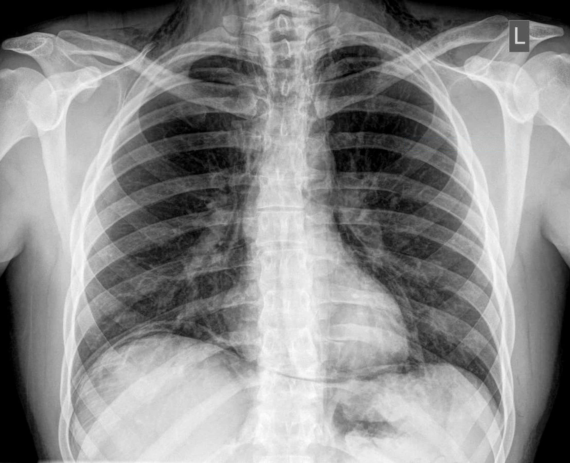
A patient presents with abdominal pain. On physical examination there was abdominal guarding and tenderness. A plain erect chest X-ray reveals air under diaphragm. Probable diagnosis is

Sengstaken-Blakemore tube is used to control bleeding in:
Explanation: Duodenum or pancreas - The majority, approximately 80%, of gastrinomas are found in the duodenum or pancreas [1]. - These tumors secrete excessive gastrin, leading to Zollinger-Ellison syndrome [1]. Stomach antrum - The stomach antrum contains G cells that produce gastrin, but primary gastrinomas rarely originate here. - While G-cell hyperplasia can occur in the antrum, it is distinct from a gastrin-secreting tumor (gastrinoma). Liver - The liver is a common site for metastases from gastrinomas, but it is not typically the primary site of tumor origin [1]. - Primary liver cancers are usually hepatocellular carcinomas or cholangiocarcinomas, which do not secrete gastrin. Small intestine (jejunum/ileum) - Gastrinomas are only rarely found in the more distal parts of the small intestine, such as the jejunum or ileum. - The duodenum and pancreas are the predominant locations for these neuroendocrine tumors due to embryological development and cell differentiation.
Explanation: ***Intestinal metaplasia*** - **Intestinal metaplasia** is a precursor lesion where gastric epithelium is replaced by intestinal-type epithelium, significantly increasing the risk for **gastric carcinoma** [1][2]. - It is a recognized **high-risk factor**, especially in cases of chronic gastritis and atrophic changes in the stomach lining [1][2]. *Ciliated metaplasia* - This condition is generally associated with **respiratory epithelium** and is not linked to gastric carcinoma risk. - It does not involve gastric epithelial changes, therefore, it does not influence **gastric cancer development**. *Pyloric metaplasia* - Pyloric metaplasia typically occurs in chronic gastritis but does not confer a significant **risk** of gastric carcinoma. - It is more related to gastric mucosa adaptation and does not show the same risk association as **intestinal metaplasia**. *Paneth cell metaplasia* - Paneth cell metaplasia is primarily seen in **intestinal disorders** and does not serve as an indicator for gastric carcinoma. - It does not reflect changes in gastric epithelium that are related to cancer risk in the stomach. **References:** [1] Kumar V, Abbas AK, et al.. Robbins and Cotran Pathologic Basis of Disease. 9th ed. The Gastrointestinal Tract, pp. 777-778. [2] Cross SS. Underwood's Pathology: A Clinical Approach. 6th ed. Common Clinical Problems From Alimentary System Disease, pp. 354-355.
Explanation: ***Dysphagia*** - **Dysphagia** (difficulty swallowing) is a prominent symptom typically associated with carcinomas of the **cardia** or **fundus** of the stomach, where the tumor directly impedes food passage [1]. - A silent carcinoma of the **body of the stomach** is less likely to cause dysphagia until it has grown extensively or metastasized, making this the exception among the listed options [1]. *Obstructive jaundice* - A silent carcinoma of the stomach body can metastasize to the **liver** or the **porta hepatis** lymph nodes, leading to compression of the bile ducts [1]. - This compression can cause **obstructive jaundice**, characterized by yellow skin and eyes due to bile flow obstruction [1]. *Ascites* - **Ascites**, the accumulation of fluid in the abdominal cavity, can occur with advanced gastric cancer due to **peritoneal carcinomatosis** (spread of cancer cells to the peritoneum) [1]. - This is a common finding in widespread metastatic disease, even from a initially "silent" primary tumor. *Krukenberg's tumours* - These are **metastatic ovarian tumors** originating from a primary gastrointestinal cancer, most commonly the stomach [1]. - A silent gastric carcinoma can present with these secondary tumors as the first noticeable symptom, especially due to **lymphatic spread** to the ovaries [1].
Explanation: ***Decreased gastric mucosal protection due to H. pylori infection.*** [1] - *Helicobacter pylori* primarily causes peptic ulcer disease by **damaging the protective mucosal layer** of the stomach and duodenum. - This bacterium produces enzymes like **urease**, which neutralizes gastric acid locally, allowing it to colonize the mucus layer and subsequently weaken its integrity, making the underlying cells vulnerable to acid. *Increased gastric acid secretion due to H. pylori infection.* - While *H. pylori* can indirectly affect gastric acid secretion (e.g., by altering gastrin and somatostatin release), its primary and most direct mechanism for ulcer formation is **mucosal damage**, not solely increased acid. [1] - Some strains directly influence **parietal cell function**, but this is a secondary effect compared to the direct mucosal assault. [1] *Increased production of gastric mucus.* - *H. pylori* infection typically leads to a **reduction** or alteration in gastric mucus production and quality, not an increase. - The bacterium thrives by **penetrating the mucus layer**, indicating that it benefits from a compromised barrier rather than an enhanced one. *Increased gastric motility due to H. pylori.* - There is no direct evidence that *H. pylori* significantly increases **gastric motility** as a primary mechanism for peptic ulcer development. - Motility disorders are not considered a hallmark of *H. pylori*-induced ulcerogenesis.
Explanation: ***Hypertrophic pyloric stenosis*** - The classic presentation includes **projectile non-bilious vomiting** and a palpable **olive-shaped mass** in the epigastrium of an infant typically between 3 weeks and 6 months of age. - The vomiting is non-bilious because the obstruction is proximal to the ampulla of Vater. *Intussusception* - While it can present with an **abdominal mass** and vomiting, the vomiting is often **bilious** and the classic stool is **'currant jelly'**, which is not mentioned here. - It usually presents with sudden onset of severe, **colicky abdominal pain** and occurs more commonly in slightly older infants (6-12 months). *Tracheoesophageal fistula* - This condition presents at birth with symptoms such as **choking, coughing**, and **cyanosis** during feeding. - It usually causes respiratory distress and feeding difficulties from the first days of life, not a palpable abdominal mass and non-bilious vomiting at 3 months. *Duodenal atresia* - This is a congenital obstruction that typically presents with **bilious vomiting** (as the obstruction is distal to the ampulla of Vater) within the first 24-48 hours of life. - Imaging usually shows a **“double bubble” sign** on abdominal X-ray, and an abdominal mass is not typically palpable.
Explanation: ***Cyclosporine*** - Cyclosporine is an **immunosuppressant** that primarily acts by inhibiting **calcineurin**, which reduces T-cell activation. It is **least likely** to cause peptic ulcers among the choices. - While it has various side effects, gastrointestinal irritation or peptic ulceration is not a prominent or direct adverse effect compared to other listed drugs. *KCl* - **Potassium chloride (KCl)**, particularly in solid tablet form, can cause **direct mucosal injury** and **ulceration** in the esophagus and stomach if it dissolves slowly or is taken without adequate water. - This is due to its **hyperosmolar** nature and potential for local high concentrations of potassium ions. *Diclofenac* - **Diclofenac** is a **non-steroidal anti-inflammatory drug (NSAID)** that inhibits **COX-1 and COX-2 enzymes**, leading to reduced prostaglandin synthesis. - Reduced prostaglandins diminish the stomach's protective mucous barrier and bicarbonate secretion, making it highly prone to **peptic ulcer formation** and gastrointestinal bleeding. *Clopidogrel* - **Clopidogrel** is an **antiplatelet drug** that inhibits **ADP-induced platelet aggregation**, increasing the risk of bleeding throughout the body, including the gastrointestinal tract. - While it doesn't directly cause ulcers through mucosal damage like NSAIDs or KCl, its antiplatelet effect significantly **increases the risk of gastrointestinal bleeding** from any existing erosions or ulcers, which can complicate peptic ulcer disease.
Explanation: ***It usually presents on the mesenteric border of small intestine*** - Meckel's diverticulum is a **true diverticulum** arising from the **anti-mesenteric border** of the ileum, typically 2 feet from the ileocecal valve. - Its mesenteric positioning would be highly atypical and contradict its embryological origin as a remnant of the **vitelline duct**. - This statement is **FALSE** - it arises from the anti-mesenteric border, making it the correct answer to this "except" question. *Bleeding is a common complication* - **Bleeding** is indeed a common complication in adults, often due to **ectopic gastric mucosa** (present in ~50% of cases) within the diverticulum causing ulceration. - This complication can manifest as **painless rectal bleeding**. - This statement is **TRUE**. *Incidental removal is often recommended in younger patients with risk factors* - Current evidence-based guidelines recommend **selective removal** based on risk factors including age <50 years, palpable abnormalities (thickening, nodularity), narrow neck, length >2cm, or presence of bands. - In younger patients with risk factors, the lifetime risk of complications justifies prophylactic removal. - In older adults or those without risk factors, the morbidity of resection may outweigh the lifetime risk of complications. - This statement is **TRUE**. *It is a remnant of omphalomesenteric duct* - Meckel's diverticulum is the most common congenital anomaly of the gastrointestinal tract, representing a persistent portion of the **embryonic vitelline (omphalomesenteric) duct**. - This duct normally connects the fetal midgut to the yolk sac and should completely regress by the 7th week of gestation. - This statement is **TRUE**.
Explanation: ***Laparoscopic repair of the perforation*** - For a **perforated peptic ulcer**, the immediate goal is to close the perforation and control contamination, which is typically achieved via **laparoscopic primary repair** using sutures and an omental patch (Graham patch). - This minimally invasive approach has advantages of reduced pain, shorter hospital stay, and faster recovery compared to open surgery, making it suitable for most stable patients. *Total gastrectomy* - **Total gastrectomy** involves the complete removal of the stomach and is a major, highly invasive procedure. - It is typically reserved for extensive gastric malignancies or diffuse, intractable ulcer disease, not for an acute, localized perforation. *Gastric bypass* - **Gastric bypass** surgery is primarily a **bariatric procedure** performed for severe obesity or severe, uncontrolled diabetes. - It is not indicated for the emergency management of a perforated peptic ulcer. *Pyloroplasty* - **Pyloroplasty** is a procedure to widen the pyloric channel and is performed to improve gastric emptying. - It is typically done in conjunction with a vagotomy for recurrent, complicated duodenal ulcers that cause obstruction, not as the primary treatment for an acute perforation.
Explanation: ***Perforated abdominal viscus*** - The presence of **abdominal guarding** and **tenderness** indicates peritoneal irritation, while **air under the diaphragm** on an erect chest X-ray (**pneumoperitoneum**) is a classic sign of a perforated hollow abdominal organ. - This combination strongly suggests a **perforated abdominal viscus**, such as a **perforated peptic ulcer** or perforated diverticulitis, leading to the leakage of air and intestinal contents into the peritoneal cavity. *Acute myocardial infarction* - Acute myocardial infarction primarily presents with **chest pain**, radiation to the arm/jaw, and shortness of breath, not typically severe abdominal pain with guarding. - While it can cause some epigastric discomfort, it would not explain the **pneumoperitoneum** seen on the chest X-ray. *Aortic dissection* - Aortic dissection typically causes **sudden, severe tearing chest or back pain**, often radiating to the back. - There is no direct link between aortic dissection and **air under the diaphragm** unless there's a co-existing, unrelated issue, which is not suggested by the primary symptoms. *None of the options* - Given the clear clinical and radiological findings of **pneumoperitoneum** and **peritoneal signs**, a perforated abdominal viscus is the most fitting diagnosis among the choices provided. - This option is incorrect as there is a highly probable diagnosis among the given choices.
Explanation: ***Bleeding varices*** - The **Sengstaken-Blakemore tube** is specifically designed with gastric and esophageal balloons to apply direct pressure and tamponade actively bleeding **esophageal** or **gastric varices**. - This device is a temporary measure used to control life-threatening hemorrhage from varices secondary to **portal hypertension** when endoscopic therapies are unsuccessful or unavailable. *Duodenal ulcer bleed* - Bleeding from a duodenal ulcer is typically managed with **endoscopic intervention** (e.g., clipping, injection, cautery) or **surgical repair**. - A Sengstaken-Blakemore tube is not suitable for controlling duodenal bleeds as it cannot reach or apply pressure to the bleeding site in the **duodenum**. *Renal trauma* - Renal trauma causes bleeding within or around the **kidney**, which is usually managed conservatively, with embolization of bleeding vessels, or surgically (e.g., nephrectomy). - The Sengstaken-Blakemore tube is an **upper gastrointestinal device** and has no role in managing bleeding from renal injuries. *Splenic injury in portal hypertension* - Splenic injury with bleeding in the context of portal hypertension typically requires **splenectomy** or **splenic artery embolization**. - While portal hypertension can be a contributing factor, the tube is not designed to control bleeding originating from a **damaged spleen**.
Get full access to all questions, explanations, and performance tracking.
Start For Free