
Endocrine Surgery — MCQs

On this page
Surgical treatment for a 40-years old lady with 3 x 3 cm. papillary carcinoma thyroid with level III enlarged lymph nodes is :
Vocal cord palsy after thyroid surgery is due to injury to:
A 25 year old female patient with previous history of neck irradiation presents with thyroid swelling for last 6 months. The patient is clinically euthyroid. On examination, the right lobe of thyroid gland is enlarged with presence of ipsilateral cervical lymphadenopathy. The most probable clinical diagnosis in this patient is
A 29-year-old woman presents with a neck mass. Fine needle aspiration shows medullary thyroid carcinoma. Her calcitonin level is normal, but genetic testing reveals a RET proto-oncogene mutation. What is the most appropriate management strategy?
A 45-year-old woman undergoes thyroidectomy for papillary thyroid cancer. Postoperatively, she develops perioral numbness and tingling in her fingers. Her calcium is 7.2 mg/dL (normal 8.5-10.5), phosphorus is 5.8 mg/dL (normal 2.5-4.5), and intact PTH is 8 pg/mL (normal 15-65). What is the most likely cause of these findings?
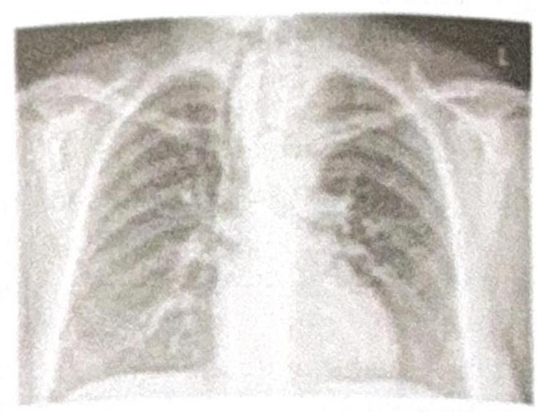
Which of the following procedures would be difficult to perform based on the given Chest X-ray?
After a total thyroidectomy, the surgeon is unable to extubate the patient, who shows cyanosis and respiratory distress. What is the most likely cause of the inability to extubate?
A 32-year-old female patient with Graves' disease with eye signs and enlarged thyroid planned for a total thyroidectomy. What can be given in the preoperative period to reduce intraoperative bleeding in the patient?
A 36-year-old woman comes to the physician for a follow-up visit after she had a PET scan that showed a nodule on the thyroid gland. She has no difficulty or pain while swallowing. She was treated for non-Hodgkin lymphoma at the age of 28 years, which included external beam radiation to the head and neck and 4 cycles of chemotherapy. She appears healthy. Vital signs are within normal limits. Physical examination shows no abnormalities. Serum studies show: Glucose 82 mg/dL Creatinine 0.7 mg/dL Thyroid-stimulating hormone 3 μU/mL Ultrasound of the neck shows a 1.2-cm (0.5-in) nodule on the left lobe of the thyroid with irregular margins and microcalcifications. A fine-needle aspiration biopsy shows Psammoma bodies and cells with clear, ground-glass, empty nuclei. Which of the following is the most appropriate next step in management?
A 27-year-old woman presents with 26 weeks of gestation with a thyroid lesion which is found to be papillary carcinoma of thyroid. Which is the best treatment for this patient?
Practice by Chapter
Thyroid Nodules
Practice Questions
Thyroid Cancer
Practice Questions
Graves' Disease
Practice Questions
Thyroiditis
Practice Questions
Primary Hyperparathyroidism
Practice Questions
Secondary and Tertiary Hyperparathyroidism
Practice Questions
Adrenal Cortical Tumors
Practice Questions
Pheochromocytoma
Practice Questions
Adrenal Incidentalomas
Practice Questions
Multiple Endocrine Neoplasia
Practice Questions
Neuroendocrine Tumors
Practice Questions
Intraoperative Monitoring in Endocrine Surgery
Practice Questions
Want unlimited practice?
Get full access to all questions, explanations, and performance tracking.
Scan to download app