
Colorectal Surgery — MCQs

On this page
Which of the following statements is true regarding Familial adenomatous polyposis (FAP)?
Which of the following is NOT true about solitary rectal ulcer syndrome?
Which of the following are associated with an increased risk of colorectal cancer?
A 74-year-old woman presents with 3 weeks of left lower quadrant abdominal pain, changes in bowel habits, and intermittent fever. Her temperature is 38°C (101°F), respirations are 19 per minute, and blood pressure is 130/80 mm Hg. Physical examination shows left lower quadrant tenderness. A CBC reveals neutrophilia. An abdominal-pelvic ultrasound examination is normal. Which of the following is the most likely diagnosis?
What type of stapler is used for Minimally Invasive Procedures in Hepatic Resection (MIPH)?
What is the primary aim of surgery in carcinoma of the rectum?
Which of the following statements is NOT true about hemorrhoids?
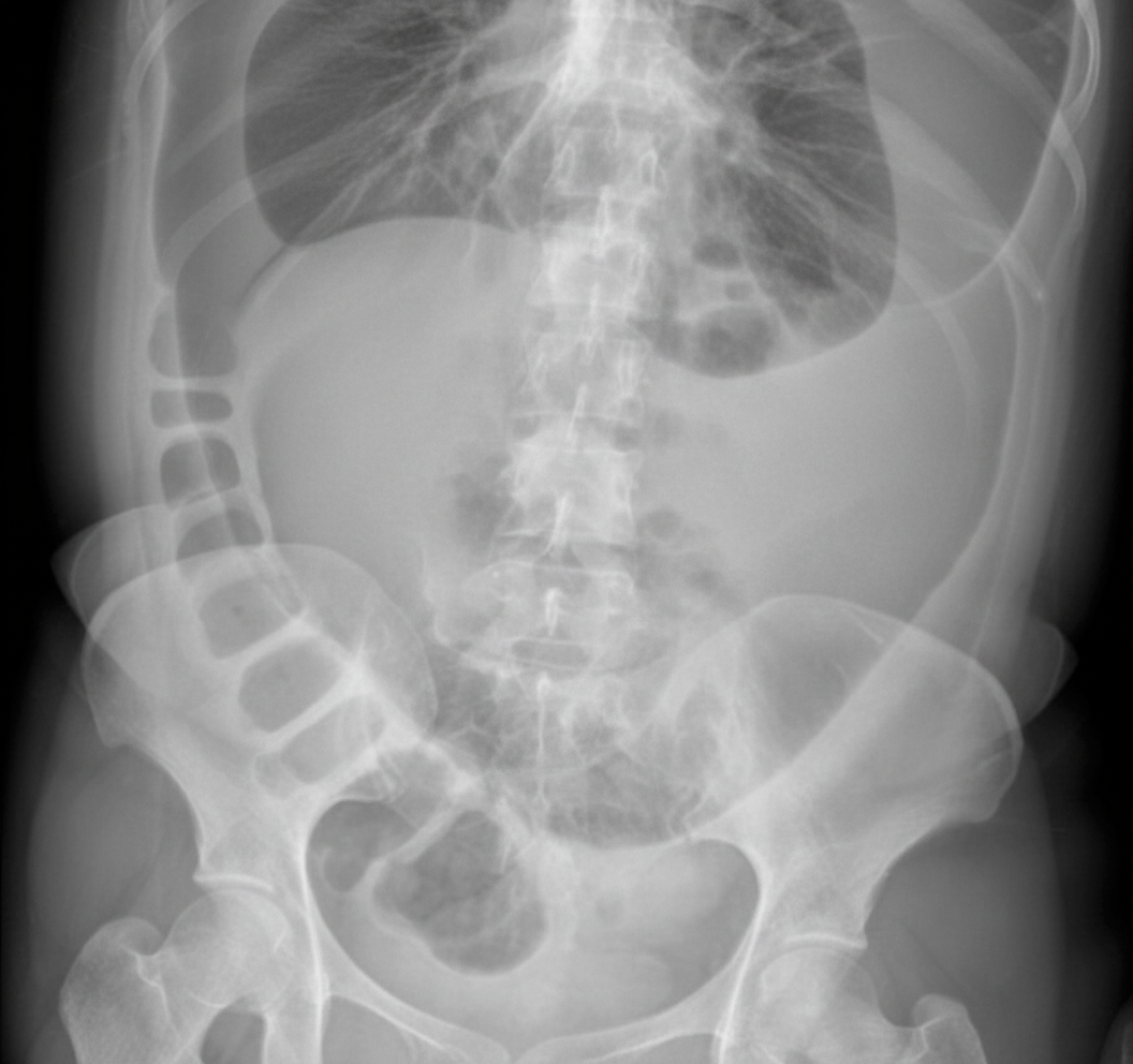
A 70-year-old male presents with colicky pain in the lower abdomen, with non-passage of feces and flatus. Abdominal X-ray findings are provided. Which of the following is the preferred treatment option for this patient?
During an operation for carcinoma of the hepatic flexure of the colon, an unexpected discontinuous 3-cm metastasis is discovered in the edge of the right lobe of liver. What should the surgeon do?
Which of the following is NOT an abdominal procedure for rectal prolapse?
Practice by Chapter
Colorectal Anatomy and Physiology
Practice Questions
Diverticular Disease
Practice Questions
Inflammatory Bowel Disease
Practice Questions
Colorectal Polyps
Practice Questions
Colorectal Cancer
Practice Questions
Anorectal Abscess and Fistula
Practice Questions
Hemorrhoids
Practice Questions
Rectal Prolapse
Practice Questions
Fecal Incontinence
Practice Questions
Intestinal Stomas Creation and Management
Practice Questions
Pelvic Floor Disorders
Practice Questions
Enhanced Recovery After Colorectal Surgery
Practice Questions
Want unlimited practice?
Get full access to all questions, explanations, and performance tracking.
Scan to download app