
Colorectal Surgery — MCQs

On this page
A complicated case of diverticular disease is defined as diverticula with which of the following complications?
In Duke's classification of cancer rectum, what does stage B2 denote?
Which of the following is false regarding cecal volvulus?
A 72-year-old male presents with complete rectal prolapse and a 10-year history of constipation. What is the recommended management for this patient?
Which condition presents with a 'bird's beak' appearance?
All of the following are true about rectal cancer EXCEPT:
Which of the following statements regarding 'Fistula in ano' is true?
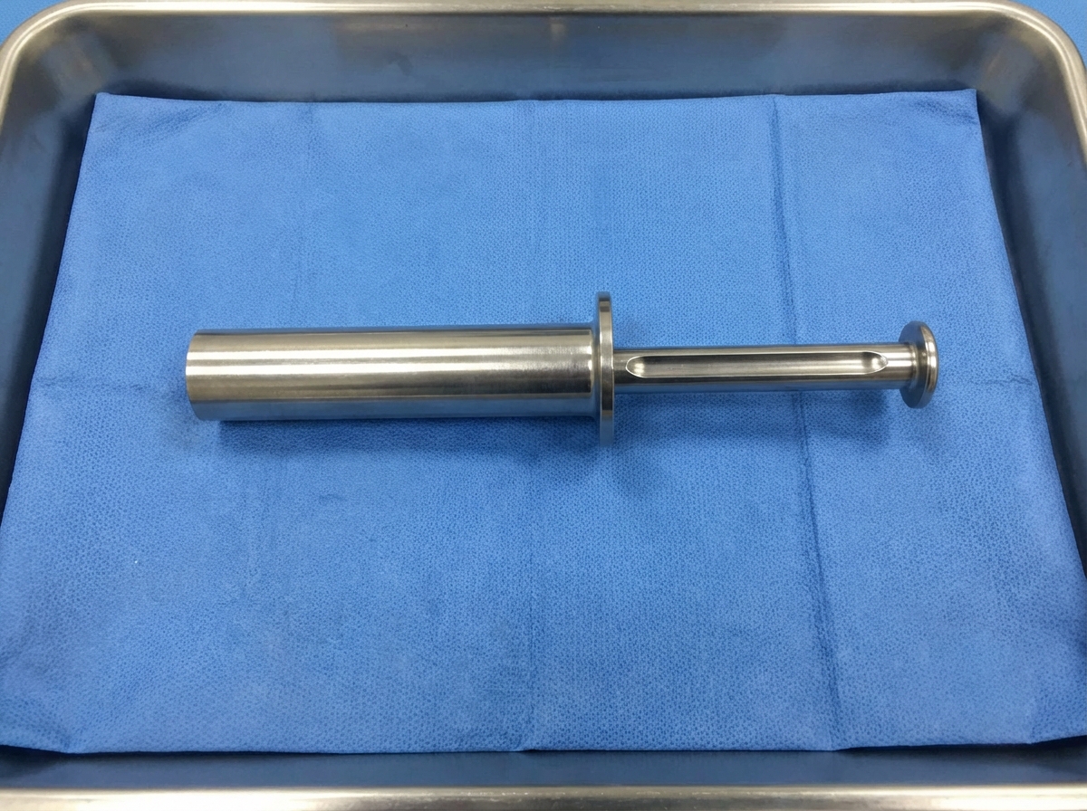
What is the name of the instrument shown?
What is meant by Ca colon stage III c?
A villous polyp of the rectum most commonly manifests as which of the following?
Practice by Chapter
Colorectal Anatomy and Physiology
Practice Questions
Diverticular Disease
Practice Questions
Inflammatory Bowel Disease
Practice Questions
Colorectal Polyps
Practice Questions
Colorectal Cancer
Practice Questions
Anorectal Abscess and Fistula
Practice Questions
Hemorrhoids
Practice Questions
Rectal Prolapse
Practice Questions
Fecal Incontinence
Practice Questions
Intestinal Stomas Creation and Management
Practice Questions
Pelvic Floor Disorders
Practice Questions
Enhanced Recovery After Colorectal Surgery
Practice Questions
Want unlimited practice?
Get full access to all questions, explanations, and performance tracking.
Scan to download app