Breast Surgery — MCQs
On this page
What condition is characterized by greenish-black discharge from the breast?
A 45-year-old female patient with a family history of breast carcinoma presented with diffuse microcalcification on mammography. Biopsy revealed intraductal carcinoma in situ. What is the most appropriate management?
A 27-year-old woman requests a mammogram due to a strong family history of early-onset breast cancer. Which of the following factors would significantly increase this patient's risk for breast cancer?
A 62-year-old woman with a family history of breast cancer (her mother died of breast cancer at age 63) is concerned about her future risk. Which of the following features is a recognized risk factor for breast cancer?
In a patient with carcinoma breast stage T4b, as per TNM classification, what defines this stage?
In a post-mastectomy patient, the suction drain is accidentally removed on the 2nd post-operative day, and the patient presents with oozing and swelling in the chest. What is the next best step in management?
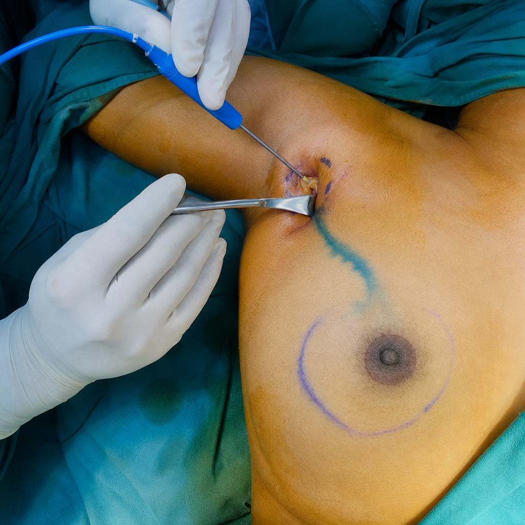
What procedure is depicted?
Inflammatory carcinoma is classified under which TNM staging category?
Adjuvant therapy after mastectomy is needed in all of the following situations except:
A 30-year-old female underwent lumpectomy for a 1.2x1 cm breast lesion with positive axillary nodes. She received adjuvant radiotherapy, tamoxifen, and chemotherapy. What is the recommended line of management for her follow-up?
Practice by Chapter
Breast Anatomy and Physiology
Practice Questions
Benign Breast Diseases
Practice Questions
Breast Cancer Screening
Practice Questions
Breast Cancer: Diagnosis and Staging
Practice Questions
Surgical Management of Breast Cancer
Practice Questions
Oncoplastic Breast Surgery
Practice Questions
Sentinel Lymph Node Biopsy
Practice Questions
Axillary Surgery
Practice Questions
Breast Reconstruction Techniques
Practice Questions
Male Breast Disorders
Practice Questions
Phyllodes Tumors
Practice Questions
Management of Ductal Carcinoma In Situ
Practice Questions
Want unlimited practice?
Get full access to all questions, explanations, and performance tracking.
Scan to download app