Breast Surgery — MCQs
On this page
Which of the following statements regarding lymphoedema following breast cancer treatment are correct? 1. Incidence has decreased due to rarely combined therapy of axillary LN dissection and radiotherapy 2. Precipitating cause like LN metastasis is a major determinant 3. The condition is often painful 4. Oedematous limb is susceptible to bacterial infection Select the correct answer using the code given below:
A 32-year-old woman presents with a 2 cm breast mass. Core needle biopsy shows invasive ductal carcinoma, ER+, PR+, HER2-. Sentinel lymph node biopsy reveals 2 positive nodes with no extracapsular extension. What is the most appropriate next step in management?
A 45-year-old woman with BRCA1 mutation has completed childbearing and requests prophylactic surgery. She has no evidence of breast or ovarian cancer. Her mother died of ovarian cancer at age 50, and her sister had breast cancer at age 35. Evaluate the optimal prophylactic surgical strategy.
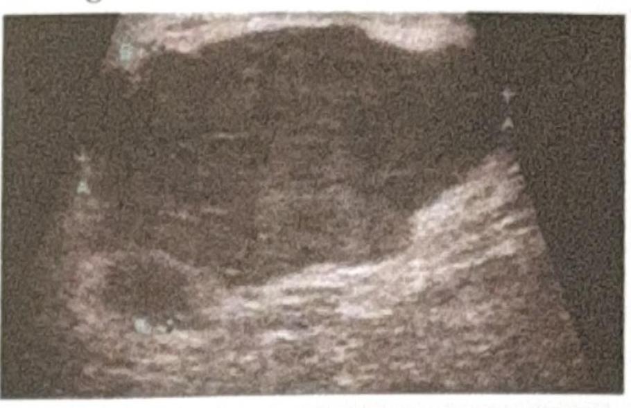
A patient presents to the OPD with a right-sided ulcerated breast lesion. Radiological imaging shows liver metastasis, as seen in the provided ultrasound image. What is the most appropriate management?
A patient presents with upper limb swelling after undergoing a modified radical mastectomy (MRM). What is the most likely cause?
Which of the following is not a relative contraindication for breast conservative surgery?
A 55-year-old female smoker presents with a breast lump and is seeking medical evaluation. On examination, a palpable mass is detected in the breast. The patient's smoking history is significant, with a 30-year smoking habit. Which of the following conditions is strongly associated with smoking in relation to breast health?
A 45-year-old woman with early-stage breast cancer is discussing treatment options with her surgeon. Which of the following statements regarding breast conservation surgery is NOT true?
A 55-year-old female patient presented with a $4 \times 3 \mathrm{~cm}$ lump in the right upper outer quadrant, with no axillary lymph node involvement. Mammography revealed BIRADS 4b staging. She underwent breast conservation surgery, and the final HPE report showed high nuclear-grade DCIS with necrosis and 10 mm margin clearance. What is the further management?
Dye for Sentinel Lymph Node Biopsy is injected in which of the following sites?
Practice by Chapter
Breast Anatomy and Physiology
Practice Questions
Benign Breast Diseases
Practice Questions
Breast Cancer Screening
Practice Questions
Breast Cancer: Diagnosis and Staging
Practice Questions
Surgical Management of Breast Cancer
Practice Questions
Oncoplastic Breast Surgery
Practice Questions
Sentinel Lymph Node Biopsy
Practice Questions
Axillary Surgery
Practice Questions
Breast Reconstruction Techniques
Practice Questions
Male Breast Disorders
Practice Questions
Phyllodes Tumors
Practice Questions
Management of Ductal Carcinoma In Situ
Practice Questions
Want unlimited practice?
Get full access to all questions, explanations, and performance tracking.
Scan to download app