Breast Surgery — MCQs
On this page
A 45-year-old female presents to the OPD with a complaint of a lump in the right breast. On examination, a 5 x 5 cm lump is palpated. Axillary lymph nodes are found to be normal in size, consistency, and texture. What is the MOST likely diagnosis?
A female patient underwent mastectomy for carcinoma of the breast. A few days after surgery, she experienced burning pain along the medial aspect of her arm. What is the most likely cause of this pain?
A 43-year-old lady presents with a 5 cm lump in her right breast and a 3 cm node in the supraclavicular fossa. According to the latest AJCC staging system, to which TNM stage does she belong?
A 40-year-old woman has the following family and personal history: her mother died of breast cancer at age 64, she smokes one pack per day, she drinks five or more cups of coffee per day, she has no children, and she takes birth control pills. Which of the following is the most significant risk factor for breast cancer in this patient?
Which of the following are risk factors for breast cancer?
Which of the following is NOT true of Paget's disease of the breast?
A 45-year-old woman presents with a weeping eczematoid lesion of her nipple. Which of the following statement is false concerning her diagnosis and management?
A 28-year-old female presents with a 1 cm hard lump indicative of infiltrating breast carcinoma. Staging is T1-N0-M0. What is the best treatment?
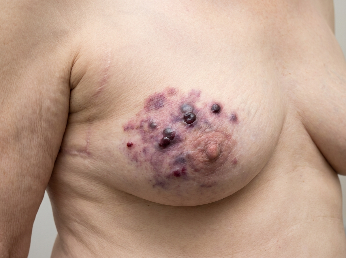
A 72-year-old woman presented with skin changes over her left breast. She had a history of breast cancer 5 years earlier and was treated with a lumpectomy and radiation therapy. What is the diagnosis?
What is the recommended duration for tamoxifen therapy in breast carcinoma?
Practice by Chapter
Breast Anatomy and Physiology
Practice Questions
Benign Breast Diseases
Practice Questions
Breast Cancer Screening
Practice Questions
Breast Cancer: Diagnosis and Staging
Practice Questions
Surgical Management of Breast Cancer
Practice Questions
Oncoplastic Breast Surgery
Practice Questions
Sentinel Lymph Node Biopsy
Practice Questions
Axillary Surgery
Practice Questions
Breast Reconstruction Techniques
Practice Questions
Male Breast Disorders
Practice Questions
Phyllodes Tumors
Practice Questions
Management of Ductal Carcinoma In Situ
Practice Questions
Want unlimited practice?
Get full access to all questions, explanations, and performance tracking.
Scan to download app