Sentinel lymph node biopsy in carcinoma breast is done if -
N3a TNM staging of head and neck tumors (AJCC 8th edition) shows:
Which of the following walls of axilla is formed by the surgical neck of humerus:
What is a late complication of elbow dislocation?
Axillary Nerve Injury is least likely in:
A patient with head and neck cancer has a contralateral lymph node of 3 cm size. What is the N staging?
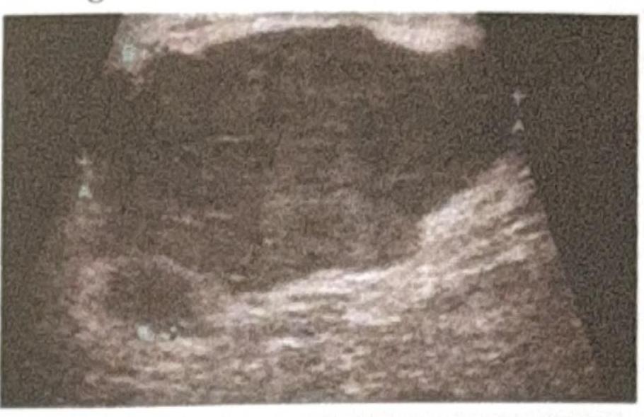
A patient presents to the OPD with a right-sided ulcerated breast lesion. Radiological imaging shows liver metastasis, as seen in the provided ultrasound image. What is the most appropriate management?

A 45-year-old woman with early-stage breast cancer is discussing treatment options with her surgeon. Which of the following statements regarding breast conservation surgery is NOT true?
Which of the following is a contraindication to breast conservation surgery?
In which of the following situations is breast conservation surgery not indicated?
Explanation: ***Breast mass but no lymph node palpable*** - Sentinel lymph node biopsy is primarily performed in patients with **clinically negative axillae** (no palpable lymph nodes) to assess for microscopic metastatic disease. - The goal is to avoid full axillary lymph node dissection if the sentinel nodes are negative, thus reducing the risk of **lymphedema** and other complications. *LN palpable* - If a lymph node is palpable, it is often considered **clinically suspicious** and may warrant a direct fine-needle aspiration (FNA) or core biopsy rather than a sentinel node biopsy. - A positive biopsy from a palpable node would typically lead directly to an **axillary lymph node dissection** or neoadjuvant therapy, as the sentinel node procedure offers less benefit in this scenario. *Breast lump with palpable axillary node* - Similar to a palpable LN, a **palpable axillary node** in the presence of a breast lump suggests established nodal involvement. - In such cases, **sentinel lymph node biopsy** is often not the initial step; rather, direct biopsy of the palpable node or upfront axillary dissection (sometimes after neoadjuvant treatment) is considered. *Metastatic CA breast* - In **metastatic breast cancer** (stage IV disease), the focus shifts to systemic treatment, and axillary lymph node dissection, including sentinel node biopsy, is generally not indicated for staging purposes. - The primary goal is palliative care or controlling systemic disease, not regional lymph node staging.
Explanation: ***Metastasis in a lymph node >6 cm*** - **N3a disease** in head and neck cancer staging (AJCC 8th edition) specifically refers to metastasis in a single lymph node larger than 6 cm in greatest dimension **without extranodal extension (ENE)**. - This applies to oral cavity, oropharynx (HPV-negative), hypopharynx, and larynx cancers. - **Note:** N3 staging also includes **N3b** (metastasis in any node with clinically overt ENE), but this question specifically asks about N3a criteria. *Metastasis in lymph nodes >2 cm* - Lymph nodes in the 2-3 cm range typically fall within **N1 or N2a categories**, depending on laterality and number of involved nodes. - **N3a disease** requires a single lymph node to exceed 6 cm in greatest dimension without ENE. *Metastasis in lymph nodes >5 cm* - A lymph node between 3-6 cm is usually classified as **N2 disease** (N2a if single ipsilateral ≤6 cm, N2b if multiple ipsilateral ≤6 cm, N2c if bilateral or contralateral ≤6 cm). - To be classified as **N3a**, the lymph node must be **>6 cm** without extranodal extension. *None of the options* - This option is incorrect because the first option accurately describes the size criterion for **N3a TNM staging** in head and neck tumors according to AJCC 8th edition guidelines. - While N3 staging has two subcategories (N3a and N3b), the size criterion of >6 cm correctly defines N3a disease.
Explanation: ***Lateral*** - The **lateral wall** of the axilla is formed by the **surgical neck of the humerus** and the coracobrachialis and biceps brachii muscles as they pass down into the arm. - This wall provides the main connection between the axilla and the arm proper. *Anterior* - The **anterior wall** is formed by the **pectoralis major** [1] and **pectoralis minor** muscles [1], along with the clavipectoral fascia. - This wall also forms the anterior axillary fold. *Posterior* - The **posterior wall** is comprised of the **subscapularis**, **teres major**, and **latissimus dorsi** muscles [1]. - It forms the posterior axillary fold. *Medial* - The **medial wall** of the axilla is formed by the **upper four or five ribs** along with the **serratus anterior muscle**. - This wall lies against the chest wall.
Explanation: **Myositis ossificans** - **Myositis ossificans** is the abnormal formation of **heterotopic bone** within muscle or other soft tissues, often developing weeks to months after joint trauma such as an elbow dislocation. - It typically presents as a painful, firm mass with restricted joint movement, especially **flexion** and **extension** at the elbow. *Median nerve injury* - **Median nerve injury** can occur at the time of the initial elbow dislocation (an **acute complication**), but it is not typically considered a late complication that develops over weeks or months. - Symptoms include numbness in the thumb, index, and middle fingers, as well as weakness in **thumb opposition** and **flexion** of the index finger. *Brachial artery injury* - **Brachial artery injury** is an **acute complication** of severe elbow dislocation, leading to compromise of distal blood flow. - Signs include absence of pulses, pallor, paresthesia, and pain in the forearm and hand, requiring immediate surgical intervention. *None of the options* - This option is incorrect because **myositis ossificans** is a well-recognized late complication of elbow dislocation.
Explanation: Improper use of crutch - **Improper crutch usage** primarily affects the **radial nerve** in the axilla due to direct compression against the humerus. - While it can cause nerve damage, the **axillary nerve** is less commonly injured by crutch use as it lies more distally and laterally, protected by the deltoid muscle. *Intramuscular injection* - Injections in the **deltoid muscle** can directly injure the **axillary nerve** due to its superficial course around the surgical neck of the humerus. [1] - This risk is higher with improper technique or very deep injections, leading to **deltoid weakness** and **sensory loss** over the lateral shoulder. *Shoulder dislocation* - **Anterior shoulder dislocations** are a common cause of **axillary nerve injury** due to the stretching or tearing of the nerve as the humeral head displaces. - The nerve wraps around the **surgical neck of the humerus**, making it vulnerable during dislocation. *Fracture proximal humerus* - Fractures of the **surgical neck of the humerus** often lead to **axillary nerve damage** because the nerve lies in close proximity to this region. - The blunt force or displacement of bone fragments can directly compress or lacerate the nerve.
Explanation: ***N2c (Single or Multiple, Bilateral or Contralateral, None > 6 cm)*** - A 3 cm **contralateral** lymph node falls under the **N2c** category according to the AJCC staging system for head and neck cancers. - **N2c** indicates involvement of **contralateral** or **bilateral lymph nodes**, with the largest node being **no greater than 6 cm**. - This is the correct staging for the described clinical scenario. *N2a (Single, Ipsilateral, 3 to 6 cm)* - This option incorrectly describes an **ipsilateral** lymph node, whereas the question specifies a **contralateral** node. - **N2a** is defined by a single **ipsilateral** lymph node between **3 and 6 cm** in greatest dimension. - The key differentiator is **laterality** (ipsilateral vs contralateral). *N1 (Single, Ipsilateral, Equal to or <3 cm)* - This option refers to an **ipsilateral** lymph node that is **3 cm or smaller**, which does not match the contralateral location provided in the question. - **N1** describes a single **ipsilateral** lymph node that is **≤ 3 cm** in greatest dimension. - This fails on both **laterality** (ipsilateral vs contralateral) and **size criteria** (the node is exactly 3 cm, at the boundary). *N3 (Single or Multiple, Ipsilateral/Bilateral/Contralateral, Any node >6 cm)* - While it includes contralateral involvement, **N3** is specifically for a lymph node **greater than 6 cm**, which is not the case for a 3 cm node. - A **N3** classification applies when **any** regional lymph node (ipsilateral, bilateral, or contralateral) exceeds **6 cm** in greatest dimension. - The described 3 cm node does not meet the **size threshold** for N3 staging.
Explanation: ***Neoadjuvant chemotherapy followed by surgery*** - The presence of **distant metastasis** (liver metastasis) indicates **Stage IV breast cancer**, where **systemic treatment is the primary goal**. - In Stage IV disease, **palliative systemic chemotherapy** is the mainstay of treatment to control distant disease and improve survival. - Surgery in metastatic breast cancer may be considered for **local control of symptomatic disease** (ulceration, bleeding, pain), typically after initiating systemic therapy. - The combination of systemic therapy followed by local surgery for the ulcerated lesion addresses both the metastatic disease and provides local symptom relief. *Simple mastectomy* - While this could provide local control of the ulcerated lesion, it does **not address the distant metastasis**. - In Stage IV disease, **systemic therapy must be prioritized** before considering any local surgical intervention. - Surgery alone without systemic treatment would be inadequate for metastatic disease. *Modified Radical Mastectomy (MRM)* - MRM involves removal of the entire breast tissue, skin, nipple-areolar complex, and level I and II axillary lymph nodes. - While this provides comprehensive local-regional control, it **does not address distant metastasis**. - In Stage IV disease, extensive locoregional surgery without systemic therapy first would be inappropriate, as the primary issue is systemic disease. *Radical mastectomy* - This extensive procedure involves removal of the breast, axillary lymph nodes, and pectoralis muscles. - It is **rarely performed today** due to significant morbidity and no survival benefit over less extensive procedures. - Like other surgical options alone, it fails to address the systemic nature of Stage IV disease.
Explanation: ***Axillary dissection*** - **Axillary dissection is NOT a routine component of breast conservation surgery** for early-stage breast cancer. - In early-stage disease, **sentinel lymph node biopsy (SLNB)** has largely replaced routine axillary dissection as it provides accurate staging with significantly less morbidity. - Axillary dissection is only performed when there is **proven extensive lymph node involvement** or when SLNB shows metastatic disease requiring further assessment. - Therefore, this statement is **NOT true** regarding routine breast conservation surgery. *Wide local excision* - **Wide local excision (lumpectomy)** is the primary surgical component of breast conservation therapy. - It involves removing the cancerous tumor along with a margin of healthy breast tissue to achieve clear margins while preserving the breast. *Sentinel lymph node biopsy* - **SLNB** is a standard procedure performed with breast conservation surgery to assess for regional lymph node metastasis. - It identifies and removes the first few lymph nodes draining the tumor, allowing accurate staging with minimal morbidity. *Post-operative radiotherapy* - **Post-operative radiotherapy** to the preserved breast is a critical and essential component of breast conservation therapy. - It significantly reduces the risk of local recurrence by treating any microscopic tumor cells that may remain after surgery.
Explanation: ***Presence of multicentric tumors*** - **Multicentric tumors** are defined as two or more discrete tumors in different quadrants of the breast, which cannot be removed with a single lumpectomy. - This condition is a contraindication for breast conservation surgery (BCS) because complete removal of all tumor foci while maintaining an acceptable cosmetic outcome is highly unlikely. *Involvement of axillary lymph nodes* - **Axillary lymph node involvement** is an important prognostic factor in breast cancer and influences adjuvant therapy decisions, but it is not a direct contraindication to BCS. - Patients with positive nodes often undergo axillary dissection or sentinel lymph node biopsy, followed by radiation and/or systemic therapy, which can be combined with BCS. *Tumor size greater than 4 cm* - While larger tumor size (e.g., >4-5 cm) can make achieving negative surgical margins and a good cosmetic outcome more challenging with BCS, it is not an absolute contraindication. - **Neoadjuvant chemotherapy** can often downstage larger tumors, making BCS a viable option for many patients. *Presence of diffuse microcalcifications* - **Diffuse microcalcifications** can sometimes indicate extensive ductal carcinoma in situ (**DCIS**) or invasive lobular carcinoma with a widespread component. - However, if the microcalcifications represent a single focus of disease that can be entirely excised with negative margins, BCS may still be an option, especially if guided by stereotactic biopsy and imaging.
Explanation: ***All of the options*** - All listed scenarios—**large pendular breast**, **SLE**, and **diffuse microcalcification**—represent situations where breast conservation surgery is generally contraindicated or challenging. - Their presence often necessitates alternative treatment approaches, such as mastectomy, to achieve optimal oncologic and cosmetic outcomes. *Large pendular breast* - While not an absolute contraindication, a **very large or pendulous breast** can make it difficult to achieve a satisfactory cosmetic outcome after breast conservation surgery. - The disproportionate breast size post-lumpectomy may lead to significant **asymmetry**, requiring further reconstructive procedures. *SLE* - Patients with **Systemic Lupus Erythematosus (SLE)** are at an increased risk of complications from radiation therapy, a mandatory component of breast conservation surgery. - They tend to experience more severe and prolonged **acute and chronic skin reactions** to radiation, which can significantly impair healing and quality of life. *Diffuse microcalcification* - **Diffuse microcalcification** within the breast can indicate widespread in situ carcinoma (e.g., DCIS) or an invasive carcinoma with extensive intraductal component. - In such cases, achieving **clear surgical margins** with breast conservation surgery can be challenging and often leads to multiple re-excisions or an increased risk of local recurrence.
Get full access to all questions, explanations, and performance tracking.
Start For Free