
Bariatric Surgery — MCQs

On this page
Which of the following is NOT a component of the Obesity Surgery-Mortality Risk Score (OS-MRS)?
Which of the following is NOT true about bariatric surgery?
All of the following regarding papillary carcinoma of the thyroid is true EXCEPT?
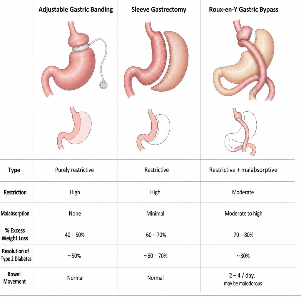
Which of the following is NOT a bariatric surgical procedure?
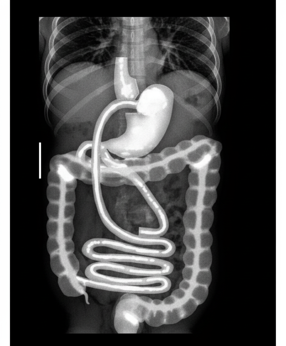
What procedure was most recently performed on this patient?
Practice by Chapter
Pathophysiology of Obesity
Practice Questions
Patient Selection and Preoperative Evaluation
Practice Questions
Restrictive Procedures
Practice Questions
Malabsorptive Procedures
Practice Questions
Sleeve Gastrectomy
Practice Questions
Roux-en-Y Gastric Bypass
Practice Questions
Biliopancreatic Diversion
Practice Questions
Adjustable Gastric Banding
Practice Questions
Revisional Bariatric Surgery
Practice Questions
Postoperative Management
Practice Questions
Nutritional Considerations
Practice Questions
Metabolic Effects of Bariatric Surgery
Practice Questions
Want unlimited practice?
Get full access to all questions, explanations, and performance tracking.
Scan to download app