
Ultrasound — MCQs

On this page
The diagnostic feature of congenital diaphragmatic hernia on prenatal ultrasonography is:
What is the most sensitive investigation for a thyroid nodule?
Which of the following is a sonographic finding of a fetus with Down's syndrome?
What is the frequency range of sound waves typically used for transabdominal ultrasonography?
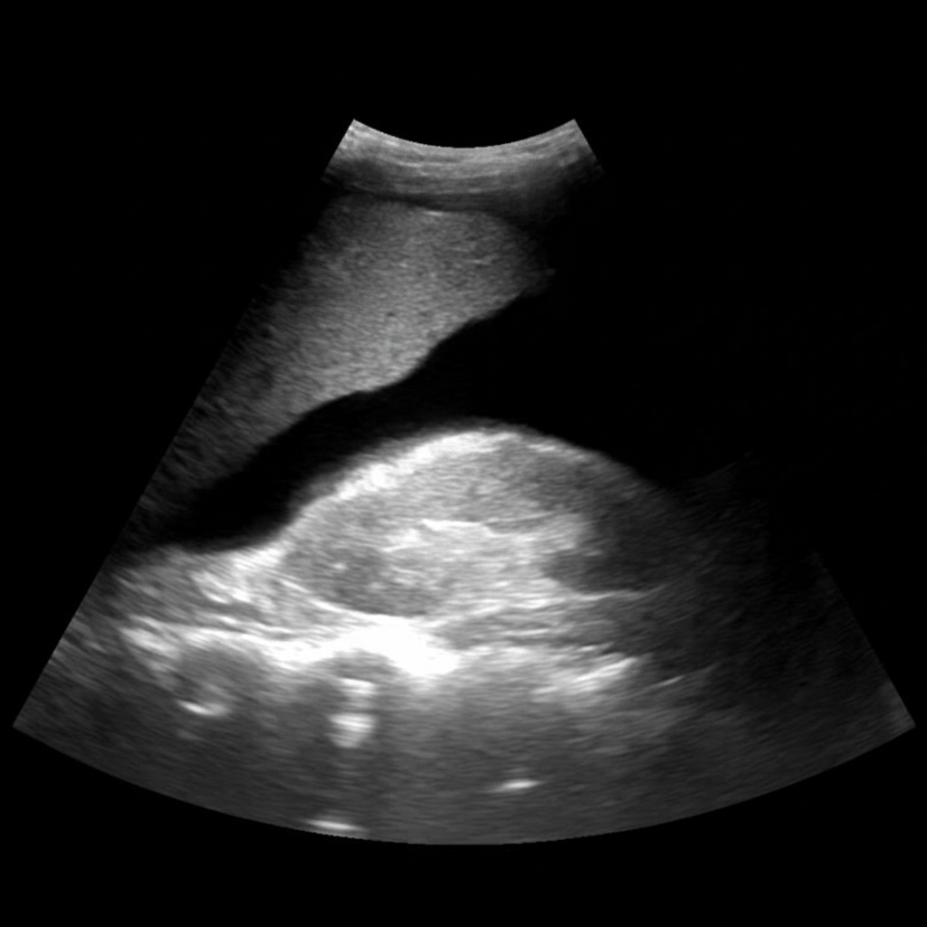
Comment on the provided ultrasound image?
Which of the following probes is used for transcranial ultrasonography?
The 'Filaria dance' sign is observed in which imaging modality?
What is the commonly used frequency range in obstetrical ultrasonography?
Endoscopic ultrasound is extremely useful in staging which tumour?
Which of the following is NOT a feature of a normal venous Doppler study?
Practice by Chapter
Physics of Ultrasound
Practice Questions
Instrumentation and Techniques
Practice Questions
Abdominal Ultrasonography
Practice Questions
Pelvic Ultrasonography
Practice Questions
Obstetric Ultrasonography
Practice Questions
Small Parts Ultrasonography
Practice Questions
Musculoskeletal Ultrasonography
Practice Questions
Vascular Ultrasonography
Practice Questions
Pediatric Ultrasonography
Practice Questions
Contrast-Enhanced Ultrasound
Practice Questions
Ultrasound-Guided Interventions
Practice Questions
Doppler Ultrasound Principles and Applications
Practice Questions
Want unlimited practice?
Get full access to all questions, explanations, and performance tracking.
Scan to download app