
Ultrasound — MCQs

On this page
The double bleb sign on ultrasound is indicative of which of the following?
Which of the following statements regarding Doppler ultrasound is incorrect?
Evaluation of cardiac valve motion and fetal heart rate is done in which mode of ultrasound?
TI-RADS is a reporting system for thyroid nodules. It is based on the following properties, except:
On ultrasound, what typically causes dirty shadowing?
What causes an acoustic shadow in ultrasound imaging?
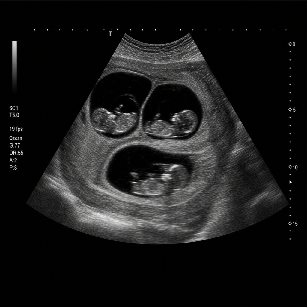
Sonographic scan shows:
What is the smallest size of gallstone that can be confidently diagnosed using ultrasonography?
Ultrasound diagnoses all the following except?
At what gestational week is accurate diagnosis of anencephaly typically seen on ultrasound?
Practice by Chapter
Physics of Ultrasound
Practice Questions
Instrumentation and Techniques
Practice Questions
Abdominal Ultrasonography
Practice Questions
Pelvic Ultrasonography
Practice Questions
Obstetric Ultrasonography
Practice Questions
Small Parts Ultrasonography
Practice Questions
Musculoskeletal Ultrasonography
Practice Questions
Vascular Ultrasonography
Practice Questions
Pediatric Ultrasonography
Practice Questions
Contrast-Enhanced Ultrasound
Practice Questions
Ultrasound-Guided Interventions
Practice Questions
Doppler Ultrasound Principles and Applications
Practice Questions
Want unlimited practice?
Get full access to all questions, explanations, and performance tracking.
Scan to download app