Following are the ultrasound parameters used in the diagnosis of intrauterine growth restriction except?
Which of the following structures cannot be effectively visualized using ultrasonography (USG)?
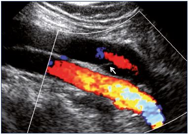
A patient presents with sudden-onset right leg pain. An investigation was done, and the obtained image is shown below. What is the investigation based on the image?

Which of the following is NOT a typical ultrasonographic finding in autosomal recessive polycystic kidney disease (ARPKD)?
Frequency of ultrasound waves in USG -
What is the echogenic lesion size criterion for chronic pancreatitis?
Investigation of choice for screening of proximal internal carotid artery stenosis is :
Which of the following techniques uses piezoelectric crystals?
Which imaging and Doppler techniques are combined in duplex ultrasonography?
What is the best way to diagnose gallbladder stones?
Explanation: ***Ponderal index*** - The **ponderal index** is a measure of proportionality (weight/length³) used to assess fetal body proportions and nutritional status, but it is calculated **after birth** using the newborn's weight and length. - It is **not an ultrasound parameter** used during prenatal diagnosis of intrauterine growth restriction (IUGR). - While it can identify asymmetric growth patterns postnatally, it has no role in prenatal IUGR assessment. *Abdominal circumference* - **Fetal abdominal circumference (AC)** is a key **biometric ultrasound parameter** that is essential in diagnosing IUGR. - It reflects fetal liver size and subcutaneous fat stores, making it the **most sensitive single parameter** for detecting IUGR. - Serial AC measurements below the 10th percentile or showing poor growth velocity are diagnostic criteria for IUGR. *Doppler velocimetry* - **Doppler velocimetry**, particularly of the **umbilical artery**, is crucial for assessing placental vascular resistance and diagnosing IUGR. - Abnormal findings include **increased pulsatility index (PI)**, **absent end-diastolic flow (AEDF)**, or **reversed end-diastolic flow (REDF)**. - These findings indicate placental insufficiency and help classify IUGR severity. *Increased diastolic velocity in middle cerebral artery* - **Increased diastolic velocity** in the middle cerebral artery (leading to **decreased PI/RI**) indicates the **brain-sparing effect** (cerebrovasodilation). - This is a fetal compensatory response to chronic hypoxemia in IUGR. - The **cerebroplacental ratio (CPR)** (umbilical artery PI / MCA PI) is a sensitive marker for adverse perinatal outcomes in IUGR.
Explanation: ***Bones and dense structures*** - **Ultrasound waves** cannot effectively penetrate through **bones or other dense structures** due to their high acoustic impedance, leading to significant reflection and scattering of the waves. - This limitation means that structures located behind bone are poorly visualized, resulting in an acoustic shadow. *Fluid-filled structures* - **Ultrasonography excels** at visualizing **fluid-filled structures** because fluid allows for excellent transmission of sound waves with minimal attenuation. - This results in well-defined anechoic (black) areas on the ultrasound image, making structures like cysts or the urinary bladder easily discernible. *Gallbladder and bile ducts* - The **gallbladder and bile ducts** are routinely and effectively visualized by USG, as they are typically fluid-filled (with bile) and surrounded by soft tissues. - USG is the primary imaging modality for detecting gallstones, cholecystitis, and evaluating biliary obstruction. *Blood vessels and blood flow* - **Doppler ultrasonography** is specifically designed to visualize **blood vessels and assess blood flow** characteristics. - It uses the Doppler effect to detect changes in frequency caused by moving red blood cells, allowing for evaluation of vessel patency, flow direction, and velocity.
Explanation: ***Ultrasound doppler*** - The image clearly displays **blood flow** (red and blue colors indicating direction) superimposed on a grayscale anatomical image, which is characteristic of a **Color Doppler ultrasound**. - A **Doppler ultrasound** is an ideal initial investigation for sudden-onset limb pain to assess for vascular pathologies like **deep vein thrombosis** or arterial occlusion. *A digital subtraction angiography* - This technique involves injecting a **contrast agent** and taking X-ray images, with computer software subtracting background bone and soft tissue to highlight blood vessels. - The image shown does not exhibit the typical appearance of a **DSA**, which would appear as radiopaque vessels against a subtracted background. *MR angiography* - **MR angiography** uses magnetic fields and radio waves to create detailed images of blood vessels, often utilizing contrast agents like gadolinium. - The image provided is clearly from an **ultrasound modality**, not an MRI, as indicated by the characteristic acoustic window and transducer artifacts. *Plethysmography* - **Plethysmography** measures changes in volume within an organ or whole body, usually resulting from fluctuations in blood flow. - It does not produce direct images of blood vessels but rather **waveform tracings** reflecting blood volume changes, making it unrelated to the visual data presented.
Explanation: **Cysts larger than 2 cm** - **ARPKD** is characterized by numerous tiny cysts (typically 1-2 mm, rarely up to 1 cm) that are microscopically dilated collecting ducts, leading to diffuse renal enlargement. - Cysts larger than 2 cm are much more typical of **autosomal dominant polycystic kidney disease (ADPKD)**, which involves macroscopic cysts of various sizes. *Enlarged kidneys* - The proliferation of dilated collecting ducts and associated interstitial fibrosis in **ARPKD** leads to significantly enlarged kidneys, which is a hallmark ultrasound finding. - This enlargement is often bilateral and can be detected prenatally or in neonates. *Increased echogenicity* - The presence of numerous tiny cysts and dense fibrous tissue throughout the renal parenchyma in **ARPKD** causes increased diffuse echogenicity on ultrasound. - This is a common and important diagnostic feature, often described as "bright" or "hyperechoic" kidneys. *Corticomedullary differentiation is lost* - In **ARPKD**, the normal distinct differentiation between the renal cortex and medulla is obliterated due to the widespread involvement of the collecting ducts and the diffuse nature of the disease. - This loss of corticomedullary differentiation is a typical finding on ultrasound for severe renal parenchymal disease, including ARPKD.
Explanation: ***>2 MHz*** - Medical diagnostic ultrasound typically uses frequencies in the **range of 2-15 MHz**, with some applications extending from 1-20 MHz. - Frequencies **above 2 MHz** are considered the standard for diagnostic ultrasonography, providing adequate **spatial resolution** and tissue penetration for imaging internal structures. - **Frequency selection** depends on the application: - **2-5 MHz**: Deep structures (abdominal, obstetric imaging) - better penetration - **5-10 MHz**: Vascular studies, cardiac imaging - **7-15 MHz**: Superficial structures (thyroid, breast, musculoskeletal) - better resolution - Higher frequencies provide better resolution but less penetration; the choice represents a trade-off based on clinical needs. *2000 Hz* - This frequency (2 kHz) falls within the **audible range** for humans (20 Hz to 20 kHz). - Such low frequencies would not provide the necessary **spatial resolution** for diagnostic imaging and lack the characteristics needed for medical ultrasound. *5000 Hz* - At 5 kHz, this is still within the **audible frequency range**. - These frequencies are far too low for medical ultrasound imaging, which requires **megahertz frequencies** to generate diagnostically useful images with adequate detail. *< 2 MHz* - Frequencies below 2 MHz, while technically ultrasound (>20 kHz), are generally **below the diagnostic range** for most clinical applications. - Although lower frequencies offer better tissue penetration, frequencies below 2 MHz provide **insufficient spatial resolution** for standard diagnostic medical imaging.
Explanation: ***> 3 mm*** - An echogenic lesion **greater than 3 mm** in size is a diagnostic criterion for **chronic pancreatitis** when observed on ultrasound imaging. - This represents one of the **major features** in ultrasound diagnosis of chronic pancreatitis, particularly when echogenic foci demonstrate acoustic shadowing (suggesting calcifications). - This criterion is part of established diagnostic frameworks and helps differentiate pathological calcifications from minor, non-specific findings. *> 4 mm* - While a **4 mm threshold** would indicate significant findings, the established diagnostic criterion for chronic pancreatitis uses **> 3 mm** as the cutoff. - Using a higher threshold would reduce sensitivity for detecting chronic pancreatitis. *> 2 mm* - A lesion larger than **2 mm** is generally considered below the established diagnostic threshold for **chronic pancreatitis**. - This size may represent early changes or incidental findings that are not yet definitive for diagnosis. *> 1 mm* - A lesion **greater than 1 mm** is too small to be a definitive criterion for **chronic pancreatitis** and could represent minor, non-pathological findings. - Such small echogenic foci lack sufficient specificity for diagnosing chronic pancreatic disease.
Explanation: ***Doppler ultrasound (USG)*** - **Doppler ultrasound** is the investigation of choice for screening carotid artery stenosis due to its **non-invasive nature**, widespread availability, and cost-effectiveness. It provides excellent anatomical information and hemodynamics, including **blood flow velocity** and presence of **plaque**. - It can effectively estimate the degree of **stenosis** based on flow characteristics and is safe for serial monitoring. *CT angiography* - **CT angiography** provides detailed anatomical imaging of the carotid arteries but involves **ionizing radiation** and **iodinated contrast agents**, making it less suitable for routine screening, especially in patients with renal impairment or contrast allergies. - While it offers higher resolution for certain plaque characteristics, it’s typically reserved for cases where ultrasound findings are inconclusive or for surgical planning. *Magnetic resonance imaging (MRI)* - **MRI** and **MR angiography (MRA)** can visualize carotid stenosis without ionizing radiation but are more expensive and time-consuming than ultrasound, and can be limited by artifacts from patient movement. - Patients with **claustrophobia** or **metallic implants** may not be suitable for MRI, making it less ideal for general screening. *Digital subtraction angiography (DSA)* - **Digital subtraction angiography (DSA)** is the **gold standard** for diagnosing carotid stenosis as it provides the most detailed and accurate images of the arterial lumen. However, it is an **invasive procedure** associated with risks such as stroke, arterial dissection, contrast nephropathy, and radiation exposure. - Due to its invasiveness and potential complications, DSA is typically reserved for cases with **discordant non-invasive findings** or prior to intervention rather than as a primary screening tool.
Explanation: ***Ultrasonography*** - **Piezoelectric crystals** are the core component of **ultrasound transducers**, which generate and detect ultrasonic waves. - These crystals convert electrical energy into mechanical vibrations (sound waves) and vice-versa, allowing for the creation of images. *NMR imaging* - **NMR imaging** (Nuclear Magnetic Resonance, now commonly known as **MRI**) uses strong magnetic fields and **radiofrequency pulses** to generate images. - It relies on the magnetic properties of atomic nuclei, particularly hydrogen, rather than piezoelectric effects. *X-ray diffraction* - **X-ray diffraction** is a technique used to study the atomic and molecular structure of materials, and it involves the interaction of **X-rays** with the electron clouds of atoms. - It does not involve piezoelectric crystals; instead, it uses an X-ray source and a detector to measure diffracted X-rays. *Xeroradiography* - **Xeroradiography** is an older imaging technique that used **xerographic plates** coated with a photoconductive material (like selenium) to capture X-ray images. - It relies on electrostatic charges and dry development rather than piezoelectric crystals to produce images.
Explanation: ***B-mode imaging and pulse-wave Doppler examination*** - **B-mode imaging** provides a real-time, two-dimensional grayscale image of the vessel structure. - **Pulse-wave Doppler** assesses blood flow direction, velocity, and characteristics within the visualized vessel. *M-mode imaging and power Doppler examination* - **M-mode imaging** is primarily used for visualizing moving structures over time (e.g., cardiac valves), not for detailed vessel anatomy. - **Power Doppler** is sensitive to the presence of blood flow but does not provide information on flow direction or velocity, which is crucial for full duplex ultrasound. *M-mode imaging and waveform analysis* - **M-mode imaging** is not the primary imaging modality for evaluating vascular structures in duplex ultrasonography. - While waveform analysis is part of Doppler interpretation, combining it with M-mode imaging does not constitute duplex ultrasonography. *A-mode imaging and pulse-wave Doppler examination* - **A-mode imaging** represents echoes as spikes on a single line, providing limited anatomical information and is not used for vascular assessment. - Although pulse-wave Doppler is a component, the primary imaging mode is incorrect for duplex ultrasonography.
Explanation: ***USG*** - **Ultrasound (USG)** is the preferred and most **sensitive imaging modality** for detecting gallbladder stones due to its non-invasive nature and high diagnostic accuracy. - It can effectively visualize stones as **echogenic foci** with **posterior acoustic shadowing** within the gallbladder lumen. *Oral cholecystography* - This method involves ingesting a contrast agent, which is then absorbed and excreted by the liver into the bile, outlining the gallbladder. - It has largely been replaced by ultrasound due to its **lower sensitivity** for small stones, **patient inconvenience**, and potential for **adverse reactions** to the contrast. *Percutaneous transhepatic cholangiography* - **PTC** is an **invasive procedure** where a needle is inserted through the skin and liver into the bile ducts to inject contrast. - It is typically reserved for cases where **other imaging modalities are inconclusive** or for **therapeutic interventions** in patients with biliary obstruction, not routine stone detection. *Intravenous cholangiogram* - This involves intravenous administration of a contrast agent that is concentrated in the bile and allows visualization of the bile ducts and gallbladder. - It has also been **largely replaced by USG and ERCP** due to the risk of **allergic reactions** to contrast, **poor visualization** in cases of liver dysfunction, and lower sensitivity than ultrasound.
Physics of Ultrasound
Practice Questions
Instrumentation and Techniques
Practice Questions
Abdominal Ultrasonography
Practice Questions
Pelvic Ultrasonography
Practice Questions
Obstetric Ultrasonography
Practice Questions
Small Parts Ultrasonography
Practice Questions
Musculoskeletal Ultrasonography
Practice Questions
Vascular Ultrasonography
Practice Questions
Pediatric Ultrasonography
Practice Questions
Contrast-Enhanced Ultrasound
Practice Questions
Ultrasound-Guided Interventions
Practice Questions
Doppler Ultrasound Principles and Applications
Practice Questions
Get full access to all questions, explanations, and performance tracking.
Start For Free