Endoluminal probe for transrectal ultrasonography operates at the frequency of:
In a fetus with Spina bifida, which of the following sign/signs may be seen on ultrasound?
Which one of the following is NOT the strength of ultrasound as a diagnostic modality?
A 45-year-old farmer presents with right upper quadrant pain and a history of exposure to livestock. An abdominal ultrasound shows a cystic lesion in the liver with internal floating membranes, described as the "Water lily sign." Based on this finding, what is the most likely Gharbi classification stage of the hydatid cyst?

Banana sign is seen in which of the following conditions?
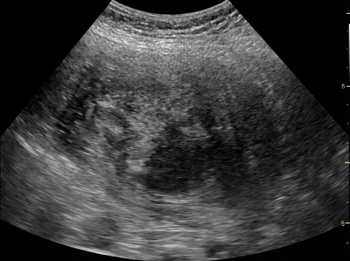
Identify the imaging modality given below.

Which of the following is best assessed by FAST USG?
Red Color on color doppler suggests?
Transrectal ultrasonography in carcinoma prostate is most useful for –
FAST USG focuses on all of the following areas except-
Explanation: ***7.5 MHz*** * **Transrectal ultrasonography (TRUS)** typically utilizes frequencies between **7 and 10 MHz** to achieve a good balance of penetration depth and resolution in the prostate and surrounding tissues. * A frequency of **7.5 MHz** is commonly employed for detailed imaging of the prostate, allowing for clear visualization of anatomical structures and detection of abnormalities like tumors or inflammation. *5.0 MHz* * A frequency of **5.0 MHz** provides greater penetration depth but offers lower resolution, which is generally not optimal for the detailed imaging required in TRUS of the prostate. * Lower frequencies are more often used for imaging deeper structures or in situations where tissue attenuation is high, which is less of a concern for the relatively superficial prostate. *2.5 MHz* * A frequency of **2.5 MHz** offers significantly higher penetration but at the cost of very low resolution, making it unsuitable for the precise diagnostic imaging of the prostate gland. * This lower frequency range is typically used for general abdominal imaging or structures requiring deep penetration, where fine detail is not the primary concern. *15.0 MHz* * A frequency of **15.0 MHz** provides excellent resolution but has very limited penetration depth, making it less practical for comprehensive transrectal imaging of the entire prostate gland. * While high frequencies can be used for very superficial structures or specific applications requiring extremely fine detail, they often cannot visualize the entire organ effectively due to rapid attenuation.
Explanation: ***All of these*** - The presence of the **banana sign**, **lemon sign**, and a **visible vertebral or overlying tissue defect** are all characteristic ultrasound findings in a fetus with spina bifida. - These signs indicate associated structural abnormalities in the **brain** and **spine** due to the neural tube defect. - All three findings are clinically significant and commonly used for prenatal diagnosis. **Banana sign** - This refers to the shape of the **cerebellum** appearing flattened and curved like a banana due to its caudal displacement into the foramen magnum in **Chiari II malformation**, which is frequently associated with spina bifida. - It is an important indicator of **hindbrain herniation**. **Lemon sign** - The lemon sign describes the shape of the fetal skull, where the frontal bones are indented, making the head appear like a **lemon** on axial view. - This sign is often seen in cases of **spina bifida** and is caused by **bifrontal bone scalloping**. **Defect seen in vertebral bodies or tissue overlying it** - This is the **direct visualization** of the open neural tube defect (myelomeningocele or meningocele) in the spine. - It involves a discontinuity in the **vertebral arches** and/or the overlying soft tissues.
Explanation: ***Short learning curve*** - While ultrasound is widely used, developing proficiency requires **extensive training** and **experience**, especially for complex evaluations. - The interpretation of ultrasound images is highly **operator-dependent**, and mastering proper technique and recognizing subtle findings demands a significant learning investment. *No radiation* - Ultrasound uses **sound waves** to generate images, meaning it does not expose patients to **ionizing radiation**. - This makes it a safer option for repeated studies, pregnant patients, and pediatric populations compared to modalities like X-rays or CT scans. *Inexpensive* - Ultrasound machines are generally **less expensive** to purchase and maintain compared to MRI or CT scanners. - The procedure itself is also typically **less costly** than other advanced imaging techniques, making it more accessible. *Allows dynamic studies to be done* - Ultrasound can capture images in **real-time**, allowing clinicians to observe organ movement, blood flow, and tissue changes during various maneuvers. - This capability is crucial for assessing joint stability, vascular patency, and the mobility of masses or fluid collections.
Explanation: ***Gharbi Stage III*** - This stage is characterized by a **cystic lesion with internal floating membranes** (daughter cysts or detached endocyst), which is consistent with the "Water lily sign" described. - The membranes are indicative of a **degenerating or ruptured endocyst** within the main cyst, signifying a more advanced stage than simple cysts. *Gharbi Stage I* - This stage represents a **simple, unilocular cyst** with an anechoic (fluid-filled) lumen, without any internal structures or membranes. - It would appear as a clearly defined, fluid-filled mass with a smooth wall, lacking the internal complexity shown in the image. *Gharbi Stage II* - Gharbi Stage II is characterized by a **cyst with internal septations**, indicating the presence of multiple daughter cysts within the main cyst. - While it involves internal structures, these are typically well-defined septa rather than freely moving, detached membranes. *Gharbi Stage IV* - This stage describes a **heterogeneous, degenerating cyst** with a solid-like appearance, often without a clear cystic lumen. - It reflects a more advanced, often calcified or completely inactivated cyst, which does not match the clear cystic structure with floating membranes seen in the image.
Explanation: ***Spina bifida*** - The **"banana sign"** is a distinctive ultrasound finding associated with **Chiari II malformation**, which is frequently seen in cases of **spina bifida**. - It refers to the characteristic **flattening and anterior bowing of the cerebellar hemispheres**, compressing them against the skull and giving them a banana-like appearance. *NCC* - **Neurocysticercosis (NCC)** is caused by tapeworm larvae in the central nervous system and is characterized by **cysts** or calcifications on imaging, not a "banana sign." - Imaging findings in NCC typically include **cystic lesions**, **edema**, and calcifications, unrelated to cerebellar shape. *Omphalocele* - **Omphalocele** is an abdominal wall defect where abdominal organs protrude through the navel, covered by a membrane. - The ultrasound findings for omphalocele involve visualization of abdominal contents outside the abdominal cavity, which is distinct from intracranial findings like the "banana sign." *Anencephaly* - **Anencephaly** is a severe birth defect where a baby is born without parts of the brain and skull, typically presenting with an **absent cranial vault** and cerebral hemispheres. - While a severe neural tube defect, anencephaly features agenesis of the brain and skull, rather than the cerebellar distortion seen as a "banana sign."
Explanation: ***USG*** - The image displays characteristic **gray-scale imaging** with an **echogenic appearance** of tissues, typical of an ultrasound. - Presence of annotations like "10 MHz G 64%" for **frequency and gain**, and "PRC" suggest ultrasound parameters. *Fluoroscopy* - Fluoroscopy provides **real-time X-ray images** and often involves the use of contrast agents, appearing as a dynamic, darker image with high contrast. - The image lacks the distinct bone and air contrast and dynamic motion typical of fluoroscopy. *X-Ray* - X-ray images depict a **static shadowgram** of dense structures like bones as white, and air as black, with sharp delineation. - The image shows a **granular texture** and fluid-filled structures that are characteristic of soft tissue imaging through ultrasound, not X-ray. *MRI* - MRI produces **cross-sectional images** with high soft tissue contrast in multiple planes (axial, sagittal, coronal). - The image shows real-time B-mode ultrasound characteristics with **probe frequency notation**, not the slice-based imaging of MRI.
Explanation: ***Pericardium*** - FAST USG is **most clinically significant** for detecting **pericardial effusions** and **cardiac tamponade** in trauma patients. - The **subxiphoid view** provides **excellent direct visualization** of the heart and pericardial space with minimal interference. - **Small volumes** of pericardial fluid (as little as 50-100 mL) are **clinically significant** and potentially life-threatening, requiring immediate intervention. - Cardiac tamponade is an **immediately reversible cause of shock** that demands urgent diagnosis and pericardiocentesis. - **Sensitivity >90%** for clinically significant pericardial effusions in the trauma setting. *Liver* - FAST assesses the **hepatorenal space (Morison's pouch)** for free fluid, not the liver parenchyma itself. - Requires **larger volumes of free fluid** (>200-500 mL) to be reliably detected in the peritoneal cavity. - Detailed assessment of actual liver injury requires **contrast-enhanced CT imaging**. *Spleen* - FAST evaluates the **splenorenal recess** for free fluid surrounding the spleen, not splenic parenchymal injury. - Detection depends on adequate volume of free fluid being present. - **CT scanning** is superior for defining splenic lacerations, hematomas, and grading injury severity. *Pleural cavity* - While Extended FAST (eFAST) can assess **pleural spaces** for effusion or pneumothorax, this is an **extension** of the standard 4-view FAST protocol. - Standard FAST focuses on the **four primary windows**: pericardial, perihepatic, perisplenic, and pelvic. - **Chest X-ray** and **CT** remain primary modalities for comprehensive thoracic assessment.
Explanation: ***Flow towards the transducer*** - In **color Doppler ultrasound**, the standard convention is that **red color** represents blood flow moving **towards** the transducer. - This is based on the **Doppler effect**, where frequency shift occurs as blood cells move closer to the transducer, which the ultrasound machine translates into a red hue. - This convention helps sonographers quickly interpret the direction of blood flow within vessels or cardiac chambers. *Flow Away from the transducer* - Flow moving **away** from the transducer is depicted in **blue** on color Doppler imaging. - This is the opposite of red, creating an intuitive visual distinction for flow direction assessment. *Arterial Blood* - Color Doppler displays the **direction of flow** relative to the transducer, not whether blood is arterial or venous. - While arteries carry oxygenated blood and veins carry deoxygenated blood, the color shown (red or blue) **only indicates movement direction**, not the blood's oxygenation status or vessel type. - Both arterial and venous blood can appear red or blue depending on their flow direction relative to the transducer position. *Venous Blood* - Similar to arterial blood, venous blood can appear as either red or blue on color Doppler depending solely on flow direction. - The color assignment is **independent of vessel type** and only reflects whether flow is toward (red) or away (blue) from the transducer.
Explanation: ***Guided prostatic biopsies*** - **Transrectal ultrasonography (TRUS)** provides real-time visualization of the prostate, allowing for precise guidance during **prostatic biopsies**. - This guidance ensures that tissue samples are taken from suspicious areas, increasing the diagnostic yield for **prostate cancer**. *To detect hypoechoic area* - While TRUS can identify **hypoechoic areas** in the prostate, which may suggest malignancy, these are **not specific** to cancer and can be caused by other conditions like inflammation. - The primary utility of TRUS in prostate cancer is not merely detection of these areas, but rather using this information for targeted sampling. *Seminal vesicle involvement* - TRUS can visualize the seminal vesicles, but its accuracy in definitively determining **seminal vesicle invasion** is limited compared to more advanced imaging like **MRI**. - **MRI** is generally preferred for assessing extraprostatic extension and seminal vesicle involvement due to its superior soft tissue contrast. *Measurement of prostatic volume* - TRUS is used to measure **prostatic volume**, which is important for calculating **PSA density** and for treatment planning in benign prostatic hyperplasia (BPH). - However, in the context of prostate cancer, while volume measurement is possible, guided biopsy is its most crucial role for diagnosis.
Explanation: ***Pleura*** - While pleural assessment is included in **extended FAST (E-FAST)** for detecting pneumothorax and hemothorax, the **traditional FAST protocol** focuses specifically on detecting **free fluid** in the **peritoneal** and **pericardial** spaces in trauma patients. - The pleura is not one of the standard **four views** of traditional FAST USG, though it is assessed in the expanded E-FAST protocol. *Splenic* - The **splenorenal recess** (left upper quadrant space between the spleen and left kidney) is one of the four key regions in FAST USG to detect **free fluid**. - Fluid accumulation in this area can indicate injury to the spleen or other abdominal organs. *Hepatic* - The **hepatorenal recess (Morison's pouch)** between the liver and right kidney is a critical view in FAST USG for identifying **free fluid**. - This is often the first place fluid collects in the peritoneum in supine patients and is the most sensitive area for detecting intraperitoneal fluid. *Pelvic* - The **pouch of Douglas** (rectovesical in males or rectouterine in females) is a standard view in FAST USG to detect **free fluid** in the pelvis. - Fluid here can indicate significant abdominal or pelvic injury.
Physics of Ultrasound
Practice Questions
Instrumentation and Techniques
Practice Questions
Abdominal Ultrasonography
Practice Questions
Pelvic Ultrasonography
Practice Questions
Obstetric Ultrasonography
Practice Questions
Small Parts Ultrasonography
Practice Questions
Musculoskeletal Ultrasonography
Practice Questions
Vascular Ultrasonography
Practice Questions
Pediatric Ultrasonography
Practice Questions
Contrast-Enhanced Ultrasound
Practice Questions
Ultrasound-Guided Interventions
Practice Questions
Doppler Ultrasound Principles and Applications
Practice Questions
Get full access to all questions, explanations, and performance tracking.
Start For Free