What is the earliest fetal anomaly detectable by ultrasound?
Ultrasound scanning of a fetus shows asymmetric growth retardation. It may be associated with which of the following pathologies?
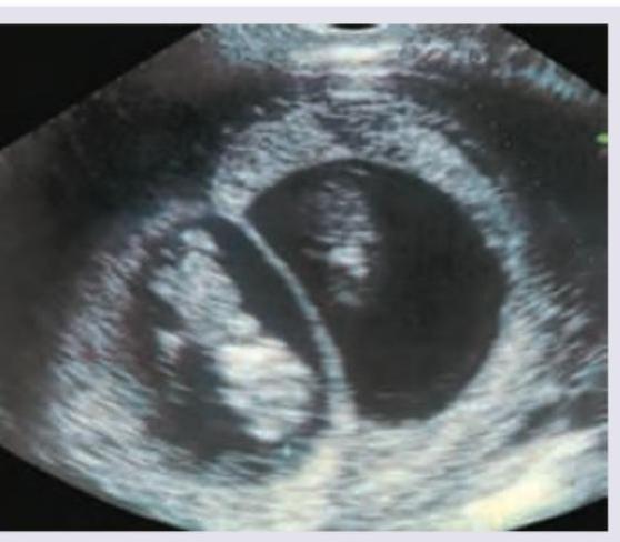
The Sonographic scan given below shows:

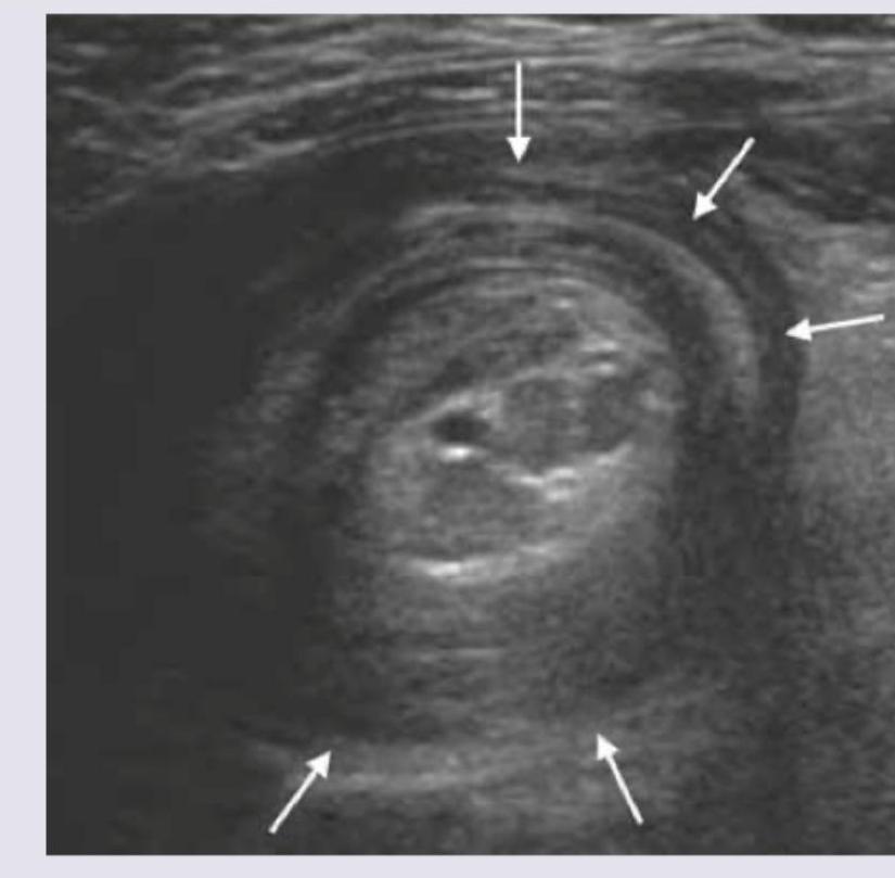
The ultrasound finding of a 7 -month-old child with abdominal pain and mass in the upper abdomen is shown below. What is the diagnosis? (NEET Pattern 2018)

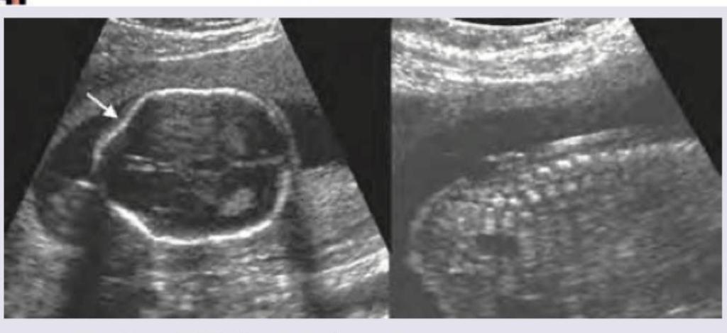
Keyhole sign on fetal ultrasound is seen in:

The first imaging modality of choice for a 35-year-old lady, presenting to surgical emergency with complaints of colicky pain in right lower quadrant of abdomen and vomiting since last 2 days is:
"Mickey Mouse Sign" during B-mode duplex ultrasound imaging comprises :
The most difficult area to visualize using duplex scanning (B-mode ultrasound), especially in an obese patient, is
Foetal anaemia can be detected non-invasively by Doppler ultrasonography on the basis of an increase in the
The sonographic finding of a cyst containing clear fluid is described as
Explanation: **Explanation:** **Anencephaly** is the correct answer because it is a lethal neural tube defect characterized by the absence of the cranial vault (acrania) and cerebral hemispheres. This structural defect is established very early in embryogenesis (failure of the rostral neuropore to close by day 25-27). With high-resolution transvaginal sonography (TVS), it can be diagnosed as early as **10–14 weeks** of gestation. The classic ultrasound findings include the "Frog-eye appearance" or "Mickey Mouse sign" due to the absence of the calvarium and bulging orbits. **Why the other options are incorrect:** * **Hydrocephalus:** Ventriculomegaly is typically not diagnosed until the **second trimester** (usually after 18 weeks), as the choroid plexus normally fills the lateral ventricles in the first trimester, making early detection unreliable. * **Achondroplasia:** This is the most common non-lethal skeletal dysplasia, but it typically manifests in the **third trimester** (after 26 weeks) when rhizomelic (proximal) limb shortening becomes sonographically evident. * **Spina Bifida:** While screening begins in the late first trimester via "intracranial translucency," a definitive diagnosis is usually made during the **level II anomaly scan (18–20 weeks)** by visualizing the vertebral defect or secondary cranial signs (Lemon/Banana signs). **High-Yield Clinical Pearls for NEET-PG:** * **Acrania-Anencephaly Sequence:** Acrania (absent skull with present brain) is the precursor to anencephaly; the unprotected brain tissue eventually degenerates due to exposure to amniotic fluid (exencephaly). * **Biochemical Marker:** Anencephaly is associated with markedly elevated **Maternal Serum Alpha-Fetoprotein (MSAFP)**. * **Associated Finding:** Polyhydramnios is common in the late second/third trimester due to the fetus's inability to swallow.
Explanation: **Explanation:** Intrauterine Growth Restriction (IUGR) is classified into two main types: Symmetric and Asymmetric. Understanding the distinction is crucial for NEET-PG. **1. Why Placental Insufficiency is Correct:** Asymmetric IUGR (also known as "head-sparing" IUGR) typically occurs in the **late second or third trimester**. It is most commonly caused by **placental insufficiency** (often secondary to maternal hypertension or pre-eclampsia). In this condition, the fetus redistributes blood flow to vital organs like the brain and heart at the expense of the liver and soft tissues. On ultrasound, this manifests as a **normal Head Circumference (HC)** but a **decreased Abdominal Circumference (AC)**, leading to an increased HC/AC ratio. **2. Why the Incorrect Options are Wrong:** * **Trisomy 18 & Congenital Rubella Syndrome (Options B & C):** These conditions typically cause **Symmetric IUGR**. Symmetric IUGR begins early in gestation (first trimester) due to a decrease in total cell number. All fetal parameters (HC, AC, and Femur Length) are proportionately small. * **Anencephaly (Option A):** This is a neural tube defect characterized by the absence of a major portion of the brain and skull. While it affects fetal measurements, it is a structural malformation rather than a pattern of growth retardation. **Clinical Pearls for NEET-PG:** * **Ponderal Index:** Used to assess the severity of asymmetric IUGR. * **Most sensitive parameter for IUGR:** Abdominal Circumference (AC), as it reflects fetal liver size and glycogen stores. * **Doppler Findings:** In placental insufficiency, look for increased resistance in the Umbilical Artery and "brain-sparing" (decreased resistance) in the Middle Cerebral Artery (MCA). * **Symmetric IUGR mnemonic:** "Early, Endogenous, Equal" (Early onset, Chromosomal/Infection causes, all parameters small).
Explanation: ***Twins*** - The ultrasound image clearly shows two distinct **gestational sacs**, separated by a membrane, each containing a developing embryo. - This presentation is characteristic of a **twin pregnancy**, specifically dichorionic-diamniotic twins, given the visible thick membrane. *Single fetus* - A single fetus would present with only one gestational sac and one developing embryo, unlike what is seen in the image. - The presence of two separate sacs rules out a singleton pregnancy. *Snowstorm appearance* - The "snowstorm appearance" is a characteristic sonographic finding in a **hydatidiform mole**, due to hydropic villi. - This image clearly depicts recognizable fetal structures within distinct gestational sacs, which are not seen in a hydatidiform mole. *Triplets* - Triplets would involve three distinct gestational sacs or three fetuses within a shared sac (depending on chorionicity/amnionicity), but the image only shows two clear sacs. - While it's possible to miss a third sac in some views, the prominent visual here is unequivocally two.
Explanation: ***Intussusception*** - The ultrasound image clearly shows a "target sign" or "doughnut sign," which is pathognomonic for **intussusception**, where one segment of the intestine telescopes into another. - This condition commonly presents in infants (around 6-36 months) with **abdominal pain**, an abdominal mass, and sometimes **currant jelly stools**. *Intestinal volvulus* - **Intestinal volvulus** involves the twisting of the intestine around its mesentery, which would typically show a "whirlpool sign" on ultrasound due to twisted mesenteric vessels, not the "target sign." - It usually presents with sudden onset of severe abdominal pain, bilious vomiting, and signs of intestinal obstruction and ischemia. *Pyloric stenosis* - **Pyloric stenosis** is characterized by hypertrophy of the pyloric muscle, which would appear as an elongated, thickened pyloric canal on ultrasound with increased pyloric muscle thickness (>3mm) and length (>15mm). - Clinical presentation involves non-bilious projectile vomiting in infants, typically between 2 and 8 weeks of age, not at 7 months with an abdominal mass and the bowel-within-bowel appearance shown. *None of above* - The classic ultrasound findings and clinical presentation strongly point to intussusception, making this option incorrect.
Explanation: ***Dandy-Walker syndrome*** - The ultrasound image shows **enlargement of the posterior fossa** with a **large cyst occupying the space normally taken by the cerebellum**, and a **keyhole sign** (arrow pointing to a defect where the cerebellar vermis should be). This is characteristic of Dandy-Walker syndrome. - Absence or **hypoplasia of the cerebellar vermis** is a hallmark feature, leading to communication of the fourth ventricle with a posterior fossa cyst. *Arnold-Chiari malformation* - Characterized by **herniation of cerebellar tonsils** through the foramen magnum and often associated with myelomeningocele. - Key ultrasound findings include a **lemon sign** (flattened frontal bones) and **banana sign** (anteriorly curved cerebellum), which are not depicted here. *Spina bifida* - This is a **neural tube defect** involving incomplete closure of the spinal column. - While it can be associated with Arnold-Chiari malformation, the primary features of spina bifida (e.g., a **sacral defect** with a mass) are not shown in these images. *Aqueductal stenosis* - Results in **dilation of the lateral and third ventricles** due to obstruction of cerebrospinal fluid flow in the aqueduct of Sylvius. - It primarily affects the supratentorial ventricular system and does not typically involve the **posterior fossa cyst** and **vermic hypoplasia** seen in the image.
Explanation: ***Ultrasound abdomen*** - Ultrasound is the **first-line imaging modality** for evaluating acute right lower quadrant pain in women of reproductive age due to its **safety (no ionizing radiation)**, availability, and cost-effectiveness. - It effectively visualizes the **appendix**, ovaries, uterus, and can detect **appendicitis**, ovarian pathology (e.g., cysts, torsion, ectopic pregnancy), and other causes of acute abdominal pain. - Follows the **ALARA principle** (As Low As Reasonably Achievable) for radiation exposure in young women. *Plain X-ray abdomen erect view* - Plain X-ray has **limited utility** for acute right lower quadrant pain as it cannot visualize soft tissue structures like the appendix or ovaries. - Primarily useful for detecting **bowel obstruction** (air-fluid levels) or **pneumoperitoneum** (free air under diaphragm), which are not suggested by this clinical presentation. *Contrast CT abdomen* - While highly sensitive for appendicitis and intra-abdominal pathologies, contrast CT involves **ionizing radiation** and **IV contrast administration**. - Should be **minimized in women of reproductive age** due to radiation risks (including potential pregnancy). - Reserved for cases where ultrasound is **inconclusive** or when detailed anatomical assessment is required. *Non-contrast CT abdomen* - Non-contrast CT exposes the patient to **ionizing radiation** without the diagnostic advantage of contrast enhancement. - **Less effective** than contrast-enhanced CT for detecting inflammatory processes and appendiceal pathology. - Not preferred as first-line imaging when ultrasound is available and appropriate.
Explanation: ***Popliteal artery, Popliteal vein and Tibial nerve*** - The \"Mickey Mouse Sign\" in the **popliteal fossa** visualizes the **popliteal artery** as the \"head\" and the **popliteal vein** and **tibial nerve** as the \"ears.\" - This specific configuration is seen on **transverse B-mode ultrasound** of the popliteal fossa and is crucial for identifying neurovascular structures for **popliteal nerve blocks** and vascular assessments. - The tibial nerve is the largest branch of the sciatic nerve in the popliteal fossa and runs alongside these vessels. *Common femoral vein, Common femoral artery and Great Saphenous vein* - This is the **most commonly referenced \"Mickey Mouse Sign\"** in ultrasound, visualized in the **groin/femoral region**. - The **common femoral artery** forms the \"head\" and the **common femoral vein** and **great saphenous vein** (at its junction) form the \"ears.\" - This sign is important for **central venous access**, **femoral vessel assessment**, and **avoiding complications** during procedures. *Anterior tibial artery, Dorsalis pedis artery and Extensor hallucis tendon* - These structures are located in the **lower leg and foot**, not in a configuration that forms the Mickey Mouse sign. - They are important for assessing **peripheral vascular status** but do not constitute this specific ultrasound landmark. *Brachial artery, Basilic vein and Biceps tendon* - These are structures found in the **upper arm**. - While important for upper extremity vascular imaging, they do not form the \"Mickey Mouse Sign\" pattern on ultrasound.
Explanation: ***aortoiliac segment*** - The **aortoiliac segment** is often challenging to visualize due to its deep location within the **pelvis** and the presence of overlying **bowel gas**, which scatters ultrasound waves. - In obese patients, increased **adipose tissue** further attenuates the ultrasound signal, making imaging of this specific segment particularly difficult. *abdominal aorta above renal vessels* - While the **abdominal aorta** can be challenging, particularly the segment above the renal vessels due to the diaphragm and lung bases, it is generally more accessible than the deep pelvic structures. - Visualization can be improved by optimizing patient position and using specific transducer angles. *carotid vessels* - **Carotid vessels** are superficial and easily accessible for duplex scanning, making them one of the easiest vascular beds to image. - There is minimal tissue attenuation, and bowel gas is not a factor. *iliofemoral segment* - The **iliofemoral segment** is more superficial than the aortoiliac segment and is generally well visualized, especially the femoral arteries in the groin. - While obesity can increase the scanning depth, it does not pose the same challenges as the deeper and often gas-obscured pelvic vessels.
Explanation: ***Peak systolic velocity of the middle cerebral artery*** - **Foetal anaemia** causes increased **cardiac output** and redistribution of blood flow to vital organs, leading to an increase in **peak systolic velocity (PSV)** in the **middle cerebral artery (MCA)**. - This increased velocity indicates **cerebral vasodilation**, a compensatory mechanism to maintain oxygen delivery to the foetal brain in situations of hypoxia due to anaemia. *SD ratio in the anterior cerebral artery* - The **SD ratio (systolic/diastolic ratio)** is more commonly used in umbilical artery flow. A higher SD ratio in cerebral arteries typically suggests increased downstream resistance, which is not characteristic of foetal anaemia. - In foetal anaemia, the cerebral arteries would typically show **decreased resistance**, leading to a lower, not higher, SD ratio. *SD ratio in the umbilical artery* - An increased **SD ratio in the umbilical artery** often indicates **placental insufficiency** or **foetal growth restriction**, not necessarily **foetal anaemia**. - While placental insufficiency can lead to foetal anaemia, the direct Doppler marker for anaemia is the MCA PSV, reflecting the foetal response to hypoxia. *Peak systolic velocity of the anterior cerebral artery* - While both anterior and middle cerebral arteries are part of the cerebral circulation, the **middle cerebral artery (MCA)** is the most widely validated and sensitive vessel for detecting changes in **foetal blood flow** due to **anaemia**. - Changes in the anterior cerebral artery's PSV are not as reliably linked to the diagnosis of foetal anaemia as those in the MCA.
Explanation: ***Anechoic*** - **Anechoic** refers to structures that do not produce echoes, appearing **black** on ultrasound. - A simple cyst with clear fluid allows sound waves to pass through without reflection, making it appear anechoic. *Isoechoic* - **Isoechoic** describes structures that have the same echogenicity as surrounding tissues. - This term is typically used for solid tissues rather than fluid-filled cysts. *Hypoechoic* - **Hypoechoic** structures produce fewer echoes than surrounding tissues, appearing darker but not completely black. - This is often seen in solid lesions or complex cysts with internal debris, not clear fluid. *Hyperechoic* - **Hyperechoic** structures produce many echoes, appearing brighter than surrounding tissues. - This can be indicative of calcifications, fat, or certain solid masses, not clear fluid.
Physics of Ultrasound
Practice Questions
Instrumentation and Techniques
Practice Questions
Abdominal Ultrasonography
Practice Questions
Pelvic Ultrasonography
Practice Questions
Obstetric Ultrasonography
Practice Questions
Small Parts Ultrasonography
Practice Questions
Musculoskeletal Ultrasonography
Practice Questions
Vascular Ultrasonography
Practice Questions
Pediatric Ultrasonography
Practice Questions
Contrast-Enhanced Ultrasound
Practice Questions
Ultrasound-Guided Interventions
Practice Questions
Doppler Ultrasound Principles and Applications
Practice Questions
Get full access to all questions, explanations, and performance tracking.
Start For Free