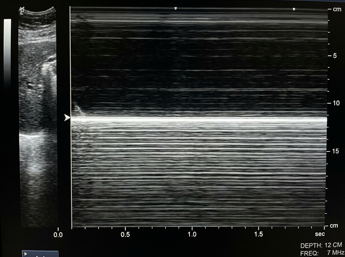
A patient presented with a history of blunt trauma to the chest and abdomen. On USG examination in M mode the following finding is seen. What is the likely diagnosis?

Which of the following is not a prerequisite for transvaginal sonography?
What is the investigation of choice for a lactating female presenting with a painful breast lump?
Nuchal Skin Fold thickness scan is typically performed between which gestational ages?
What is the ideal gestational age for performing ultrasound to assess fetal organ defects?
Distal acoustic shadow is seen in?
What is the investigation of choice to visualize gallbladder pathologies?
All are true about antenatal umbilical artery Doppler except?
Pericardial effusion is best diagnosed by?
What is the initial investigation for an amoebic liver abscess?
Explanation: ***Pneumothorax*** - In M-mode ultrasound, pneumothorax shows the **"barcode sign"** or **"stratosphere sign"** - horizontal parallel lines without the normal lung sliding motion due to air between chest wall and lung. - Following **blunt chest trauma**, pneumothorax is common and the absence of **lung sliding** on ultrasound is highly sensitive for detecting it. *Pleural effusion* - Would show **anechoic fluid collection** in the pleural space with **respiratory variation** and possible **atelectatic lung** floating within. - M-mode would demonstrate **fluid movement** and **compressed lung tissue**, not the static barcode pattern seen here. *Bronchial obstruction* - Ultrasound has **limited utility** in diagnosing bronchial obstruction as **air-filled bronchi** cannot be well visualized on USG. - Would require **CT scan** or **bronchoscopy** for diagnosis, and wouldn't produce the characteristic M-mode findings described. *Lung abscess* - Appears as a **thick-walled cavity** with **internal echoes** representing pus and debris, often with **air-fluid levels**. - M-mode would show **complex echogenic patterns** within the abscess cavity, distinctly different from the uniform barcode sign of pneumothorax.
Explanation: **Explanation:** In Transvaginal Sonography (TVS), the ultrasound probe is placed in the vaginal fornix, directly adjacent to the pelvic organs (uterus and ovaries). Because the probe is in close proximity to these structures, it uses higher-frequency transducers which provide superior resolution compared to transabdominal scans. **1. Why "Full Bladder" is the Correct Answer (Not a Prerequisite):** A **full bladder** is a prerequisite for **Transabdominal Sonography (TAS)**, where it acts as an "acoustic window" to push bowel loops away and transmit sound waves to the pelvic organs. In TVS, however, a full bladder is a hindrance. It pushes the uterus and ovaries too far away from the high-frequency probe (which has limited depth penetration) and can cause patient discomfort during probe manipulation. Therefore, a full bladder is contraindicated for TVS. **2. Analysis of Other Options:** * **Consent:** Essential for any invasive or semi-invasive procedure. Since TVS involves internal probe insertion, informed verbal or written consent is mandatory. * **Empty Bladder:** This is a strict prerequisite for TVS. It ensures the pelvic organs are as close to the vaginal vault as possible for optimal imaging. * **Lithotomy Position:** The patient must be in a lithotomy or modified lithotomy position (often using a pelvic wedge) to allow the sonographer the necessary range of motion to tilt the probe and visualize the adnexa. **High-Yield Clinical Pearls for NEET-PG:** * **TVS vs. TAS:** TVS offers better resolution (high frequency) but less depth; TAS offers better "field of view" (low frequency) but less resolution. * **First Trimester:** TVS can detect a gestational sac as early as **4.5–5 weeks**, whereas TAS usually requires **5.5–6 weeks**. * **Beta-hCG Discriminatory Zone:** A gestational sac should be visible via TVS when serum Beta-hCG levels reach **1500–2000 mIU/mL**.
Explanation: **Explanation:** The correct answer is **Ultrasound (USG)**. In a lactating female, the breast tissue undergoes physiological changes, becoming extremely dense due to glandular hyperplasia and milk production. **Why Ultrasound is the Investigation of Choice:** 1. **Sensitivity in Dense Tissue:** USG is superior to mammography in evaluating dense breasts, as it can easily differentiate between solid masses and fluid-filled structures. 2. **Safety:** It involves no ionizing radiation, making it safe for both the mother and the breastfeeding infant. 3. **Diagnostic Utility:** The most common cause of a painful lump in a lactating woman is a **Galactocele** (milk-filled cyst) or a **Breast Abscess**. USG is highly sensitive for identifying these collections and can simultaneously facilitate ultrasound-guided needle aspiration for diagnosis and treatment. **Why other options are incorrect:** * **Mammogram:** It is the primary screening tool for older women, but in lactating females, the high breast density significantly reduces its sensitivity (high false-negative rate). Additionally, it involves radiation which is generally avoided unless malignancy is strongly suspected. * **MRI:** While highly sensitive, MRI is expensive, not readily available, and often requires gadolinium contrast, which is generally avoided during lactation unless absolutely necessary. * **CT Scan:** CT has a limited role in primary breast imaging due to poor soft tissue resolution of the breast parenchyma and high radiation dose. **Clinical Pearls for NEET-PG:** * **Galactocele:** The most common benign breast lesion in lactating women; appears on USG as a well-circumscribed mass with variable internal echoes. * **Mastitis vs. Abscess:** USG is critical to differentiate between cellulitis/mastitis (managed with antibiotics) and an abscess (requires drainage). * **BI-RADS:** Remember that the Breast Imaging-Reporting and Data System applies to USG findings as well to standardize management.
Explanation: **Explanation:** The **Nuchal Skin Fold (NF)** thickness is a second-trimester ultrasound marker used to screen for chromosomal abnormalities, primarily Down Syndrome (Trisomy 21). **1. Why 18-22 weeks is correct:** The Nuchal Fold is measured during the **Anomaly Scan (Level II scan)**, typically between **18 and 22 weeks** of gestation. It is measured in the transcerebellar plane of the fetal head (axial view). A value of **≥ 6 mm** is considered thickened and is one of the most sensitive "soft markers" for Down Syndrome in the second trimester. **2. Analysis of Incorrect Options:** * **A & B (6-14 weeks):** These are too early for Nuchal *Fold* measurement. However, **11-14 weeks** is the specific window for measuring **Nuchal Translucency (NT)**. While both assess the neck area, NT measures the fluid-filled space behind the neck in the first trimester, whereas NF measures skin thickness in the second trimester. * **D (28-32 weeks):** By the third trimester, the Nuchal Fold measurement loses its diagnostic sensitivity and specificity for aneuploidy screening due to normal physiological changes in fetal soft tissue. **3. High-Yield Clinical Pearls for NEET-PG:** * **Nuchal Translucency (NT):** Measured at 11–13.6 weeks; abnormal if **> 3 mm** (associated with Trisomy 21 and Turner Syndrome). * **Nuchal Fold (NF):** Measured at 18–22 weeks; abnormal if **≥ 6 mm**. * **Most Sensitive Marker:** Among all second-trimester soft markers for Down Syndrome, a thickened Nuchal Fold has the highest likelihood ratio. * **Anatomical Landmarks for NF:** Cavum septum pellucidum, Cerebellum, and Cisterna magna.
Explanation: **Explanation:** The assessment of fetal organ defects (structural anomalies) is best performed during the **second trimester**, specifically between **16-20 weeks**. By **16-18 weeks**, organogenesis is complete, and the fetus has grown sufficiently for detailed anatomical visualization. At this stage, there is an optimal ratio of fetal size to amniotic fluid volume, allowing the ultrasound waves to clearly delineate internal structures like the cardiac chambers, kidneys, and neural tube. **Analysis of Options:** * **A & B (8-12 weeks):** These represent the first trimester. While ultrasound here is excellent for dating, confirming viability, and detecting gross anomalies (like anencephaly), most organs are too small and underdeveloped for a comprehensive structural survey. * **C (12-14 weeks):** This is the window for the **NT (Nuchal Translucency) scan**. While some major defects can be spotted, it is too early for a full "Level II" or anomaly scan, as many structures (like the cerebellum or cardiac outflow tracts) are not yet fully evaluable. * **D (16-18 weeks):** This is the traditional window for detecting major malformations. Although many centers now prefer 18-20 weeks for better cardiac detail, 16-18 weeks remains the classic textbook answer for the earliest reliable detection of most organ defects. **High-Yield NEET-PG Pearls:** * **Best time for Dating Scan:** 8-12 weeks (CRL is the most accurate parameter). * **NT Scan Window:** 11 weeks to 13 weeks 6 days. * **Anomaly Scan (Level II):** Ideally 18-20 weeks (but 16-18 weeks is the threshold for organ defect assessment). * **Amniocentesis:** Usually performed after 15 weeks to avoid fetal complications.
Explanation: **Explanation:** **Acoustic Shadowing** is a common ultrasound artifact that occurs when the ultrasound beam encounters a structure with a very high **attenuation coefficient** or high **acoustic impedance**. When the beam hits such a structure, most of the sound waves are either reflected or absorbed, leaving a signal-void (black) area distal to the object. **Why "All of the Above" is correct:** * **Calculi (Gallstones/Renal stones):** These are dense structures that reflect and absorb sound waves significantly. They typically produce "clean" shadows (sharp, black borders). * **Bone:** Bone has the highest acoustic impedance in the body. It reflects the majority of the ultrasound beam at the soft tissue-bone interface, preventing deeper penetration and creating a dense distal shadow. * **Air (Gas):** Air has extremely low acoustic impedance compared to soft tissue, causing a massive mismatch. This results in nearly total reflection of the sound beam. Air often produces "dirty" shadows (shadows containing internal echoes or reverberations). **Clinical Pearls for NEET-PG:** 1. **Clean vs. Dirty Shadowing:** * **Clean Shadow:** Characterized by an absence of echoes. Seen in **Calculi** and **Bone**. * **Dirty Shadow:** Characterized by internal echoes/reverberations. Seen in **Air/Bowel Gas** and **Emphysematous Cholecystitis**. 2. **Acoustic Enhancement:** The opposite of shadowing. It occurs distal to fluid-filled structures (e.g., simple cysts, gallbladder) because fluid attenuates sound much less than surrounding tissue. 3. **Edge Shadowing:** A specific type of shadowing seen at the curved edges of cystic structures due to refraction of the beam.
Explanation: **Explanation:** **Ultrasonography (USG)** is the investigation of choice (IOC) for gallbladder (GB) pathologies due to its high sensitivity and specificity, particularly for detecting gallstones (cholelithiasis) and acute cholecystitis. The underlying medical concept relies on the fact that the GB is a superficial, fluid-filled structure; bile provides an excellent acoustic window, allowing USG to detect stones as small as 1–2 mm with characteristic posterior acoustic shadowing. It is non-invasive, radiation-free, cost-effective, and allows for the assessment of the **Sonographic Murphy’s Sign**. **Why other options are incorrect:** * **CT Scan:** While excellent for detecting complications (like perforation or gangrenous cholecystitis) and staging GB carcinoma, it is less sensitive than USG for detecting radiolucent gallstones and involves ionizing radiation. * **Plain X-Ray:** Only about 10–15% of gallstones are radiopaque (contain enough calcium to be seen). It is no longer a standard screening tool for GB disease. * **MRCP:** This is the gold standard for visualizing the biliary tree and detecting **Choledocholithiasis** (stones in the CBD), but it is too expensive and time-consuming for primary GB evaluation. **High-Yield Clinical Pearls for NEET-PG:** * **IOC for Gallstones/Acute Cholecystitis:** USG. * **Gold Standard for Acute Cholecystitis:** HIDA Scan (Radionuclide scan). * **Gold Standard for Choledocholithiasis:** MRCP (Diagnostic) / ERCP (Therapeutic). * **WES Triad (Wall-Echo-Shadow):** A classic USG finding indicating a gallbladder packed with stones. * **Porcelain Gallbladder:** Intramural calcification of the GB wall; seen on X-ray/CT and associated with an increased risk of GB carcinoma.
Explanation: **Explanation:** The umbilical artery Doppler is a critical tool for assessing placental resistance and fetal well-being. The **S/D ratio (Systolic/Diastolic ratio)** reflects the resistance to blood flow within the placenta. **Why Option C is the correct answer (False statement):** Smoking causes vasoconstriction and placental damage, which **increases placental vascular resistance**. This leads to a decrease in diastolic flow, thereby **increasing the S/D ratio**, not decreasing it. Any factor that compromises placental health (like smoking, hypertension, or pre-eclampsia) typically increases this ratio. **Analysis of other options:** * **Option A:** In Intrauterine Growth Restriction (IUGR), placental resistance increases, leading to **decreased end-diastolic flow (EDF)**. This is a hallmark sign used to monitor fetuses with growth restriction. * **Option B:** In a healthy pregnancy, the umbilical artery is a low-resistance vessel (low S/D), while the fetal Middle Cerebral Artery (MCA) is a high-resistance vessel (high S/D). Therefore, the **Umbilical S/D is normally lower than the MCA S/D**. A reversal of this (Cerebro-placental ratio < 1) indicates "Brain Sparing Effect." * **Option D:** **Absent or Reversal of End-Diastolic Flow (AREDF/REDF)** in the umbilical artery is a grave sign. It indicates extreme placental resistance and is a precursor to fetal hypoxia, acidosis, and impending demise, necessitating urgent delivery. **High-Yield Clinical Pearls for NEET-PG:** 1. **Normal Trend:** As pregnancy advances, placental resistance decreases; therefore, the S/D ratio normally **decreases** with increasing gestational age. 2. **Brain Sparing Effect:** In hypoxia, blood is shunted to the brain, causing **vasodilation of the MCA** (decreased MCA resistance/PI). 3. **Ductus Venosus:** The most sensitive Doppler indicator of fetal cardiac failure or imminent death is the appearance of a **reversed 'a' wave** in the Ductus Venosus.
Explanation: **Explanation:** **Echocardiography (Option B)** is the gold standard and investigation of choice for diagnosing pericardial effusion. It is highly sensitive, non-invasive, and can detect as little as **15–20 mL** of fluid. It allows for the visualization of an "echo-free space" between the epicardium and parietal pericardium. Crucially, it can assess hemodynamic significance, such as signs of cardiac tamponade (e.g., right ventricular diastolic collapse). **Why other options are incorrect:** * **Chest X-ray (Option A):** While a "Water-bottle" or "Money-bag" heart appearance is classic, it is non-specific and only becomes apparent when at least **200–250 mL** of fluid has accumulated. * **ECG (Option C):** May show low voltage QRS complexes or **Electrical Alternans** (pathognomonic for large effusions/tamponade), but these are late findings and lack the sensitivity required for a primary diagnosis. * **Angiography (Option D):** This is an invasive procedure used primarily for coronary anatomy or pressures; it is not used for diagnosing pericardial fluid. **High-Yield Clinical Pearls for NEET-PG:** * **Investigation of Choice:** Echocardiography. * **Most Sensitive Imaging:** MRI (can detect even smaller amounts and characterize fluid), but Echo remains the practical first-line gold standard. * **Swinging Heart Syndrome:** Seen on Echo in large effusions, leading to Electrical Alternans on ECG. * **Beck’s Triad (Tamponade):** Hypotension, JVD, and muffled heart sounds. * **Ewart’s Sign:** Dullness to percussion at the left lung base due to compression by a large pericardial sac.
Explanation: **Explanation:** **A. USG (Correct):** Ultrasonography is the **initial investigation of choice** for a suspected amoebic liver abscess (ALA) due to its high sensitivity (75-95%), low cost, wide availability, and lack of ionizing radiation. On USG, an ALA typically appears as a well-defined, round or oval hypoechoic lesion, usually located in the right lobe (posterosuperior segment), with internal echoes and no significant wall enhancement. It is also used to guide therapeutic aspiration if required. **B. Exploratory Laparotomy:** This is an invasive surgical procedure and is never the initial investigation. It is reserved for complications like a ruptured abscess leading to peritonitis when percutaneous drainage or medical management fails. **C. CT Scan:** While Contrast-Enhanced CT (CECT) is more sensitive than USG for detecting small abscesses and evaluating complications (like rupture), it is not the *initial* step. It is considered the "gold standard" for localization but is reserved for cases where USG is inconclusive. **D. Technetium-99 Scan:** Nuclear imaging (using Tc-99m sulfur colloid) shows a "cold" spot in ALA (as it is avascular). While it helps differentiate ALA from a Pyogenic Abscess (which may show "hot" spots on Gallium scans), it is not a primary diagnostic tool. **High-Yield Clinical Pearls for NEET-PG:** * **Most common site:** Right lobe of the liver (due to the bulk of blood flow from the superior mesenteric vein). * **Classic appearance:** "Anchovy sauce" pus (chocolate brown, odorless). * **Diagnosis:** Combination of **USG + Serology** (ELISA for Entamoeba histolytica antibodies) is the most definitive non-invasive approach. * **Drug of Choice:** Metronidazole. * **Indications for Aspiration:** Large abscess (>10 cm), risk of rupture (left lobe abscess), or failure to respond to medical therapy within 48-72 hours.
Physics of Ultrasound
Practice Questions
Instrumentation and Techniques
Practice Questions
Abdominal Ultrasonography
Practice Questions
Pelvic Ultrasonography
Practice Questions
Obstetric Ultrasonography
Practice Questions
Small Parts Ultrasonography
Practice Questions
Musculoskeletal Ultrasonography
Practice Questions
Vascular Ultrasonography
Practice Questions
Pediatric Ultrasonography
Practice Questions
Contrast-Enhanced Ultrasound
Practice Questions
Ultrasound-Guided Interventions
Practice Questions
Doppler Ultrasound Principles and Applications
Practice Questions
Get full access to all questions, explanations, and performance tracking.
Start For Free