
Ultrasound — MCQs

On this page
'Triphasic waveform' on colour Doppler is of
A 45-year-old female presents with a 2 cm thyroid nodule. Which TIRADS category has >95% risk of malignancy?
Characteristic of venous blood flow of lower limb in duplex Doppler is?
Which of the following diagnostic tests is most useful in confirming the presence of ascites?
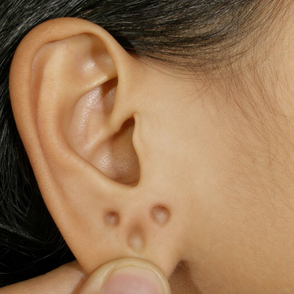
What does the sign depicted in the image represent?
What is the sonographic appearance of a hydatid cyst?
In which fetal anomalies are the 'banana sign' and 'lemon sign' observed?
Which of the following is a soft tissue marker of Down syndrome on USG?
Which piezoelectric crystal is most widely used in ultrasonography probes?
Which of the following structures cannot be effectively visualized using ultrasonography (USG)?
Practice by Chapter
Physics of Ultrasound
Practice Questions
Instrumentation and Techniques
Practice Questions
Abdominal Ultrasonography
Practice Questions
Pelvic Ultrasonography
Practice Questions
Obstetric Ultrasonography
Practice Questions
Small Parts Ultrasonography
Practice Questions
Musculoskeletal Ultrasonography
Practice Questions
Vascular Ultrasonography
Practice Questions
Pediatric Ultrasonography
Practice Questions
Contrast-Enhanced Ultrasound
Practice Questions
Ultrasound-Guided Interventions
Practice Questions
Doppler Ultrasound Principles and Applications
Practice Questions
Want unlimited practice?
Get full access to all questions, explanations, and performance tracking.
Scan to download app