Deltoid ligament is attached to all, except which structure?
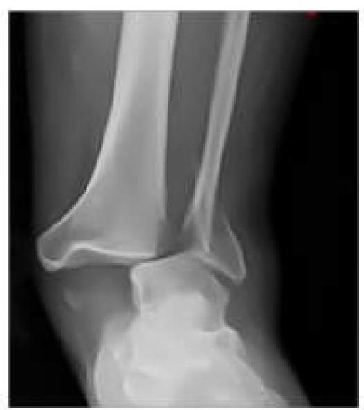
An RTA patient presented to the emergency department with severe pain in the ankle. An X-ray was performed, given below. What is the best next step in management?
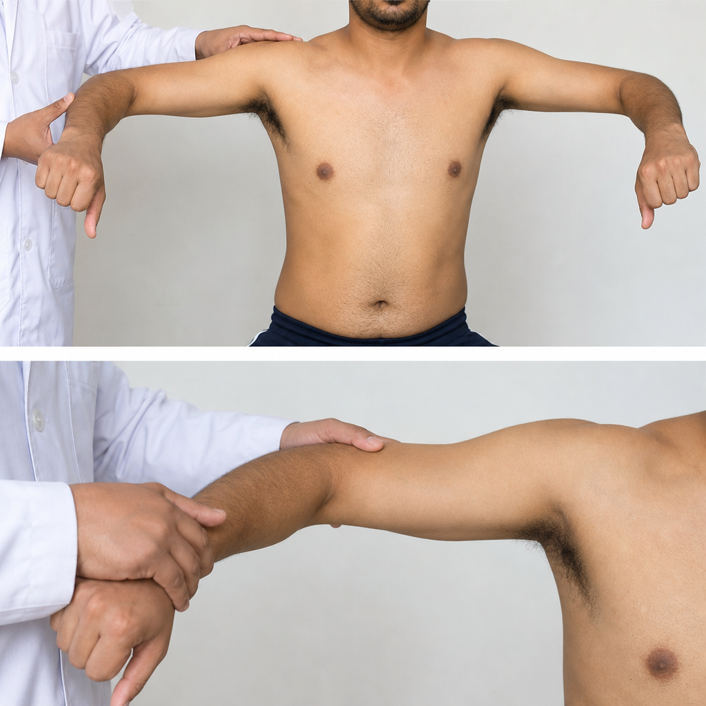
Rupture of extensor pollicis longus tendon occurs in all of the following except -
Which of the following is an intra-articular tendon?
In a child, non-functioning kidney is best diagnosed by
Transrectal ultrasonography in carcinoma prostate is most useful for –
Which is not echogenic while doing ultrasonography:
Which of the following ultrasound features of a thyroid nodule is not suggestive of malignancy?
Which of the following liver metastases appear hypoechoic on ultrasound?
At how many days from the last menstrual period can fetal heart activity be detected earliest with transvaginal sonography?
Explanation: ***Medial cuneiform*** - The **deltoid ligament** is located on the **medial side of the ankle** and primarily connects the **tibia** to several tarsal bones. It does not attach to the medial cuneiform. - The **medial cuneiform** is a midfoot bone primarily involved in the **tarsometatarsal joint** and is not a direct attachment site for the deltoid ligament. *Medial malleolus* - The **medial malleolus**, the distal end of the **tibia**, serves as the **proximal attachment point** for all four parts of the deltoid ligament. - This strong connection is crucial for **stabilizing the ankle joint** medially. *Navicular bone* - The **tibionavicular part** of the deltoid ligament attaches to the **tuberosity of the navicular bone**. - This attachment helps **limit excessive abduction** and **eversion** of the foot. *Sustentaculum tali* - The **tibiocalcaneal part** of the deltoid ligament attaches to the **sustentaculum tali** on the calcaneus. - This attachment provides significant stability to the **subtalar joint**.
Explanation: ***Neurovascular Assessment and Closed reduction with slab application*** - The X-ray shows an **ankle dislocation without an obvious fracture**, making **closed reduction** the appropriate initial treatment. - A **slab (splint)** is preferred over a full cast initially for acute injuries to accommodate for swelling, reducing the risk of compartment syndrome, and allowing for serial neurovascular checks. *Neurovascular Assessment and Closed reduction with cast application* - While closed reduction is correct, applying a **full cast** immediately after an acute injury carries a risk of **compartment syndrome** due to potential swelling that cannot be accommodated by a rigid cast. - A cast would typically be applied after the initial swelling has subsided, usually a few days to a week after initial reduction and splinting. *Neurovascular Assessment and Immediate surgery* - **Immediate surgery** is generally reserved for **open fractures/dislocations**, dislocations that cannot be reduced closed (irreducible dislocations), or those with significant associated fractures that require surgical fixation to stabilize the joint. - In this case, the dislocation appears to be isolated and amenable to closed reduction, making surgery not the immediate next step. *Neurovascular Assessment and Immediate open reduction* - **Open reduction** is performed when closed reduction fails or is contraindicated, for example, due to soft tissue interposition or highly unstable fracture patterns. - Since closed reduction has not yet been attempted, immediate open reduction is premature and unnecessary for an apparently simple dislocation.
Explanation: ***De Quervain's disease*** - This condition involves **tenosynovitis** of the **extensor pollicis brevis** and **abductor pollicis longus** tendons, not a rupture of the extensor pollicis longus. - The pathology is an inflammation and thickening of the tendon sheaths, distinct from a tendon tear. *Rheumatoid arthritis* - **Chronic inflammation** in rheumatoid arthritis can lead to weakening and eventual rupture of tendons, including the **extensor pollicis longus**, often due to synovitis eroding the tendon. - The condition creates an environment where tendons are vulnerable to **attrition** and damage, making rupture a recognized complication. *Drummers* - Repetitive, high-force movements involved in drumming can cause significant **stress** and microscopic damage to tendons, including the **extensor pollicis longus**. - Over time, this cumulative trauma can lead to inflammation, degeneration, and eventual **rupture** due to overuse. *Colles' fracture* - A **Colles' fracture** of the distal radius can cause a delayed rupture of the **extensor pollicis longus (EPL)** tendon. - This occurs due to attrition of the tendon as it rubs over the **roughened fracture site** or due to *avascular necrosis* of the tendon as it passes through a narrow osteofibrous tunnel.
Explanation: ***Popliteus*** - The **popliteus tendon** originates within the knee capsule (intra-articular) before emerging to insert onto the posterior tibia. - It plays a crucial role in **unlocking the knee joint** from full extension and contributes to posterior stability. *Anconeus* - The **anconeus muscle** is located on the posterior aspect of the elbow, extending from the lateral epicondyle of the humerus to the ulna. - It is an **extra-articular muscle** that assists in elbow extension and stabilization. *Semitendinosus* - The **semitendinosus** is one of the hamstring muscles, located in the posterior thigh. - Its tendon contributes to the **pes anserinus**, inserting on the medial aspect of the tibia distal to the knee joint, making it an extra-articular tendon. *Sartorius* - The **sartorius** is the longest muscle in the body, running obliquely across the anterior aspect of the thigh. - Its tendon also contributes to the **pes anserinus**, inserting medially to the knee joint, and is considered extra-articular.
Explanation: ***DTPA renogram*** - A **DTPA renogram** (diethylene triamine pentaacetic acid scan) is a nuclear medicine study that assesses **renal blood flow** and **glomerular filtration rate (GFR)**. - It is highly effective in determining if a kidney is non-functioning because it directly measures the **uptake and excretion of a radiotracer** by the kidney, providing quantitative data on its functional capacity. *Creatinine clearance* - **Creatinine clearance** is a measure of overall kidney function, reflecting the GFR of **both kidneys combined**. - It cannot specifically identify a non-functioning individual kidney, as the other kidney might compensate for the non-functioning one, leading to a near-normal overall creatinine clearance. *Ultrasonography* - **Ultrasonography** is excellent for evaluating **renal anatomy**, such as size, shape, and presence of cysts, hydronephrosis, or stones. - While it can show structural abnormalities, it provides limited direct information about the **functional status** of the kidney, and a structurally normal kidney can still be non-functional. *IVU (Intravenous Urography)* - **Intravenous Urography (IVU)** uses contrast dye injected intravenously to visualize the kidneys, ureters, and bladder, assessing both anatomy and some aspects of function. - If a kidney is non-functioning, it would show **no uptake or excretion of the contrast dye**, but IVU involves radiation exposure and nephrotoxic contrast, making DTPA renogram often preferred in children for functional assessment.
Explanation: ***Guided prostatic biopsies*** - **Transrectal ultrasonography (TRUS)** provides real-time visualization of the prostate, allowing for precise guidance during **prostatic biopsies**. - This guidance ensures that tissue samples are taken from suspicious areas, increasing the diagnostic yield for **prostate cancer**. *To detect hypoechoic area* - While TRUS can identify **hypoechoic areas** in the prostate, which may suggest malignancy, these are **not specific** to cancer and can be caused by other conditions like inflammation. - The primary utility of TRUS in prostate cancer is not merely detection of these areas, but rather using this information for targeted sampling. *Seminal vesicle involvement* - TRUS can visualize the seminal vesicles, but its accuracy in definitively determining **seminal vesicle invasion** is limited compared to more advanced imaging like **MRI**. - **MRI** is generally preferred for assessing extraprostatic extension and seminal vesicle involvement due to its superior soft tissue contrast. *Measurement of prostatic volume* - TRUS is used to measure **prostatic volume**, which is important for calculating **PSA density** and for treatment planning in benign prostatic hyperplasia (BPH). - However, in the context of prostate cancer, while volume measurement is possible, guided biopsy is its most crucial role for diagnosis.
Explanation: ***Bile*** - Bile is largely composed of **water**, which allows ultrasound waves to pass through it with minimal reflection, appearing **anechoic** (black) on ultrasound. - This property makes the gallbladder lumen, when filled with bile, appear anechoic, which is crucial for identifying structures like gallstones. *Bone* - **Bone** is highly dense and reflects a significant portion of ultrasound waves, making it appear very **echogenic** (bright) on ultrasonography. - Due to its high reflectivity, bone often produces a strong **acoustic shadow** behind it, obscuring deeper structures. *Gas* - **Gas** (air) is a strong reflector of ultrasound waves and appears brightly echogenic, often with a characteristic **dirty shadowing** or **reverberation artifact**. - The presence of gas can significantly hinder visualization of underlying tissues due to its strong reflection and scatter of the ultrasound beam. *Gall stones* - **Gallstones** are solid concretions that are highly reflective of ultrasound waves, appearing as bright, **echogenic foci** within the gallbladder lumen. - A classic ultrasound sign of gallstones is an echogenic structure with strong **posterior acoustic shadowing**.
Explanation: ***Hyperechogenicity*** - A **hyperechoic** thyroid nodule appears brighter than the surrounding parenchyma on ultrasound, typically indicating a benign lesion, such as a **colloid nodule**. - This feature suggests a higher reflection of sound waves, characteristic of tissues rich in **fluid or colloid material**. *Hypoechogenicity* - **Hypoechoic** nodules appear darker than the surrounding thyroid tissue, which is a strong indicator of malignancy due to their often dense cellular structure. - This feature is associated with a higher risk of thyroid cancer and often prompts further investigation with **fine-needle aspiration (FNA)**. *Microcalcification* - The presence of **microcalcifications** (tiny, bright spots) within a thyroid nodule is one of the most specific ultrasound signs of **papillary thyroid carcinoma**. - These calcifications, often punctate, represent psammoma bodies, which are a histopathological hallmark of this common thyroid cancer. *Nonhomogeneous* - A **nonhomogeneous** (heterogeneous) echotexture within a thyroid nodule, characterized by irregular internal architecture, can be suggestive of malignancy. - This often indicates disorganized cellular growth, fibrosis, or cystic degeneration with solid components, which are features seen in various thyroid cancers.
Explanation: **Breast cancer** - Liver metastases from **breast cancer** frequently present as **hypoechoic lesions** on ultrasound, due to the tumor's cellular composition and vascularity. - This appearance helps differentiate them from other more commonly echogenic or mixed metastatic patterns. *RCC* - **Renal cell carcinoma (RCC)** metastases to the liver often appear **hyperechoic** or **mixed echogenicity** on ultrasound due to their rich vascularity. - This is a distinct characteristic, different from the predominantly hypoechoic nature seen with breast cancer metastases. *Colon cancer* - Liver metastases from **colon cancer** are typically **echogenic** or **mixed echogenicity** on ultrasound, sometimes with a hypoechoic rim ("target sign"). - Their presentation is generally not purely hypoechoic, making them distinguishable from breast cancer metastases. *Mucinous adenocarcinoma* - Liver metastases from **mucinous adenocarcinoma** can be quite variable, but they often appear **complex**, possibly with **cystic components** or mixed echogenicity, rather than uniformly hypoechoic. - The mucin content can create a distinct internal architecture on ultrasound that differs from solid hypoechoic lesions.
Explanation: **Explanation:** The detection of fetal heart activity is a critical milestone in early pregnancy ultrasound. In a normal pregnancy, the fetal heart begins to beat at approximately **5 to 6 weeks of gestation**. **1. Why 46 days is correct:** Using **Transvaginal Sonography (TVS)**, fetal cardiac activity can be detected when the embryo reaches a Crown-Rump Length (CRL) of 2–5 mm. Chronologically, this occurs at approximately **6 to 6.5 weeks** from the Last Menstrual Period (LMP). * 6 weeks = 42 days * 6.5 weeks = **45.5 days (rounded to 46 days)**. By 46 days, the heart tube has undergone folding and begins rhythmic contractions detectable by high-frequency TVS probes. **2. Analysis of Incorrect Options:** * **35 days (5 weeks):** At this stage, only the gestational sac is usually visible. The yolk sac appears around 5.5 weeks, but cardiac activity is generally not yet detectable. * **38 days (5.5 weeks):** This is the earliest the yolk sac is consistently seen. While the heart tube starts beating, it is often below the resolution threshold of most ultrasound machines. * **53 days (7.5 weeks):** By this time, cardiac activity is easily visible even on Transabdominal Sonography (TAS). This is too late to be considered the "earliest" detection point. **3. High-Yield Clinical Pearls for NEET-PG:** * **Order of appearance (TVS):** Gestational Sac (4.5–5 weeks) → Yolk Sac (5.5 weeks) → Embryo with Heartbeat (6–6.5 weeks). * **Discriminatory CRL:** If the CRL is **>7 mm** and no heartbeat is detected on TVS, it is diagnostic of pregnancy failure (per Society of Radiologists in Ultrasound criteria). * **TVS vs. TAS:** TVS can detect pregnancy milestones approximately **1 week earlier** than Transabdominal Sonography. * **Mean Sac Diameter (MSD):** A heartbeat should be visible when the MSD is >25 mm on TVS.
Get full access to all questions, explanations, and performance tracking.
Start For Free