Christmas tree appearance of urinary bladder is seen in
In which condition is the Lambda-Panda sign typically observed?
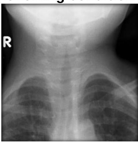
Steeple sign is seen in which of the following conditions?

Which of the following statements about lipoma is radiologically true?
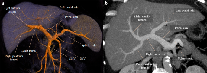
Identify the structure branching into the liver as seen in the provided CT scan of the abdomen.

Caldwell’s view is primarily used to visualize which sinus?
Water’s view is used to obtain diagnostic information of:
Which of the following statements about MRI is incorrect?
What is the primary use of the Rhese view in radiographic imaging?
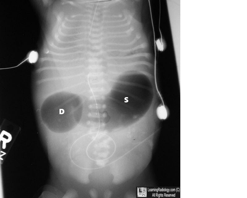
What condition is indicated by the double bubble sign on an abdominal X-ray?

Explanation: ***Neurogenic bladder dysfunction*** - The "Christmas tree appearance" refers to the **trabeculated and irregular bladder wall** with associated **bladder diverticula** seen on a voiding cystourethrogram (VCUG). - This appearance is characteristic of chronic, high-pressure bladder emptying due to **neurological impairment**, leading to hypertrophy of the detrusor muscle. *Autonomous bladder function* - An autonomous bladder (or isolated detrusor contraction) typically results from **complete spinal cord lesions** above the sacral micturition center after spinal shock. - While it is a type of neurogenic bladder, it doesn't specifically correlate with the "Christmas tree appearance," which is broader and signifies any form of chronic bladder outlet obstruction or spasticity. *Nocturnal enuresis* - This refers to **involuntary urination during sleep** and is primarily a functional disorder, especially in children, without structural bladder changes. - It is not associated with the characteristic radiological findings of bladder wall hypertrophy or diverticula. *Urinary stress incontinence* - Characterized by **involuntary leakage of urine** during physical activity that increases intra-abdominal pressure. - This condition is typically due to **urethral hypermobility** or **intrinsic sphincter deficiency** and does not involve structural changes to the bladder wall itself that would cause a "Christmas tree appearance."
Explanation: ***Sarcoidosis*** - The **Lambda-Panda sign** is a characteristic finding in sarcoidosis on **gallium-67 scintigraphy**. - **Lambda sign**: Bilateral hilar and right paratracheal lymph node uptake forming a pattern resembling the Greek letter lambda (λ). - **Panda sign**: Symmetric uptake in the **lacrimal and parotid glands**, creating a facial pattern resembling a panda. - These signs are highly specific for sarcoidosis and reflect the **non-caseating granulomatous** inflammation characteristic of the disease. *Tuberculosis* - While tuberculosis can cause lymphadenopathy, it typically does not present with the specific **Lambda-Panda sign** on gallium-67 scintigraphy. - **Granulomas** in tuberculosis are caseating, differentiating it from the non-caseating granulomas of sarcoidosis. *Histoplasmosis* - Histoplasmosis is a fungal infection that can cause pulmonary and disseminated lesions, but the **Lambda-Panda sign** is not characteristic of its presentation. - Diagnosis relies on fungal cultures, **antigen detection**, or serology. *Leishmaniasis* - Leishmaniasis is a parasitic disease that manifests as cutaneous, mucocutaneous, or visceral forms, which do not typically involve the specific sites to produce the **Lambda-Panda sign**. - Diagnosis is primarily by **microscopic identification** of amastigotes in tissue samples.
Explanation: ***Acute laryngotracheobronchitis*** - The **steeple sign** on an anteroposterior (AP) neck radiograph is a classic finding in acute laryngotracheobronchitis, also known as **croup**. - This sign refers to the **subglottic narrowing** of the trachea, resembling a church steeple, due to edema caused by viral infection. *Acute epiglottitis* - Acute epiglottitis is characterized by the **thumb sign** on a lateral neck radiograph, where the swollen epiglottis appears enlarged. - This condition involves inflammation primarily of the epiglottis, not the subglottic region. *Laryngeal papillomatosis* - Laryngeal papillomatosis is characterized by **wart-like growths** (papillomas) on the vocal cords and larynx, often leading to hoarseness. - Radiographically, it typically appears as irregular soft tissue masses, not the diffuse subglottic narrowing seen in croup. *Bilateral abductor paralysis* - Bilateral abductor paralysis involves the inability of both vocal cords to abduct, leading to a **fixed, narrowed glottic opening**. - This condition presents as a smooth, constant narrowing at the level of the vocal cords rather than the subglottic, conical narrowing of the steeple sign.
Explanation: ***Low attenuation on CT scan*** - Lipomas, being composed of **fat**, appear as areas of **low attenuation** (typically -50 to -150 Hounsfield Units) on computed tomography (CT) scans. - This low attenuation is a **key diagnostic characteristic** that helps differentiate lipomas from other soft tissue masses. *Hyperechoic on ultrasound* - Lipomas typically appear **isoechoic to hypoechoic** on ultrasound, not consistently hyperechoic. - They may have a thin echogenic capsule, but the internal contents are usually similar to or less echogenic than adjacent subcutaneous fat. *Hyperintense on fat-suppressed sequences* - This is **incorrect** - lipomas show **signal dropout** (become dark/hypointense) on fat-suppressed sequences (STIR, fat-sat T1/T2). - Signal suppression on fat-saturated sequences is actually a **diagnostic feature** confirming the fatty nature of the lesion. - Note: Lipomas ARE hyperintense on standard T1-weighted imaging due to fat content. *Hyper-intense on T2-weighted MRI* - Lipomas typically show **intermediate to slightly hyperintense signal** on T2-weighted MRI, but not markedly hyperintense like fluid. - They are less bright than fluid-filled structures or highly vascular lesions on T2-weighted sequences.
Explanation: ***Portal Vein*** - The image clearly labels the **Portal Vein** as the large vessel entering the liver and branching into the right and left portal veins. - The portal vein is crucial for carrying nutrient-rich, deoxygenated blood from the gastrointestinal tract and spleen to the liver for processing. *Superior Vena Cava* - The **superior vena cava** is located in the chest and drains blood from the upper body, not directly into the liver. - It would not be visible branching within the liver parenchyma on an abdominal CT scan in this manner. *Inferior Vena Cava* - The **inferior vena cava** is a large vein that runs along the posterior abdominal wall, draining blood from the lower body. - While it passes through the liver, it does not branch *into* the liver parenchyma in the same way the portal vein does. *Splenic Vein* - The **splenic vein** is shown in the image as a vessel that contributes to the formation of the portal vein. - It drains the spleen and merges with the superior mesenteric vein to form the portal vein, but it does not branch *into* the liver directly as depicted for the portal vein.
Explanation: ***Frontal sinus*** - **Caldwell's view**, a posteroanterior radiographic projection with 15-degree caudal angulation, is optimized for visualizing the **frontal sinuses**, providing a clear image above the orbital rims. - This view is particularly useful for assessing conditions like **frontal sinusitis** or **fractures** affecting the frontal region. - It also provides good visualization of the **ethmoid air cells**. *Maxillary sinus* - The **Waters' view** (occipitomental projection) is primarily used for the visualization of the **maxillary sinuses**, providing an unobstructed view of their floor and walls. - While Caldwell's view might show portions of the maxillary sinuses, it is not optimized for their comprehensive assessment due to superimposition of other structures. *Ethmoidal sinus* - While Caldwell's view does provide visualization of the **ethmoidal air cells**, the **frontal sinuses** remain the primary target of this projection. - The **lateral view** can also be used for assessing the ethmoidal air cells, as it demonstrates their anterior and posterior groups. *Sphenoid sinus* - The **sphenoid sinus** is best visualized using the **submentovertex (base) view** or **lateral view**. - Caldwell's view does not provide adequate visualization of the sphenoid sinus due to its deep posterior location in the skull base.
Explanation: ***Maxillary sinus*** - **Water's view** (occipitomental projection) is specifically designed to visualize the **maxillary sinuses** and highlight any **fluid levels** or **mucosal thickening** within them. - In this view, the **petrous ridges are projected below the floors of the maxillary sinuses**, allowing for clear assessment and optimal visualization. *Ethmoidal sinuses* - While partially visible on Water's view, the **ethmoidal sinuses** are best visualized with a **Caldwell view** (occipitofrontal projection) or a **CT scan**. - The complex bony structures surrounding them make detailed assessment difficult with Water's view alone. *Frontal sinus* - The **frontal sinuses** are best evaluated with a **Caldwell view** (occipitofrontal projection), which projects the petrous ridges at the lower third of the orbits. - Water's view offers a suboptimal projection for comprehensive assessment of the frontal sinuses. *Sphenoid sinus* - The **sphenoid sinus** is centrally located and often obscured by other structures on plain radiographs like Water's view. - It is best visualized with a **lateral skull view** or, more comprehensively, with a **CT scan**.
Explanation: ***MRI is better for calcified lesions*** - **Magnetic Resonance Imaging (MRI)** is generally **poor** at visualizing **calcified lesions** like gallstones, kidney stones, or bone fragments. - **Computed Tomography (CT) scans** are the **modality of choice** for detecting and characterizing calcifications due to their ability to directly measure tissue density. *MRI is contraindicated in patients with pacemakers* - This statement is generally correct, as the strong magnetic fields and radiofrequency pulses can interfere with pacemaker function, leading to **device malfunction** or **patient harm**. - While "MRI-conditional" pacemakers exist, standard pacemakers are a **relative or absolute contraindication** for MRI. *MRI is useful for evaluating bone marrow* - **MRI** is highly effective for visualizing and characterizing the **bone marrow**, allowing for the detection of tumors, infections, and other marrow-related pathologies. - It can differentiate between various marrow components, such as **fatty marrow** and **hematopoietic marrow**, and detect early changes not visible on other imaging modalities. *MRI is useful for localizing small lesions in the brain* - **MRI** offers superior **soft tissue contrast** compared to CT, making it highly effective for detecting and precisely localizing even **small lesions** within the brain. - Its ability to visualize different tissue types and pathology makes it crucial for diagnosing conditions like **multiple sclerosis plaques, tumors, and ischemic strokes**.
Explanation: ***Optic foramen*** - The Rhese view, or **parieto-orbital oblique projection**, is specifically designed to isolate and visualize the **optic canal**, which houses the **optic nerve** and **ophthalmic artery**. - This view helps in detecting fractures, tumors, or erosions affecting the **optic foramen** and optic nerve. *Superior orbital fissure* - While located near the optic foramen, the **superior orbital fissure** is typically better visualized with other radiographic views such as the **Caldwell view** or specific CT scans. - The Rhese view's angulation is optimized for the optic canal, not the **superior orbital fissure**. *Infraorbital foramen* - The **infraorbital foramen** is located on the maxilla, inferior to the orbit, and is not the primary target of the Rhese view. - Views like the **Waters view** or specialized facial bone projections are more appropriate for visualizing the **infraorbital foramen**. *Sella turcica* - The **sella turcica**, which houses the pituitary gland, is best visualized using lateral skull projections or dedicated CT/MRI scans of the brain and pituitary region. - The Rhese view does not provide an optimal projection for examining the **sella turcica**.
Explanation: ***Duodenal atresia*** - The **double bubble sign** on an abdominal X-ray is pathognomonic for **duodenal atresia**, characterized by two air-filled bubbles: one in the stomach and one in the proximal duodenum, separated by the pylorus. - This congenital anomaly results from a complete obstruction of the duodenum, preventing the passage of air and fluid past this point. *Duodenal stenosis* - While also an obstruction of the duodenum, **duodenal stenosis** is an incomplete obstruction, meaning some gas will pass beyond the duodenum. - This would result in gas being present in the distal bowel, which is not seen with a classic "double bubble" where the bowel distal to the duodenum is gasless. *Volvulus* - **Volvulus** involves the twisting of a loop of intestine, leading to obstruction and potentially ischemia. - While it can cause obstruction, it typically presents with a "corkscrew" appearance on an upper GI series or signs of diffuse bowel distension, not the isolated double bubble. *All of the options* - The double bubble sign is highly specific to **duodenal atresia** due to the complete obstruction it signifies. - Other conditions like duodenal stenosis and volvulus cause different radiological patterns, making this option incorrect.
Radiographic Anatomy of Skull and Face
Practice Questions
Radiographic Anatomy of Spine
Practice Questions
Radiographic Anatomy of Chest
Practice Questions
Radiographic Anatomy of Abdomen
Practice Questions
Radiographic Anatomy of Extremities
Practice Questions
Cross-sectional Anatomy: Brain and Head
Practice Questions
Cross-sectional Anatomy: Neck
Practice Questions
Cross-sectional Anatomy: Thorax
Practice Questions
Cross-sectional Anatomy: Abdomen and Pelvis
Practice Questions
Vascular Anatomy
Practice Questions
Developmental Anatomy Variations
Practice Questions
Anatomic Landmarks for Interventional Procedures
Practice Questions
Get full access to all questions, explanations, and performance tracking.
Start For Free