Which of the following is the best initial imaging study for evaluating a suspected fracture?
Stenver's view is used for -
What type of lesions in the skull calvarium can be identified on this X-ray?

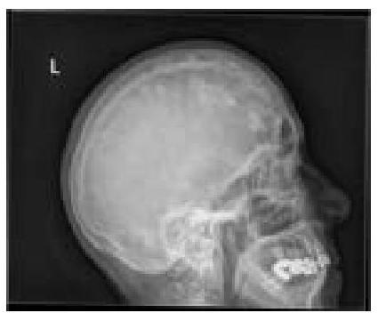
What is the diagnosis based on the following X-ray?
Which of the following provides excellent details about the chemodectomas?
Which substance has the highest Hounsfield Unit (HU) value?
In normal X-ray of shoulder, which is the superior most structure?
Radiological view which best shows maxillary sinus and orbit is -
At what age are the first four carpal bones (capitate, hamate, triquetrum, and lunate) typically visible on an X-ray?
Which radiographic view is best for visualizing the sphenoid sinus?
Explanation: ***X-ray*** - X-rays are generally the **first-line imaging study** for suspected fractures due to their widespread availability, low cost, and ability to clearly visualize bone structures. - They effectively show **bone alignment**, fracture patterns, and presence of **displacement**. *CT scan* - While excellent for detailed bony anatomy, **CT scans** expose patients to higher doses of **radiation** and are typically reserved for complex fractures or when standard X-rays are inconclusive. - They provide **cross-sectional views** and are useful for evaluating intra-articular fractures or bone fragments. *MRI* - **MRI** is superior for visualizing **soft tissues** like tendons, ligaments, and cartilage, and detecting occult fractures not seen on X-ray. - It is not the initial imaging choice for suspected fractures because it is **more expensive**, time-consuming, and less readily available than X-rays. *Ultrasound* - **Ultrasound** is useful for evaluating certain soft tissue injuries and can detect **ligamentous tears** or effusions. - However, its ability to clearly visualize and definitively diagnose most bone fractures is **limited**, especially deep or complex ones.
Explanation: ***Internal auditory canal*** - The **Stenvers position** (Stenvers view) is a specific radiographic projection designed to visualize the **petrous portion of the temporal bone**, with a clear profile of the internal auditory canal. - It is used to evaluate the **internal auditory canals** for abnormalities such as **acoustic neuromas** or other lesions affecting the vestibulocochlear nerve. *Inferior orbital foramen* - The inferior orbital foramen is typically visualized with standard facial bone views or specific orbital projections, not the Stenvers view. - It is a small opening on the floor of the orbit, transmitting the **infraorbital nerve and vessels**. *Sella turcica* - The sella turcica, which houses the pituitary gland, is best visualized using a **lateral skull view** or CT/MRI. - Stenvers view is not designed to provide optimal imaging of the sella turcica. *Superior orbital foramen* - The superior orbital foramen (more commonly referred to as the **superior orbital fissure**) is best seen on standard orbital or skull views. - It transmits cranial nerves III, IV, VI, and the ophthalmic division of V, and is not the primary focus of the Stenvers view.
Explanation: ***Brain metastases (Skull metastases)*** - The image shows multiple **lytic lesions** in the skull calvarium, which are characteristic of metastatic disease that has spread to bone - Metastases from primary cancers (lung, breast, kidney, thyroid, prostate) commonly involve the skull and appear as **punched-out or moth-eaten lytic lesions** - While plain X-rays cannot visualize brain parenchyma, they can detect **bony destruction** caused by metastatic deposits in the skull - These appear as well-defined osteolytic lesions without sclerotic margins *Multiple myeloma* - Multiple myeloma typically presents with **multiple punched-out lytic lesions** in the skull that can appear very similar to metastases - However, multiple myeloma is a **primary bone marrow malignancy** rather than metastatic disease - Key differentiator: myeloma lesions are usually more uniform in size and distribution - Clinical context (monoclonal protein, anemia, renal dysfunction) helps distinguish from metastases *Osteosarcoma* - Osteosarcoma is a **primary bone tumor** that usually causes a mixture of lytic and blastic (bone-forming) lesions - Typically presents with **sunburst or spiculated periosteal reaction** and soft tissue mass - Usually occurs as a **solitary aggressive lesion** in younger patients, not multiple scattered lesions - Rarely occurs in the skull compared to long bones *Osteomyelitis* - Osteomyelitis is an **infection of the bone** that causes bone destruction and reactive new bone formation - Shows features of **bone destruction, periosteal reaction**, and possibly sequestra (dead bone fragments) - Typically presents as a **focal process** with surrounding inflammatory changes - Does not produce the multiple discrete lytic lesions pattern seen in metastatic disease
Explanation: ***Renal Tuberculosis*** - Characterized by **calcifications** in the renal parenchyma and collecting system visible on X-ray, often appearing as **moth-eaten** or **amputated calyces** - Associated with **sterile pyuria**, **acidic urine**, and **caseous necrosis** leading to characteristic radiographic findings of calcified granulomas *Uterine Fibroid* - Appears as a **well-defined soft tissue mass** arising from the pelvis, often with **popcorn calcifications** if degenerating - Located in the **uterine region** rather than the renal area, and typically presents with menstrual abnormalities and pelvic pressure *Bladder Carcinoma* - Presents as an **irregular filling defect** or **mass** within the bladder on contrast studies, rarely with calcifications - Associated with **hematuria** and typically shows **soft tissue density** rather than the dense calcifications seen in renal TB *Bladder Stone* - Appears as a **rounded, smooth radiopaque density** in the pelvis corresponding to the bladder location - Usually has a **homogeneous density** with well-defined borders, unlike the irregular calcifications of renal tuberculosis
Explanation: ***MRI*** - **Magnetic Resonance Imaging (MRI)** is generally considered the best modality for evaluating **chemodectomas** due to its superior soft tissue contrast. - It can accurately delineate the tumor's extent, its relationship to surrounding structures, and detect any **vascular invasion** or **nerve involvement**. *Xray* - **X-rays** provide limited detail for soft tissue tumors like chemodectomas. - They primarily visualize bone structures and are not effective in characterizing small or non-calcified soft tissue masses. *Ultrasound* - **Ultrasound** can detect the presence of a mass and assess its vascularity, but its utility for chemodectomas is limited by its lower resolution and dependence on operator skill. - It may not provide sufficient detail for precise anatomical localization or differentiation from other neck masses. *CT angiography (can provide useful information regarding vascular involvement)* - **CT angiography** is excellent for visualizing the **vascular supply** to the tumor and assessing its relationship with critical blood vessels. - While useful for surgical planning, it typically offers less detailed soft tissue characterization compared to MRI for the primary tumor itself.
Explanation: ***Bone*** - **Bone**, particularly cortical bone, has the **highest density** and thus the highest Hounsfield Unit (HU) value among the given options, typically ranging from +300 to +1000 HU. - Its high density causes **significant attenuation** of X-rays, leading to a bright appearance on CT scans. *Water* - **Water** is defined as having a Hounsfield Unit (HU) value of **0**, serving as the reference point for the scale. - Substances less dense than water have negative HU values, while those more dense have positive values. *Fat* - **Fat** has a negative Hounsfield Unit (HU) value, typically ranging from **-50 to -100 HU**, indicating it is less dense than water. - This low density causes less X-ray attenuation, making fat appear dark on CT scans. *Soft tissue* - **Soft tissues** such as muscle or organs generally have HU values slightly higher than water, ranging from **+20 to +60 HU**. - Their density is greater than fat but significantly less than bone.
Explanation: ***Coracoid process*** - On a standard **X-ray projection of the shoulder**, especially an **AP view**, the **coracoid process** of the scapula is typically the most superior bony structure visible. - Its anterior projection often situates it cranially to other humeral landmarks. *Greater tubercle* - The **greater tubercle** is part of the proximal humerus and serves as an attachment site for rotator cuff muscles. - While prominent, it usually lies infero-lateral to the coracoid process in most standard shoulder X-ray views. *Surgical neck of the humerus* - The **surgical neck** is located distal to the head and tubercles of the humerus. - It is positioned significantly inferior to the superior aspect of the shoulder joint. *Head of the humerus* - The **head of the humerus** articulates with the glenoid fossa of the scapula. - While superior to much of the humerus, the coracoid process typically extends more superiorly.
Explanation: ***Water's view*** - The **Water's view**, or occipitomental view, is ideal for visualizing the **maxillary sinuses** as it projects the petrous ridges below the maxillary sinuses. - This projection also provides a clear view of the **orbits**, zygomatic arches, and nasal cavity. *Caldwell view* - The Caldwell view (occipitofrontal view) is primarily used to visualize the **frontal sinuses** and ethmoid air cells. - In this view, the **petrous ridges** overlap the lower half of the orbits, obscuring some orbital details. *Lateral view* - A lateral view of the skull is useful for assessing the **sphenoid sinus**, sella turcica, and overall skull alignment. - It does not provide detailed, unobstructed views of the maxillary sinuses or the entire orbit crucial for assessing symmetry and pathology in these specific areas. *Towne view* - The Towne view (half-axial or anteroposterior axial view) is primarily used to visualize the **occipital bone**, foramen magnum, and condyles. - It is not designed to provide clear views of the maxillary sinuses or orbits, as these structures are typically superimposed or distorted in this projection.
Explanation: ***Correct: 4 years*** - By **4 years of age**, all four carpal bones—**capitate**, **hamate**, **triquetrum**, and **lunate**—are typically visible on X-ray. - The **capitate** and **hamate** appear first (around 2-4 months of age). - The **triquetrum** appears around **2-3 years**. - The **lunate** is the last of these four to appear, typically visible by **3-4 years**. - This age marks a significant progression in **skeletal maturation** and provides key milestones for assessing bone age. *Incorrect: 2 years* - At **2 years of age**, typically only the **capitate**, **hamate**, and **triquetrum** are visible on X-ray. - The **lunate** has usually not yet ossified sufficiently to be radiographically apparent at this age. - Since the question asks when all four specified bones are visible, 2 years is too early. *Incorrect: 5 years* - By **5 years of age**, all four carpal bones (capitate, hamate, triquetrum, lunate) would have already been well-established and visible for over a year. - Additionally, the **scaphoid** would likely be appearing or already visible by this age. - This is later than when all four bones first become visible together. *Incorrect: 7 years* - At **7 years of age**, not only these four carpal bones but also additional carpal bones like the **scaphoid**, **trapezium**, and **trapezoid** would be clearly visible. - The visibility of the four bones specified in the question would be well established much earlier than this age. - This represents a much more advanced stage of carpal bone maturation.
Explanation: ***Lateral view*** - The **lateral view** provides an excellent profile image of the **sphenoid sinus**, allowing for clear visualization of its anterior and posterior walls, and its relationship with the sella turcica. - This projection is particularly useful for assessing the **depth** and **anteroposterior extent** of the sphenoid sinus. *Water's view* - The Water's view (occipitomental projection) is primarily used to visualize the **maxillary sinuses**, and the **orbits**. - While it offers some indirect information about the posterior ethmoid cells, it is not ideal for direct assessment of the sphenoid sinus due to **superimposition** of other facial structures. *Caldwell view* - The Caldwell view (occipitofrontal projection) is optimized for viewing the **frontal sinuses** and **ethmoid sinuses**. - The sphenoid sinus is often **obscured** by overlying structures in this projection, making it difficult to assess. *Towne's view* - The Towne's view (occipital/half-axial projection) is primarily used to visualize the **occipital bone**, **petrous pyramids**, and the **foramen magnum**. - This projection does not provide adequate visualization of the sphenoid sinus due to the angle and superimposition of the skull base.
Radiographic Anatomy of Skull and Face
Practice Questions
Radiographic Anatomy of Spine
Practice Questions
Radiographic Anatomy of Chest
Practice Questions
Radiographic Anatomy of Abdomen
Practice Questions
Radiographic Anatomy of Extremities
Practice Questions
Cross-sectional Anatomy: Brain and Head
Practice Questions
Cross-sectional Anatomy: Neck
Practice Questions
Cross-sectional Anatomy: Thorax
Practice Questions
Cross-sectional Anatomy: Abdomen and Pelvis
Practice Questions
Vascular Anatomy
Practice Questions
Developmental Anatomy Variations
Practice Questions
Anatomic Landmarks for Interventional Procedures
Practice Questions
Get full access to all questions, explanations, and performance tracking.
Start For Free