Which of the following structures is best visualised using Schuller's view and Law's view?
Transverse and vertical study of the skeleton can be done in?
"Hair on End" appearance is seen in:
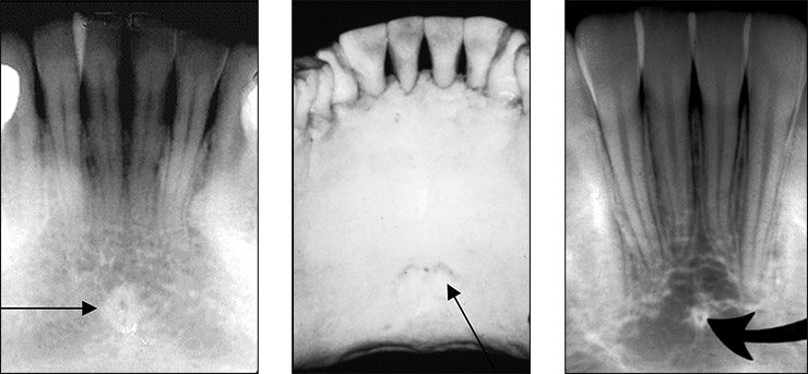
The radiopaque structure shown in the box is most likely:

Which of these structures appears radiopaque?
Which of the following is most radiolucent?
Preferred imaging modality for choanal atresia is
In pantomogram positioning, which anatomical landmark is used for initial patient alignment?
Best view for visualizing sella turcica on X-Ray:
Which of the following will cause posterior impression on barium swallow:
Explanation: ***Mastoid air cells*** - **Schuller's view** is an **oblique lateral radiograph** specifically designed to visualize the mastoid air cells, the **tegmen mastoideum**, and the **sigmoid sinus**. - **Law's view**, another lateral oblique projection, also provides excellent visualization of the mastoid air cells and the surrounding bony structures. *Sphenoid sinus* - The **sphenoid sinus** is best visualized with lateral and submentovertex (SMV) views, not Schuller's or Law's views. - These projections are critical for assessing sinus opacification, **mucoperiosteal thickening**, or presence of fluid levels. *Carotid canal* - The **carotid canal** requires specialized views like the **Stenvers view** or advanced imaging techniques like **CT scans** and **MRI** for optimal visualization. - Standard skull radiographs like Schuller's and Law's views do not adequately demonstrate the intricacies of the carotid canal. *Foramen ovale and spinosum* - The **foramen ovale** and **foramen spinosum** are located at the base of the skull and are best visualized using the **submentovertex (SMV)** or **Hirtz view**, or with cross-sectional imaging. - These foramina transmit the **mandibular nerve**, lesser petrosal nerve, accessory meningeal artery, and middle meningeal vessels, respectively.
Explanation: ***PA ceph*** - A **posteroanterior (PA) cephalometric radiograph** allows for the evaluation of the skeletal structures in both **transverse (width) and vertical (height) dimensions** of the face and skull. - It is particularly useful for assessing **facial symmetry**, transverse discrepancies, and the vertical relationships of the upper and lower jaws. - This is the **gold standard** for evaluating both transverse and vertical skeletal relationships simultaneously. *SMV* - The **submentovertex (SMV)** projection primarily visualizes the **cranial base**, zygomatic arches, and sphenoid sinuses. - It is mainly used for assessing the **width of the cranial base** and specific structural morphology, but does not provide comprehensive transverse and vertical skeletal analysis in the way PA ceph does. *OMV* - The **occipitomental view (OMV)**, also known as **Waters view**, is a standard radiographic projection primarily used for evaluating **paranasal sinuses** and **facial bones**. - While it shows some anteroposterior and limited transverse information, it is **not the primary projection** for comprehensive transverse and vertical skeletal study. - PA ceph is superior for systematic transverse and vertical skeletal assessment. *Lateral Ceph* - A **lateral cephalometric radiograph** primarily provides a two-dimensional view of the skull and facial bones in the **sagittal plane**. - It is excellent for assessing **anteroposterior (AP) and vertical skeletal relationships**, but it **does not show transverse dimensions or asymmetry** since it's a side view.
Explanation: ***Thalassemia*** - The **"hair on end" appearance** on skull X-rays is caused by **extramedullary hematopoiesis** in the bone marrow, as the body tries to compensate for severe anemia. - This persistent, high-volume erythropoiesis leads to the **expansion** of the **bone marrow spaces**, especially in the skull, resulting in the characteristic radiating spicules. *Rickets* - Rickets is a disorder caused by **vitamin D deficiency**, leading to impaired bone mineralization. - It presents with **bowing of legs**, **rachitic rosary**, and **widening of growth plates**, not "hair on end" appearance. *Scurvy* - Scurvy is due to a **deficiency of vitamin C**, which is essential for collagen synthesis. - It manifests as **bleeding gums**, **perifollicular hemorrhages**, and **impaired wound healing**, not changes in skull bone marrow. *Hemochromatosis* - Hemochromatosis is characterized by **excessive iron accumulation** in various organs, leading to organ damage. - It can cause **liver cirrhosis**, **diabetes**, and **skin pigmentation**, but does not present with the "hair on end" skull manifestation.
Explanation: ***Genial tubercle*** - The radiopaque structure in the image, located in the midline of the mandible below the incisor roots, is characteristic of the **genial tubercle**. - These are small, bony projections on the lingual surface of the mandible that serve as attachment points for the **genioglossus** and **geniohyoid muscles**. - Also known as **mental spines**, though technically the genial tubercle is the more anatomically precise term for these specific structures. *Lingual foramen* - The lingual foramen is a small opening located near the genial tubercles, but it appears as a **radiolucent** (darker) dot within the radiopaque genial tubercles on a radiograph, not a radiopaque structure itself. - It allows passage for a small branch of the **lingual artery**. *Periapical cyst (PCD)* - A periapical cyst typically presents as a well-defined **radiolucency** (dark area) around the apex of a tooth root, indicating bone destruction due to infection. - It would not appear as a dense radiopaque structure in the mandibular midline. *Mental spine* - While "mental spine" is sometimes used as a synonym for genial tubercles, **genial tubercle** is the more precise anatomical term for these specific bony prominences. - The term "mental spine" can be ambiguous as it may refer to the broader midline prominence rather than the specific tubercles.
Explanation: ***Nasal septum*** - The **nasal septum** is composed of bone and cartilage, which readily absorb X-rays due to their density. - This absorption causes less X-ray penetration to the film, resulting in a **radiopaque** (white or light) appearance on a radiograph. *Mental foramen* - The **mental foramen** is an opening in the bone, allowing nerves and vessels to pass through. - As an opening, it appears as a **radiolucent** (dark) area on a radiograph because X-rays pass through it more easily. *Maxillary sinus* - The **maxillary sinus** is an air-filled cavity within the maxilla. - Air does not absorb many X-rays, making the maxillary sinus appear **radiolucent** (dark) on an X-ray image. *Nasal fossa* - The **nasal fossa** (or nasal cavity) is primarily an air-filled space within the skull. - Due to the presence of air, which offers little resistance to X-rays, the nasal fossa appears **radiolucent** (dark) on a radiograph.
Explanation: ***Air space*** - **Air** has a very low density, allowing almost all X-rays to pass through it, resulting in a **dark (radiolucent)** appearance on an X-ray image. - In dental radiography, air spaces such as the **maxillary sinus** or nasal cavity appear as distinct dark areas. *Enamel* - **Enamel** is the **most highly mineralized** tissue in the human body, containing approximately 96% inorganic material. - Due to its high mineral content, enamel absorbs a large amount of X-rays, making it appear very **bright (radiopaque)** on an X-ray. *Bone* - **Bone tissue** is highly mineralized, though less so than enamel, and contains significant amounts of calcium and phosphate. - It absorbs a substantial portion of X-rays, appearing **radiopaque (white or light gray)** on a radiograph, indicating its density. *Dentin* - **Dentin** is less mineralized than enamel but more so than pulp, composed of about 70% inorganic material. - It appears **less radiopaque than enamel** but still significantly radiopaque (light gray) compared to soft tissues or air.
Explanation: ***CT scan*** - **Computed Tomography (CT) scan** is the preferred imaging modality for diagnosing **choanal atresia** due to its superior ability to visualize **bony anatomy** and the precise location and extent of the obstruction. - It effectively differentiates between **bony** and **membranous atresia** and helps in surgical planning. *X-ray* - **X-rays** provide limited detail of the complex **nasal and choanal anatomy** and are not sufficient to accurately diagnose choanal atresia or characterize the obstruction. - While it might show some signs, it lacks the **spatial resolution** needed for definitive diagnosis and surgical guidance. *MRI* - **Magnetic Resonance Imaging (MRI)** is excellent for visualizing **soft tissue structures** but is less optimal for evaluating bony defects characteristic of most choanal atresia cases. - It can be used as an adjunct to assess associated **craniofacial anomalies** but is not the primary diagnostic tool for the atresia itself. *PET scan* - **Positron Emission Tomography (PET) scan** is primarily used for assessing **metabolic activity**, particularly in oncology, and has no role in the diagnosis of **choanal atresia**. - It does not provide the anatomical detail required to visualize the obstruction in the posterior nasal airway.
Explanation: ***Midsagittal plane positioning*** - The **midsagittal plane** is the **first and most critical anatomical landmark** used for initial patient alignment in pantomogram positioning - A vertical light beam indicator is used to align the **midsagittal plane perpendicular to the floor**, ensuring the patient's head is centered in the machine - This prevents **horizontal distortion** and ensures symmetrical imaging of both sides of the dental arches - Proper midsagittal alignment must be established **before** other positioning steps *Frankfort horizontal plane alignment* - The **Frankfort horizontal plane** (from the inferior border of the orbit to the superior border of the external auditory meatus) is the **second positioning step** - A horizontal light beam is used to ensure this plane is **parallel to the floor**, preventing vertical distortion - This alignment is performed **after** midsagittal plane positioning is established *Bite block placement* - The **bite block** is positioned **after** the midsagittal and Frankfort planes are aligned - It serves to **stabilize** the patient's head position and maintain the established alignment throughout the exposure - The bite block also slightly **separates the dental arches** to prevent superimposition of upper and lower teeth *Canine-to-canine bite positioning* - This refers to the **width of the bite groove** on the bite block, which typically spans from canine to canine - While important for optimal arch coverage, this is a **final positioning check** rather than an initial alignment landmark - It ensures the **focal trough** properly encompasses the dental arches
Explanation: ***Lateral view*** - A **lateral skull X-ray** provides the clearest profile view of the sella turcica, allowing for assessment of its size, shape, and cortical margins. - This projection effectively separates the structures of the sella from overlying bone, making it ideal for visualizing the pituitary fossa. *Open mouth view* - Primarily used to visualize the **odontoid process** (dens) of the C2 vertebra and atlantoaxial alignment. - It does not offer a clear or unobstructed view of the sella turcica. *Town's view* - Also known as an **anteroposterior (AP) axial view of the skull**, it is used to visualize the occipital bone and posterior fossa. - This view does not provide adequate visualization of the sella turcica due to superimposed structures. *AP view* - An **anteroposterior (AP) view of the skull** shows the frontal bone, orbits, and maxillary sinuses. - The sella turcica is significantly obscured by other cranial structures in this projection, making it unsuitable for detailed assessment.
Explanation: ***Aberrant right subclavian artery*** - An **aberrant right subclavian artery** (ARSA) arises from the **descending aorta** and passes **behind the esophagus** to reach the right arm. - This posterior course causes an **extrinsic impression** on the posterior wall of the esophagus, visible on barium swallow. *ASD* - An **Atrial Septal Defect (ASD)** involves a hole between the atria and does not directly impinge on the esophagus. - ASD primarily affects **cardiac hemodynamics** and may lead to right heart enlargement, but not esophageal compression. *VSD* - A **Ventricular Septal Defect (VSD)** is a hole between the ventricles of the heart, leading to shunting of blood. - VSDs typically cause **cardiac enlargement** but do not involve structures that would cause a posterior esophageal impression. *Pulmonary vascular hypertension* - **Pulmonary vascular hypertension** leads to enlargement of the **pulmonary arteries** and right side of the heart. - While significant cardiac enlargement can cause esophageal displacement, it typically results in **anterior or right-sided deviation**, not a direct posterior impression.
Radiographic Anatomy of Skull and Face
Practice Questions
Radiographic Anatomy of Spine
Practice Questions
Radiographic Anatomy of Chest
Practice Questions
Radiographic Anatomy of Abdomen
Practice Questions
Radiographic Anatomy of Extremities
Practice Questions
Cross-sectional Anatomy: Brain and Head
Practice Questions
Cross-sectional Anatomy: Neck
Practice Questions
Cross-sectional Anatomy: Thorax
Practice Questions
Cross-sectional Anatomy: Abdomen and Pelvis
Practice Questions
Vascular Anatomy
Practice Questions
Developmental Anatomy Variations
Practice Questions
Anatomic Landmarks for Interventional Procedures
Practice Questions
Get full access to all questions, explanations, and performance tracking.
Start For Free