What is the radiological sign shown in the image?

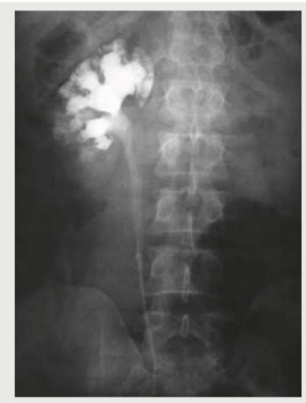
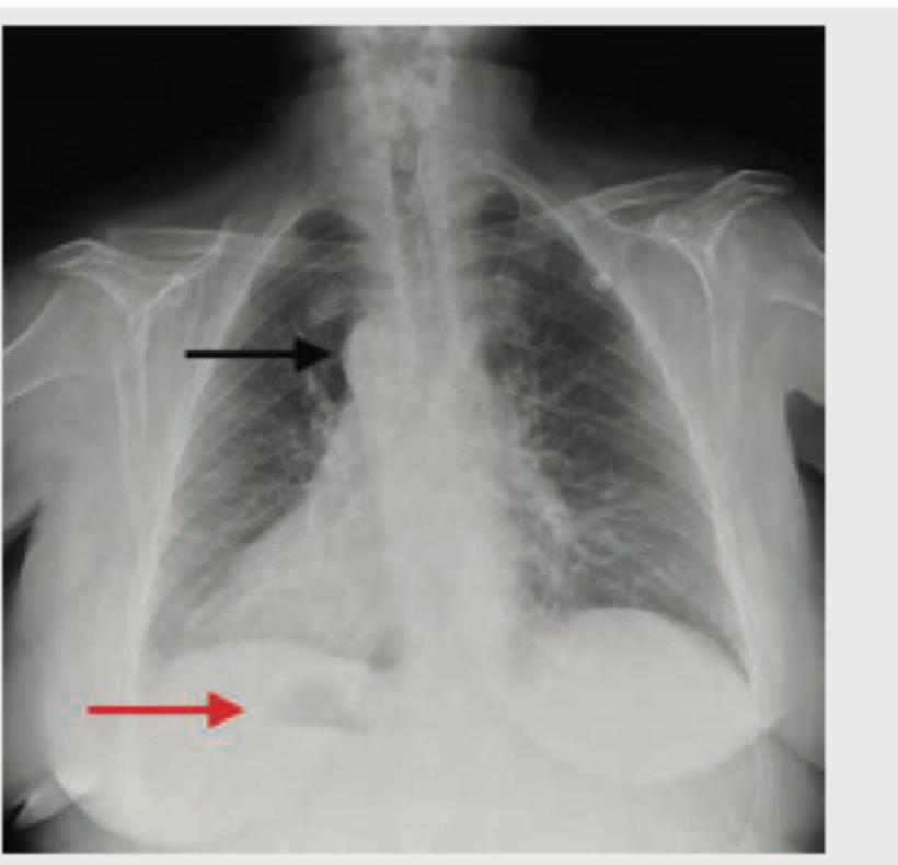
What does the following radiograph show?

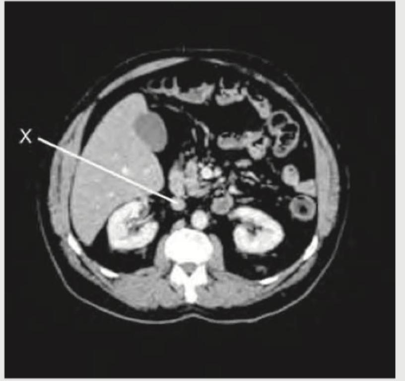
The following CT abdomen shows:

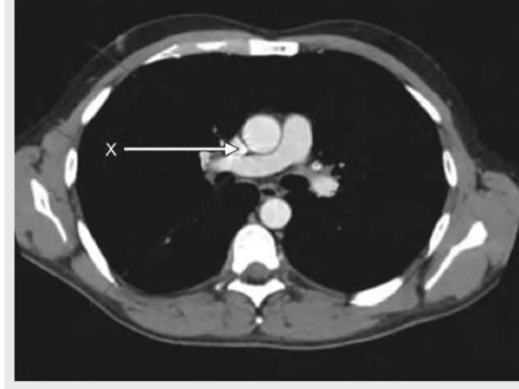
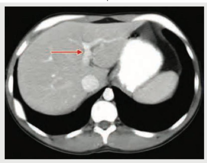
The X mark in the following CT chest shows:

What is the radiological sign demonstrated in the image provided?

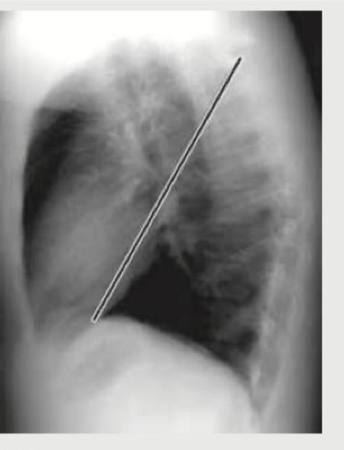
The lateral view chest X-ray shows:

Identify the structure shown in CT abdomen section. (Recent NEET Pattern 2018-19)

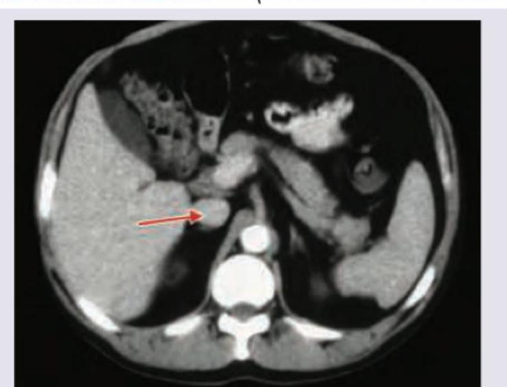
Identify the marked structure in the CT abdomen section shown below? (Recent NEET Pattern 2018-19)

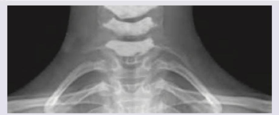
A 25-year-old female presents with neck pain and tingling sensation in her left arm. An X-ray of the cervicothoracic region is obtained. What is the radiological finding shown in the image?

Radiologic views used for fracture Mandible (body and Ramus) are all EXCEPT:
Explanation: ***Adder head*** - The image displays an **intravenous pyelogram (IVP)**, and the "adder head" sign refers to the characteristic appearance of the dilated ureter and renal pelvis (renal collecting system) due to an **ureterocele**. - An **ureterocele** is a congenital dilatation of the distal part of the ureter as it enters the bladder, causing a filling defect and proximal hydronephrosis resembling a **snake's head** on an IVP. *Flower vase* - This is not a recognized radiological sign for the pathology shown. - The appearance of the collecting system, specifically the ureter proximal to the ureterocele, does not resemble a flower vase. *Spider leg* - The "spider leg" or "spidery" appearance is associated with **polyarteritis nodosa (PAN)** in renal angiography, where multiple microaneurysms and stenoses of small renal arteries give a spidery pattern. - The image here is an IVP showing the collecting system, not an angiogram of renal vessels. *Flower bouquet* - This is not a recognized radiological sign for the pathology shown. - The configuration of the collecting system in this IVP does not depict a flower bouquet.
Explanation: ***Scurvy*** - The radiograph shows a **dense provisional zone of calcification (white line of Frankel)**, **rarefaction of the metaphysis (Trummerfeld zone)**, and **epiphyseal separation**, which are classic signs of scurvy. - Scurvy results from **vitamin C deficiency**, impairing collagen synthesis crucial for bone matrix formation. *Rickets* - Rickets typically presents with **widened, cupped, and frayed metaphyses** due to impaired mineralization of cartilage in the growth plates. - This image does not show the characteristic widening and fraying of the growth plates seen in rickets. *Osteoid osteoma* - An osteoid osteoma is a **benign bone tumor** characterized by a small radiolucent **nidus** surrounded by dense sclerotic bone. - The findings in the radiograph (metaphyseal changes, epiphyseal separation) are not consistent with osteoid osteoma. *Haemophilia* - Hemophilia causes **recurrent hemarthrosis (bleeding into joints)**, leading to joint destruction, synovial hypertrophy, and subchondral bone cysts. - While it can affect bone health, the specific changes seen in this radiograph (e.g., dense provisional zone of calcification) are not typical of hemophilia.
Explanation: ***Inferior vena cava*** - The structure labeled 'X' is a large, **thin-walled vessel** located to the **right of the vertebral body** and anterior to the right kidney, which is characteristic of the inferior vena cava. - The **inferior vena cava (IVC)** is typically more ovoid or flattened compared to the aorta, especially at this level, and drains into the right atrium. *Aorta* - The **aorta** is typically located to the **left of the vertebral body** and is generally more rounded and has thicker, more muscular walls than the IVC, appearing as a more uniformly circular structure. - The structure labeled 'X' is on the right side, making it unlikely to be the aorta. *Ureter* - Ureters are much **smaller in diameter** and are located more peripherally in the retroperitoneum, often seen near the psoas muscles. - The structure indicated by 'X' is a large, central vessel, not consistent with a ureter. *Superior mesenteric artery* - The **superior mesenteric artery (SMA)** originates from the aorta and is typically seen anterior to the aorta, often surrounded by fat and vessels of the superior mesenteric vein. - The structure 'X' is a major vein, not an artery, and is positioned more posteriorly and to the right of the midline compared to where the SMA would typically be found.
Explanation: ***Ascending aorta*** - The arrow points to a circular, bright white structure in the anterior mediastinum, which is the **ascending aorta** enhanced with contrast. - Its position anterior to the trachea and superior to the main pulmonary artery is characteristic of the ascending aorta at this CT level. *Descending aorta* - The **descending aorta** is positioned more posteriorly and to the left of the vertebral body at this level; the structure indicated is clearly in the anterior mediastinum. - It would appear as a separate, contrast-enhanced circular structure behind the heart and often slightly to the left of midline. *Superior vena cava* - While also in the anterior mediastinum, the **superior vena cava (SVC)** is typically a thinner-walled, more ovoid structure and lies to the right of the ascending aorta. - The indicated structure is distinctly thick-walled and circular, typical of an artery, not a vein. *Inferior vena cava* - The **inferior vena cava (IVC)** is located much lower in the chest and abdomen, and it is not visible at this axial CT level through the great vessels. - At the level of the great vessels, the IVC would be out of the displayed field or would appear more inferiorly, entering the right atrium.
Explanation: ***Correct Option: Gas under diaphragm*** - The image clearly shows a crescent-shaped lucency (gas) beneath the diaphragm, indicated by the red arrow, which is characteristic of **pneumoperitoneum**. - This finding is a critical sign of a **perforated viscus** within the abdomen, such as a perforated peptic ulcer, perforated diverticulitis, or other hollow viscus perforation. - **Free air under the diaphragm** is best seen on an erect chest X-ray or erect abdominal film and is a surgical emergency. *Incorrect Option: Bat wing sign* - The **"bat wing"** or **"butterfly" sign** refers to bilateral perihilar infiltrates seen in conditions like **pulmonary edema** due to congestive heart failure or acute respiratory distress syndrome (ARDS). - This pattern is not present in the provided image; the lung fields appear relatively clear except for the presence of gas under the diaphragm. *Incorrect Option: Situs inversus* - **Situs inversus** is a congenital condition where the major visceral organs are reversed or mirrored from their normal positions, manifesting as the **heart shadow** on the right side and liver on the left side. - The image shows the heart on the normal left side, and there is no evidence of reversed organ positioning. *Incorrect Option: Money bag appearance* - The **"money bag appearance"** is a recognized radiological sign seen in conditions like **hydronephrosis** on intravenous pyelogram (IVP) or in **hiatal hernia** where the gastric fundus appears as a rounded sac above the diaphragm. - This finding is not demonstrated in the provided image, which clearly shows free gas beneath the diaphragm rather than a soft tissue mass or fluid-filled structure.
Explanation: ***Major fissure*** - The image clearly shows an oblique line running from the mid-thoracic region superiorly and posteriorly to the diaphragm anteriorly, which is characteristic of the **major (oblique) fissure** on a lateral chest X-ray. - This fissure separates the **upper and middle lobes from the lower lobe** in the right lung, and the upper lobe from the lower lobe in the left lung. *Minor fissure* - The minor (horizontal) fissure is typically seen as a **horizontal line** on the lateral view, running from the mid-axillary line to meet the major fissure anteriorly. - It is located between the **upper and middle lobes** of the right lung only and is not depicted by the annotated line. *Azygos fissure* - An azygos fissure is an **anatomical variant** caused by the atypical migration of the azygos vein during development, creating a deep fissure in the superior aspect of the **right upper lobe**. - It would appear as a curvilinear line forming a "tear-drop" shape and is not represented by the oblique line shown. *Transverse fissure* - "Transverse fissure" is another name for the **minor (horizontal) fissure**. - As described earlier, its appearance and location on a lateral chest X-ray are distinct from the oblique line indicated.
Explanation: ***Portal vein*** - The arrow points to a vessel receiving blood from the splenic and superior mesenteric veins, which is characteristic of the **portal vein** entering the **liver parenchyma**. - The portal vein is typically seen anterior to the **inferior vena cava** and posterior to the **common hepatic artery** at this level. *Inferior vena cava* - The **inferior vena cava (IVC)** is a large, retroperitoneal vessel located posterior to the liver and to the right of the aorta. - The structure indicated by the arrow is clearly within the liver substance, not in the typical position of the IVC. *Splenic vein* - The **splenic vein** runs horizontally behind the body of the pancreas and joins with the superior mesenteric vein to form the portal vein. - The vessel shown is within the liver, distal to the formation of the portal vein. *Superior mesenteric vein* - The **superior mesenteric vein (SMV)** typically runs vertically in the mesentery and joins the splenic vein to form the portal vein. - The indicated structure is within the liver hilum, not in the anatomical location of the SMV.
Explanation: **Superior mesenteric artery** - The arrow points to a circular, **contrast-filled vessel** anterior to the aorta and posterior to the pancreatic head, characteristic of the **superior mesenteric artery (SMA)** - The SMA originates from the **anterior aspect of the aorta at L1 level** and supplies the midgut structures - In arterial phase CT, the SMA shows **bright contrast enhancement** and appears as a round structure between the aorta and pancreatic uncinate process *Incorrect: Aorta* - The **aorta** is the larger, more posterior vessel with a crescent or oval shape in axial sections - While the SMA branches from the aorta, the marked structure is clearly anterior to the main aortic lumen *Incorrect: Inferior vena cava* - The **IVC** is located to the **right of the aorta** in axial CT sections - The marked structure is **anterior and to the left**, not in the expected IVC position - The IVC would show less enhancement in arterial phase imaging *Incorrect: Superior mesenteric vein* - The **SMV** typically runs to the **right and slightly anterior** to the SMA - In arterial phase CT, the SMV shows **less intense opacification** compared to the arterial structures - The marked structure shows arterial-phase enhancement, indicating it is an artery, not a vein
Explanation: ***Cervical rib*** - The image displays an extra rib arising from the **C7 cervical vertebra**, which is characteristic of a cervical rib. - This **supernumerary rib** extends towards the sternum or first thoracic rib, a classic radiological finding. *Costochondritis* - **Costochondritis** is an inflammation of the cartilage connecting the ribs to the sternum, which is typically a clinical diagnosis, not visible on X-ray. - An X-ray would not show inflammatory changes in cartilage or soft tissue, making this diagnosis unlikely based on imaging alone. *Fracture of 2nd rib* - A **fracture of the 2nd rib** would appear as a discontinuity or break in the normal bony architecture of the second rib. - The image does not show any signs of a broken rib; instead, it shows an **extra, well-formed rib-like structure** originating from the cervical spine. *Spondylolisthesis* - **Spondylolisthesis** involves the anterior displacement of one vertebral body over another, usually in the lumbar spine. - This condition is also not visible in the provided image, which focuses on the cervicothoracic junction and shows an **anatomic variation** rather than vertebral slippage.
Explanation: ***Submentovertex*** - The **Submentovertex (SMV) view** is primarily used to assess the **zygomatic arches**, base of the skull, and sphenoid sinuses, not typically for mandibular body or ramus fractures. - While it can provide some information about the medial aspects of the mandible, it offers **limited structural detail** crucial for diagnosing fractures in the body and ramus. *Lower occlusal* - **Lower occlusal films** are useful for visualizing the **anterior mandible**, including the symphysis and parasymphysis regions, and the lingual aspect of the body. - They can provide detailed views of these specific areas but are not the primary view for comprehensive assessment of the entire body or ramus. *Lateral obliques* - **Lateral oblique views** are highly effective for visualizing the **body, angle, and ramus** of the mandible, providing a good representation of these regions without superimposition from the contralateral side. - This projection allows for assessment of fracture displacement and angulation in the lateral and postero-lateral aspects of the mandible. *Orthopantomogram* - An **Orthopantomogram (OPG)**, also known as a panoramic radiograph, provides a comprehensive view of the **entire mandible** and maxilla on a single film. - It is an excellent screening tool for identifying fractures in the **condyle, ramus, angle, body, and symphysis** of the mandible due to its broad coverage.
Radiographic Anatomy of Skull and Face
Practice Questions
Radiographic Anatomy of Spine
Practice Questions
Radiographic Anatomy of Chest
Practice Questions
Radiographic Anatomy of Abdomen
Practice Questions
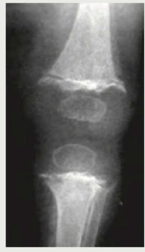
Radiographic Anatomy of Extremities
Practice Questions
Cross-sectional Anatomy: Brain and Head
Practice Questions
Cross-sectional Anatomy: Neck
Practice Questions
Cross-sectional Anatomy: Thorax
Practice Questions
Cross-sectional Anatomy: Abdomen and Pelvis
Practice Questions
Vascular Anatomy
Practice Questions
Developmental Anatomy Variations
Practice Questions
Anatomic Landmarks for Interventional Procedures
Practice Questions
Get full access to all questions, explanations, and performance tracking.
Start For Free