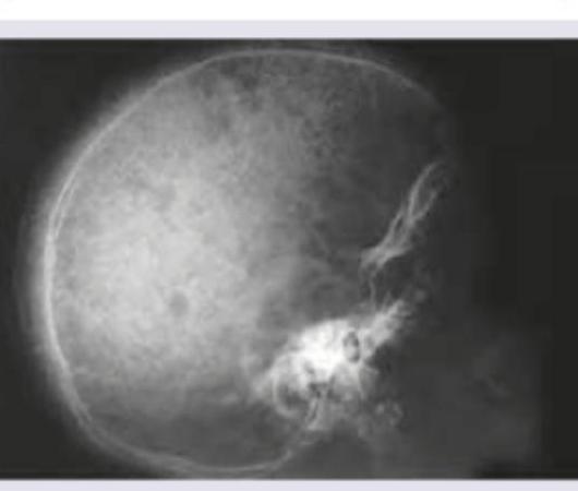
Identify the defect shown in the X-ray skull:

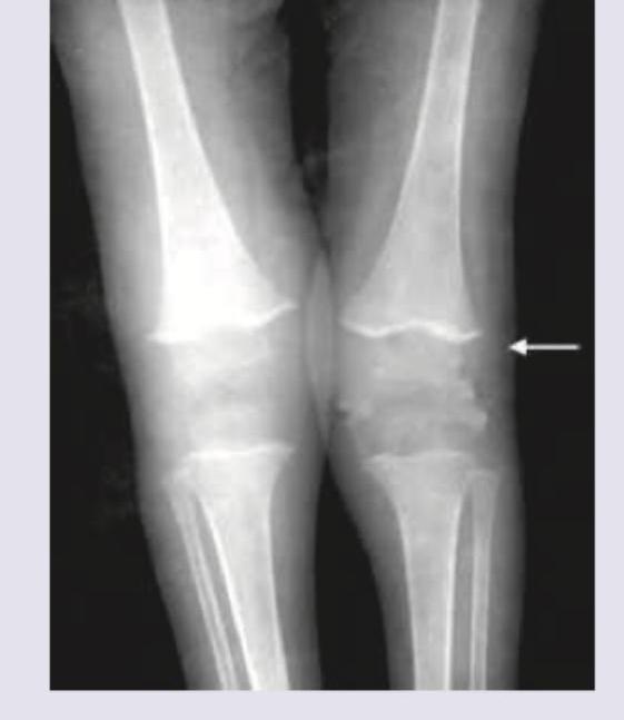
The X-ray of the patient shows?

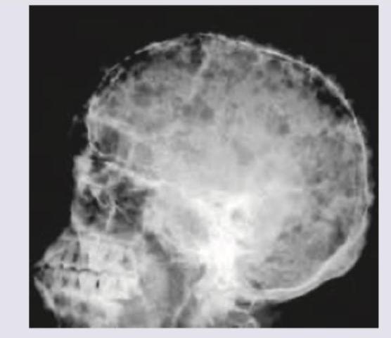
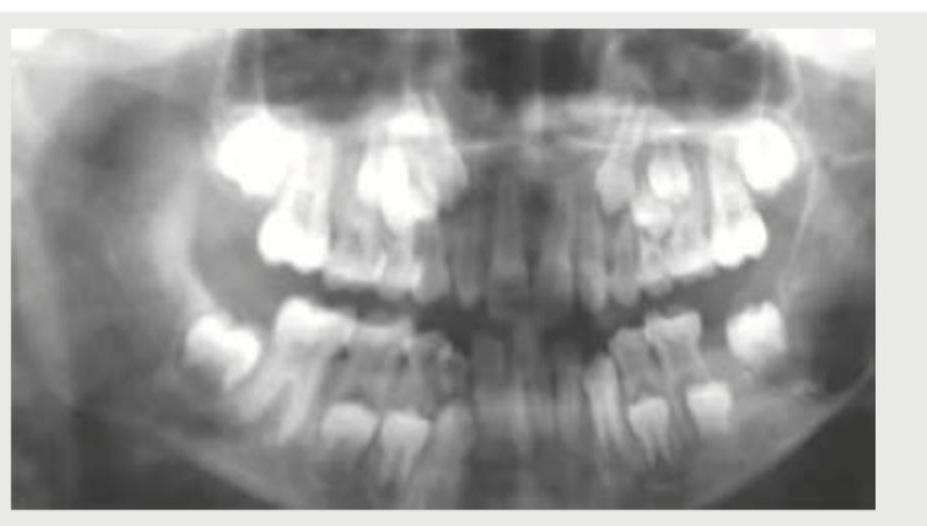
What is shown in this skull X-ray?

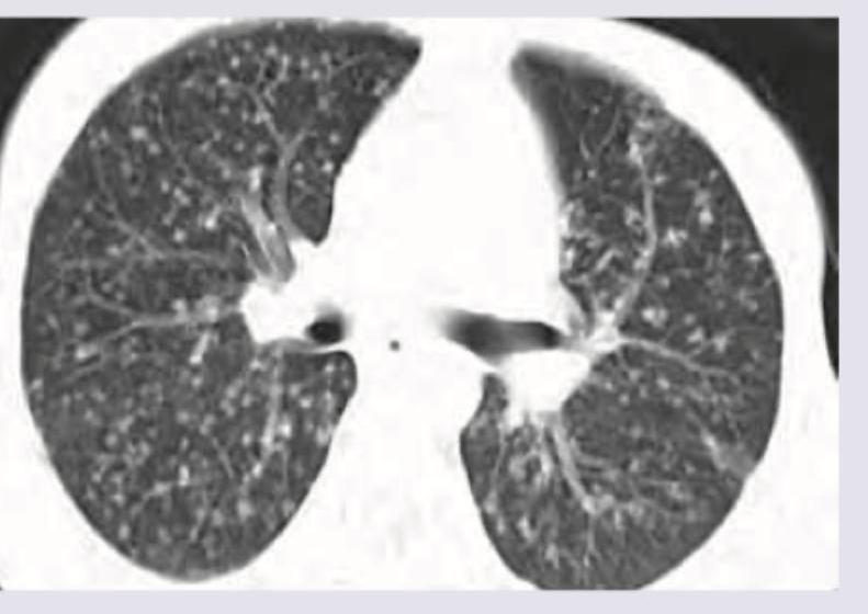
The following pattern in CT scan shown below can occur due to spread from:

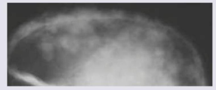
Which of the following will be the most probable diagnostic finding in the patient with X-Ray skull shown below?

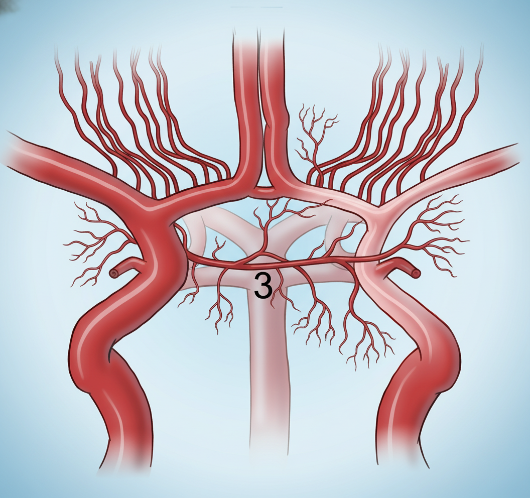
Which artery is labeled as '3' in the given angiogram?

The following image shows:

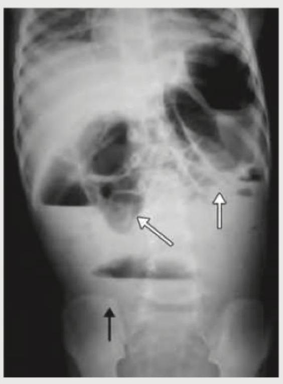
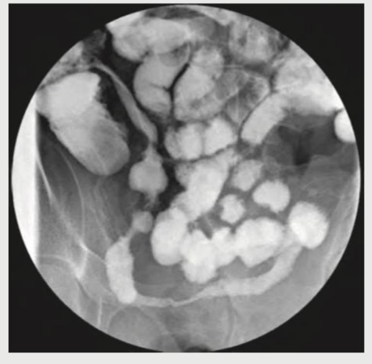
The following X-ray abdomen is diagnostic of:

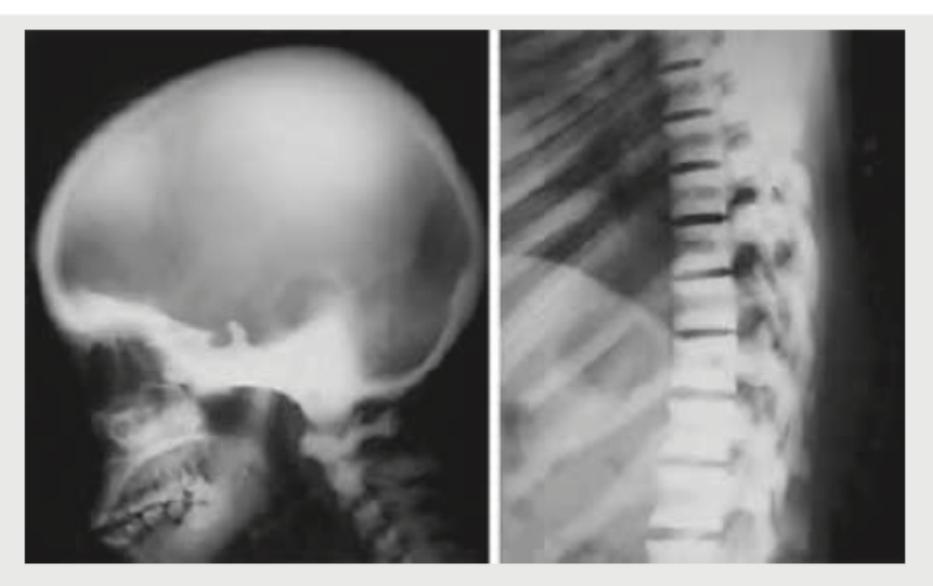
Comment on the diagnosis based on the skull and vertebral changes shown in the radiological image below:

What is the radiological sign demonstrated in the image provided?

Explanation: ***Pepper pot skull*** - The image exhibits a diffuse punctate or granular demineralization of the skull, resembling a "pepper pot." This appearance is characteristic of **hyperparathyroidism**, where excessive parathyroid hormone leads to increased osteoclastic activity and trabecular bone loss, leaving behind small, lucent areas. - This pattern results from widespread erosions of the **cortical bone** and trabeculae throughout the skull, creating a mottled, finely granular or punctate radiolucency. *Crew cut appearance* - A "crew cut" or "hair-on-end" appearance is typically seen in patients with severe, chronic **hemolytic anemias**, such as **thalassemia major** and **sickle cell anemia**. - It results from extreme marrow hyperplasia, causing expansion of the diploic space and thinning of the outer cortical bone, leading to spiculation perpendicular to the skull surface. *Punched out lytic lesions* - "Punched out lytic lesions" are well-demarcated, rounded radiolucencies without a sclerotic border, classically associated with **multiple myeloma**. - These lesions represent areas of osteolysis caused by plasma cell infiltration and are typically larger and more discrete than the fine granular appearance seen in a "pepper pot skull." *Widened sella turcica* - A widened sella turcica is usually indicative of an intrasellar mass, most commonly a **pituitary adenoma**, or can be due to hydrocephalus. - This finding focuses specifically on the sella region and does not describe the diffuse, granular demineralization seen throughout the entire skull vault in the image.
Explanation: ***Scurvy*** - The X-ray images display classical findings of scurvy, including a **dense metaphyseal line** (white line of Frankel), **lucent zone beneath the metaphysis** (Trümmerfeld zone), and **epiphyseal separation** due to capillary fragility. - The findings are particularly evident at the distal ends of the femur and proximal tibia, consistent with **subperiosteal hemorrhages** and impaired osteoid formation characteristic of **vitamin C deficiency**. *Rickets* - Rickets is characterized by **widening, cupping, and fraying of the metaphyses**, often accompanied by **growth plate widening** and bowing of long bones due to defective mineralization of bone matrix. - These features are not the predominant findings in the provided X-rays, which show distinct abnormalities related to hemorrhage and bone fragility. *Hemarthrosis* - Hemarthrosis refers to bleeding into a joint space, often characterized by **joint effusions** and possibly **bone erosions** if chronic, typically seen in conditions like hemophilia. - While subperiosteal hemorrhages are present in scurvy, the X-ray findings are broader than just intra-articular bleeding and include specific metaphyseal changes. *Sun burst appearance* - A "sunburst appearance" is a classic radiographic finding often associated with aggressive **bone tumors** like **osteosarcoma**, indicating **spiculated periosteal reaction** extending perpendicularly from the bone cortex. - This pattern is absent in the provided X-rays, which show signs of metabolic bone disease rather than a primary bone tumor.
Explanation: ***Pepper pot skull*** - The X-ray image displays a classic "pepper pot" appearance, characterized by diffuse **punctate lucencies** evenly distributed throughout the skull vault, resembling numerous small holes. - This finding is highly suggestive of **hyperparathyroidism**, where excessive parathyroid hormone leads to increased bone resorption, visible as demineralization and multiple small lytic lesions. *Artifacts* - **Artifacts** are extraneous marks or distortions on an image that do not represent true anatomical structures. - The observed pattern is a consistent, diffuse change in bone density, indicating a **pathological condition** rather than a technical error or imaging artifact. *Silver-beaten appearance* - A "silver-beaten" or **convolutional impression** refers to the undulating pattern on the inner table of the skull caused by increased **intracranial pressure** in children. - This appearance is characterized by broad, shallow indentations, which is distinct from the multiple small, sharply defined lucencies seen in pepper pot skull. *Hair-on-end appearance* - The **hair-on-end** or "crew-cut" appearance shows **perpendicular bony trabeculae** radiating outward from the skull vault, creating a spiculated pattern. - This finding is classically associated with **chronic hemolytic anemias** (thalassemia, sickle cell disease) due to marrow hyperplasia, not the diffuse lucencies seen here.
Explanation: ***Pulmonary artery*** - The CT scan demonstrates a **miliary pattern** with numerous small (1-3mm), discrete nodules showing **random distribution** throughout both lungs - This random distribution is the hallmark of **hematogenous spread via the pulmonary arteries** - When pathogens or tumor cells reach the pulmonary arteries (from right heart or systemic venous circulation), they disseminate uniformly throughout lung parenchyma - Classic examples include **miliary tuberculosis** and **hematogenous metastases** (thyroid, renal, melanoma) *Lymphatics* - Lymphatic spread produces a **perilymphatic distribution**, NOT a miliary/random pattern - Nodules appear along lymphatic pathways: peribronchovascular bundles, interlobular septa, and subpleural regions - Creates a characteristic beaded or nodular appearance along lymphatic routes - Seen in **sarcoidosis** and **lymphangitic carcinomatosis**, which do NOT present with miliary patterns *Bronchial vein* - Bronchial veins drain deoxygenated blood from bronchial walls to the systemic circulation (azygos/hemiazygos system) - This is NOT a recognized pathway for producing miliary lung nodules in standard radiology literature - Miliary patterns specifically refer to pulmonary arterial hematogenous dissemination *All of the above* - Incorrect: The miliary pattern is specifically caused by **pulmonary artery spread only** - The random distribution seen in the image is pathognomonic for hematogenous spread via pulmonary arteries, not lymphatic or bronchial venous routes
Explanation: ***CD1A positivity*** - The skull X-ray shows multiple punched-out lytic lesions without sclerotic borders, characteristic of **Langerhans Cell Histiocytosis (LCH)**. - **Langerhans cells** in LCH express surface markers like **CD1a** and S100 protein, which are diagnostic when detected via **immunohistochemistry on tissue biopsy**. - CD1a positivity is the **gold standard** for confirming LCH diagnosis. *Elevated SAP* - **Serum Alkaline Phosphatase (SAP)** is typically elevated in conditions with increased osteoblastic activity or liver/biliary disease. - LCH lesions are generally **lytic** and not associated with increased osteoblastic activity, thus SAP elevation is not a specific or common finding. *Bence Jones proteins* - **Bence Jones proteins** (monoclonal light chains) are found in the urine of patients with **Multiple Myeloma**, which can also cause lytic bone lesions. - However, the image shows more discrete, "punched-out" lesions typical of LCH, whereas myeloma lesions are often more diffuse or "salt-and-pepper" in appearance, and LCH does not secrete Bence Jones proteins. *Elevated HbA2* - **Elevated HbA2** is a characteristic finding in **beta-thalassemia trait**, a genetic blood disorder. - There is no clinical or radiological indication in the provided image or question to suggest a connection to thalassemia.
Explanation: ***Posterior cerebral artery*** - The arrow labeled '3' points to the **posterior cerebral artery (PCA)**, which originates from the **basilar artery** and supplies the posterior cerebral hemispheres. - This artery is a key component of the **Circle of Willis**, responsible for perfusing the **occipital lobes**, parts of the temporal lobes, and midbrain. - The PCA courses laterally around the midbrain after branching from the basilar artery terminus. *Anterior cerebral artery* - The ACA arises from the internal carotid artery and courses **medially** above the optic chiasm. - It supplies the **medial surfaces** of the frontal and parietal lobes, not the posterior structures shown at position 3. *Middle cerebral artery* - The MCA is the **largest branch** of the internal carotid artery and courses **laterally** through the Sylvian fissure. - It supplies the **lateral convexities** of the cerebral hemispheres, not the posterior circulation shown at position 3. *Basilar artery* - The basilar artery is formed by the **confluence of the vertebral arteries** and runs along the **ventral pons**. - It is a **midline structure** that gives rise to the PCAs, rather than being the lateral branch shown at position 3.
Explanation: ***Orthopantomogram*** - The image displays a panoramic view of the upper and lower jaws, including all teeth, the temporomandibular joints, and surrounding bone structures, which is characteristic of an **Orthopantomogram (OPG)** or panoramic radiograph. - This type of radiograph is commonly used in dentistry to assess the overall dental status, detect **impacted teeth**, and evaluate **bone pathology** across the entire oral cavity. *Water's view* - A **Water's view** (occipitomental view) is a radiographic projection typically used to visualize the maxilla, sinuses (especially the maxillary sinuses), and orbits. - It does not provide a panoramic view of the entire dental arches as seen in the image. *Invertogram* - An **invertogram** is a specialized lateral radiographic view of the abdomen and pelvis, particularly positioned to show the anus and rectum. - It is primarily used to assess **anorectal malformations** in infants and has no relation to dental imaging. *Gynogram* - The term **gynogram** is not a standard or recognized medical imaging technique. - Imaging studies related to gynecological conditions typically involve ultrasound, MRI, or CT scans of the pelvis, not a specific "gynogram" X-ray.
Explanation: ***Perforation peritonitis*** - The X-ray image shows widespread **free air under the diaphragm** and dilated loops of bowel with **air-fluid levels**, which are classic signs of **perforation peritonitis**. - **Free intraperitoneal air** (pneumoperitoneum) is a strong indicator of a perforated hollow viscus, leading to peritonitis. *Toxic megacolon* - Characterized by **extreme dilatation of the colon** (typically >6 cm in transverse colon) with loss of haustral markings on plain film, without free air. - While it can lead to perforation, the primary finding here is *free air*, not just colitis and colonic dilation. *Intestinal obstruction* - Identified by multiple **dilated bowel loops proximal to the obstruction**, with **air-fluid levels**, and a relative absence of gas distally. - While there are dilated loops and air-fluid levels, the most prominent finding of **free air under the diaphragm** is not typically associated with an uncomplicated obstruction. *Chronic mesenteric insufficiency* - This condition involves **reduced blood flow to the intestines**, leading to chronic abdominal pain, especially postprandial, and weight loss. - Its radiographic signs are often non-specific or involve bowel wall thickening and pneumatosis, not free air or acute findings of perforation.
Explanation: ***Thalassemia*** - The skull X-ray shows prominent **"hair-on-end" appearance** due to widening of the diploic space and thinning of the outer table, characteristic of chronic erythroid hyperplasia in thalassemia. - The vertebral bodies show a **"fishmouth" or "codfish" deformity**, a sign of severe osteopenia and bone marrow expansion commonly seen in thalassemia. *Rickets* - Rickets primarily affects the **growth plates** in children, leading to wide, irregular growth plates, cupping, and fraying of metaphyses, which are not seen here. - While it causes bone softening, the specific cranial and vertebral changes observed, like **"hair-on-end"** or "fishmouth" vertebrae, are not typical of rickets. *Osteopetrosis* - Osteopetrosis is characterized by **increased bone density** (sclerotic bones) due to defective osteoclast function, leading to abnormally thick and dense bones. - The radiographs show **osteopenia** and bone marrow expansion, which is the opposite of what is seen in osteopetrosis. *Hyperparathyroidism* - Hyperparathyroidism typically causes **subperiosteal bone resorption**, especially in the phalanges, and sometimes **brown tumors**. - While it can lead to generalized osteopenia, the specific "hair-on-end" skull and "fishmouth" vertebrae are not hallmark features; **salt-and-pepper skull** may be present, which differs from the findings here.
Explanation: ***Cobblestone appearance*** - The image shows a **barium study** of the small bowel, revealing an irregular, nodular mucosal pattern, which is characteristic of the **cobblestone appearance**. - This appearance is typically seen in **Crohn's disease** due to deep ulcerations crisscrossing with edematous, inflamed mucosa. - The combination of **linear ulcers** and **transverse fissures** separated by areas of edematous mucosa creates this distinctive pattern. *Belt sign* - The **belt sign** is a radiological finding on **CT imaging** associated with **blunt abdominal trauma**, particularly **seat belt injuries**. - It refers to bowel wall thickening, mesenteric hematoma, or abdominal wall bruising in the distribution of a seat belt. - This sign is not observed in the gastrointestinal barium study pattern shown in this image. *String sign* - The **string sign** is also seen in Crohn's disease but refers to a **severely narrowed or stenotic bowel lumen**, appearing as a thin, continuous string of barium. - This occurs due to chronic inflammation leading to fibrotic strictures. - While Crohn's can cause both findings, the image prominently displays the nodular, irregular mucosal pattern rather than a simple string-like narrowing. *Coffee bean sign* - The **coffee bean sign** is a classic radiographic finding in **sigmoid volvulus** on plain abdominal X-ray. - A massively dilated, gas-filled loop of bowel creates an appearance resembling a coffee bean. - This sign is distinct from the intestinal mucosal pattern observed in the provided barium study.
Radiographic Anatomy of Skull and Face
Practice Questions
Radiographic Anatomy of Spine
Practice Questions
Radiographic Anatomy of Chest
Practice Questions
Radiographic Anatomy of Abdomen
Practice Questions
Radiographic Anatomy of Extremities
Practice Questions
Cross-sectional Anatomy: Brain and Head
Practice Questions
Cross-sectional Anatomy: Neck
Practice Questions
Cross-sectional Anatomy: Thorax
Practice Questions
Cross-sectional Anatomy: Abdomen and Pelvis
Practice Questions
Vascular Anatomy
Practice Questions
Developmental Anatomy Variations
Practice Questions
Anatomic Landmarks for Interventional Procedures
Practice Questions
Get full access to all questions, explanations, and performance tracking.
Start For Free